Introduction
Lecanora Ach. s. lat. is a group that comprises c. 1000 species of crustose lichens with a lecanorine apothecial margin, unicellular colourless ascospores in mostly 8-spored asci and green algae as photobionts (Zahlbruckner Reference Zahlbruckner, Engler and Prantl1926). Eigler (Reference Eigler1969) distinguished several groups within the genus, based on chemical and morphological characters, some of which were later segregated as genera (e.g. Trapelia M. Choisy (Hertel Reference Hertel1969), Bryonora Poelt, (Poelt Reference Poelt1983), Myriolecis Clem., Protoparmeliopsis M. Choisy (Zhao et al. Reference Zhao, Leavitt, Zhao, Zhang, Arup, Grube, Pérez-Ortega, Printzen, Śliwa and Kraichak2016) and many others) while other groups (e.g. in Lecanoropsis M. Choisy) were not. Some other groups were only recently recognized as genera based on molecular data, rather than through morphological characters (e.g. Palicella Rodr.-Flakus & Printzen (Rodriguez-Flakus & Printzen Reference Rodriguez-Flakus and Printzen2014)). Some species within Lecanora s. lat. are indeed difficult to assign to one of the established groups because their phenotypic characters do not exactly match the current circumscription for these groups (see e.g. the discussion in Tripp et al. (Reference Tripp, Morse, Keepers, Stewart, Pogoda, White, Hoffman, Kane and McCain2019) regarding the phenotypic circumscription of the Lecanora dispersa-group).
One of the distinctive morpho-groups for which molecular data is currently only sparsely available is the Lecanora saligna-group, first recognized at the genus level as Lecanoropsis M. Choisy (Choisy Reference Choisy1949) and coinciding more or less with ‘group 3’ of Printzen (Reference Printzen2001). Species in the Lecanora saligna-group are characterized by a corticolous, esorediate and mostly endosubstratal (or effuse episubstratal) thallus, the production of usnic and/or isousnic acid, apothecia with a corticate, lecanorine margin that sometimes is only visible at the base, ellipsoid simple ascospores and bacilliform or reniform conidia. Among the North American species treated by Printzen (Reference Printzen2001), L. albellula (Nyl.) Th. Fr., L. coniferarum Printzen, L. densa (Śliwa & Wetmore) Printzen, L. latens Printzen, L. laxa (Śliwa & Wetmore) Printzen, L. mughicola Nyl., L. saligna (Schrad.) Zahlbr., L. subintricata (Nyl.) Th. Fr. and L. varia (Hoffm.) Ach. were assigned to the group. However, L. densa, L. laxa and L. varia were later placed in the Lecanora varia-group by Pérez-Ortega et al. (Reference Pérez-Ortega, Spribille, Palice, Elix and Printzen2010), based on molecular phylogenetic reconstructions. The same authors discussed L. anopta Nyl. as sharing some characters with members of the L. saligna-group, but its phylogenetic status within this group was yet to be clarified. Van den Boom & Brand (Reference van den Boom and Brand2008) further included L. quercicola Coppins & P. James, L. sarcopidoides (A. Massal.) Hedl. and L. subravida Nyl. in the L. saligna-group and described four new species (L. calabrica M. Brand & van den Boom, L. coppinsii M. Brand & van den Boom, L. pseudosarcopidoides M. Brand & van den Boom and L. subsaligna M. Brand & van den Boom) and one new variety (L. albellula var. macroconidiata M. Brand & van den Boom), all of them containing usnic and/or isosunic acids, plus other secondary metabolites associated with the usnic acid metabolic pathway. Furthermore, conidial and pycnidial characters were found by these authors to be of great importance in distinguishing species within the L. saligna-group, albeit without molecular support and based on a relatively small number of samples.
Zhao et al. (Reference Zhao, Leavitt, Zhao, Zhang, Arup, Grube, Pérez-Ortega, Printzen, Śliwa and Kraichak2016) published the first comprehensive phylogeny of Lecanora s. lat., based on DNA sequences from six loci, which provided further evidence for the separation of the genera Myriolecis Clem. (or Polyozosia A. Massal., see Kondratyuk et al. (Reference Kondratyuk, Lőkös, Jang, Hur and Farkas2019), formerly the L. dispersa-group), Protoparmeliopsis M. Choisy, (formerly the L. muralis-group) and Rhizoplaca Zopf. Lecanora saligna and L. anopta were the only species of the L. saligna-group included in this study but, due to missing data, were only part of a data set based on two gene regions. In the analyses by Zhao et al. (Reference Zhao, Leavitt, Zhao, Zhang, Arup, Grube, Pérez-Ortega, Printzen, Śliwa and Kraichak2016) they appeared monophyletic but poorly supported. This raised the question of whether species assigned to the group based on morphological and chemical characters really belong there. There are further problems in the species delimitation and nomenclature of the group. For example, van den Boom & Brand (Reference van den Boom and Brand2008) described L. pseudosarcopidoides as a new species. However, its morphology and chemistry seem to match L. anopta, a taxon not treated by these authors but already shown to be rather variable by Hedlund (Reference Hedlund1892; see also Pérez-Ortega et al. (Reference Pérez-Ortega, Spribille, Palice, Elix and Printzen2010)). Furthermore, whether the ‘European’ species reported from North America by Printzen (Reference Printzen2001) are conspecific with their Old World namesakes or whether they represent independent species, as in the case of L. densa, L. laxa and L. varia (Śliwa & Wetmore Reference Śliwa and Wetmore2000; Printzen Reference Printzen2001) has not yet been explored.
This study sheds light on the L. saligna-group using an integrative approach, considering classical taxonomic methods and inferences from phylogenetic reconstructions. By including as many species as possible and multiple collections per species, we assess the monophyly of the group and the phylogenetic relationships within it. We also examine whether morphological characters coincide with putative species-level clades inferred from molecular sequence data. Finally, we explore whether the reported disjunctions between Europe and North America are real, or simply artifacts caused by unclear species delimitations based solely on morphological characters and a lack of molecular data.
Materials and Methods
Taxon sampling, morphological and chemical data
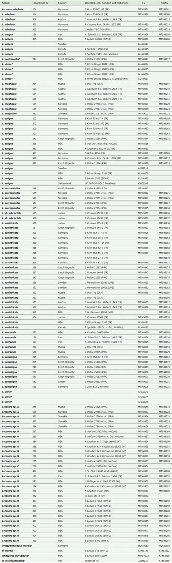
Ninety recently collected specimens belonging to the Lecanora saligna-group, and closely related groups, were either collected during field trips by the authors or obtained on loan from the following herbaria: BRY–C, FR, GLM, H, MIN, NY, PRA, UPS, hb. Cezanne–Eichler, hb. McCune and hb. Spribille (Table 1). We tried to study samples of all species included in the L. saligna-group by van den Boom & Brand (Reference van den Boom and Brand2008) and Printzen (Reference Printzen2001), but no specimens of L. calabrica, L. coniferarum, L. coppinsii and L. quercicola were available for this study. Based on the phylogenetic reconstructions of Zhao et al. (Reference Zhao, Leavitt, Zhao, Zhang, Arup, Grube, Pérez-Ortega, Printzen, Śliwa and Kraichak2016) and Pérez-Ortega et al. (Reference Pérez-Ortega, Spribille, Palice, Elix and Printzen2010), and preliminary analyses using a wide range of taxa from Lecanoraceae species from the L. varia-group, Protoparmeliopsis and Rhizoplaca were selected as possible outgroup taxa.
List of Lecanora specimens used in the phylogenetic analysis, with voucher information and GenBank Accession numbers. Taxa selected as outgroup are marked with an asterisk (*). ‘Lecanomics ID’ refers to the online database https://lecanomics.org.

External morphological characters were studied using a Euromex NZ 1902-P stereomicroscope. Anatomical characters were investigated using a Leitz DIAPLAN microscope on sections of thalli, apothecia and pycnidia prepared with a Zeiss HYRAX KS 34 cryotome and mounted in water, Lugol's solution and lactophenol cotton blue. Measurements of morphometric characters are given as: (lowest single measurement–) lowest mean value–highest mean value (–highest single measurement). Secondary metabolites were analyzed by thin-layer chromatography in solvents A, B’ and C using a CAMAG horizontal developing chamber and Macherey-Nagel 10 × 10 cm precoated TLC plates SIL G-25 UV254 (Culberson & Kristinsson Reference Culberson and Kristinsson1970; Arup et al. Reference Arup, Ekman, Lindblom and Mattson1993). Substances were identified using Orange et al. (Reference Orange, James and White2001) and Schumm & Elix (Reference Schumm and Elix2015).
Molecular methods
For all 90 specimens of species previously assigned to the Lecanora saligna-group, DNA sequences were generated for two loci: the nuclear ribosomal internal transcribed spacer region (ITS1, 5.8S and ITS2) and the mitochondrial small subunit (mtSSU) of ribosomal RNA.
For specimens collected for this study, DNA was extracted from apothecia using the DNeasy Plant Mini Kit (Qiagen) or the GF-1 Plant DNA Extraction Kit (GeneOn), following the manufacturers’ protocols. Remnants of substratum were cleaned from apothecia under a dissecting microscope prior to DNA extraction. PCR was performed for the ITS locus using primers ITS1F: 5′–CTTGTTCATTTAGAGGAAGTAA–3′ (Gardes & Bruns Reference Gardes and Bruns1993) and ITS4a: 5′–CGCCGTTACTGGGGCAATCCCTG–3′ (Larena et al. Reference Larena, Salazar, Gonzalez, Julian and Rubio1999), and for the mtSSU locus using primers mrSSU1: 5′–AGCAGTGAGGAATATTGGTC–3′ and mrSSU3r: 5′–ATGTGGCACGTCTATAGCCC–3′ (Zoller et al. Reference Zoller, Scheidegger and Sperisen1999). All PCR products were amplified using PCR Ready-To-Go beads (GE Healthcare) in 25 μl reactions containing 5 μl DNA, 18 μl of double distilled H2O and 1 μl of each primer. For both markers, cycling conditions were as follows: 1) denaturation for 5 min at 94 °C; 2) four cycles of 30 s at 94 °C, 30 s at 54 °C and 60 s at 72 °C; 3) 30 cycles of 30 s at 94 °C, 30 s at 48 °C and 60 s at 72 °C; 4) a final extension step for 10 min at 72 °C. PCR products were kept at 4 °C until processed further. Success of PCR reactions was assessed by gel electrophoresis on 1% agarose gels containing SYBR Green dye. Successful PCR reactions were then sequenced by Macrogen Europe B.V. (Amsterdam, the Netherlands). Forward and reverse sequence reads were assembled in Geneious Prime 2019.2.1 (https://www.geneious.com) and edited manually.
Alignments and phylogenetic analysis
Twenty GenBank sequences were selected and added to the data matrix as outgroup taxa: Rhizoplaca melanophthalma (DC.) Leuckert, R. chrysoleuca (Sm.) Zopf and Protoparmeliopsis muralis (Schreb.) M. Choisy. In addition, seven specimens from the L. varia-group were added: Lecanora varia (Hoffm.) Ach. and L. densa (Śliwa & Wetm.) Printzen. Sequences of Lecanora conizaeoides Nyl. ex Cromb., which also belongs to the L. varia-group, were newly generated. These potential outgroups were selected from species that were shown to be closely related to the L. saligna-group by Zhao et al. (Reference Zhao, Leavitt, Zhao, Zhang, Arup, Grube, Pérez-Ortega, Printzen, Śliwa and Kraichak2016).
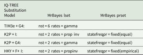
Consensus sequences for each locus were aligned through GUIDANCE2 (Landan & Graur Reference Landan and Graur2008; Penn et al. Reference Penn, Privman, Ashkenazy, Landan, Graur and Pupko2010; Sela et al. Reference Sela, Ashkenazy, Katoh and Pupko2015), removing unreliably aligned positions with a score below 0.999. Additionally, pairwise distance values were calculated using MEGA-X (Kumar et al. Reference Kumar, Stecher, Li, Knyaz and Tamura2018) (Table 2; Supplementary Material Fig. S1, available online). Maximum likelihood (ML) single-locus trees were reconstructed with W-IQ-TREE (Trifinopoulos et al. Reference Trifinopoulos, Nguyen, von Haeseler and Minh2016) in order to assess incongruencies, using the default settings of an ultrafast bootstrap analysis, 1000 bootstrap repetitions and automatic substitution model selection. Three gene partitions, ITS1, 5.8S and ITS2, were specified for the ITS region; the mtSSU was treated as a single gene partition. After assessing incongruencies, matrices were concatenated, the data matrix was partitioned and a new unrooted ML tree was created by W-IQ-TREE using the default settings of ultrafast bootstrap analysis, 1000 bootstrap repetitions and automated substitution model selection. IQ-TREE (http://www.iqtree.org/) selected the following substitution models: TIM3e + G4 for ITS1, K2P + I for 5.8S, K2P + G4 for ITS2 and HKY + F + I for mtSSU.
Mean pairwise distance values for the taxa of the clades belonging to the undescribed Lecanora species. A complete distance table of specimens of the undescribed Lecanora species is given in Supplementary Material Fig. S1 (available online).

Bayesian tree inference was carried out using MrBayes 3.2 (Ronquist et al. Reference Ronquist, Teslenko, van der Mark, Ayres, Darling, Höhna, Larget, Liu, Suchard and Huelsenbeck2012). Substitution models obtained in W-IQ-TREE were translated into the following models supported by MrBayes: GTR, nset = 6; HKY, nset = 2; K2P, nset = 2; statefreqpr = fixed (Table 3). Rhizoplaca melanophthalma was chosen as outgroup. Parameters of substitution models were unlinked between data partitions. Two independent runs started with randomly generated trees were set to run for 40 000 000 generations each, with four Markov chains incrementally heated by a factor of 0.15. To infer convergence of the Markov chains, the average standard deviation of bipartition frequencies among runs was calculated every 200 generations, discarding the first 50% of the trees sampled as burn-in and including only those bipartitions with a frequency of at least 30%. The analyses were stopped when the standard deviation between split frequencies dropped below 0.01, indicating that stationarity (statistical plateau) had been reached. The resulting trees were generated in TreeGraph2 (Stöver & Müller Reference Stöver and Müller2010). Bayesian posterior probabilities ≥ 0.95 were added to the ML tree.
IQ-TREE substitution models (http://www.iqtree.org/), and likelihood parameter and prior settings for MrBayes 3.2 (Ronquist et al. Reference Ronquist, Teslenko, van der Mark, Ayres, Darling, Höhna, Larget, Liu, Suchard and Huelsenbeck2012) used in the phylogenetic analysis of Lecanora species.

DNA sequences were submitted to GenBank (Table 1). Complete label data, together with specimen photographs and micrographs, images of TLC plates and sequence chromatograms, are available on the Lecanomics project homepage (https://lecanomics.org).
The project ‘Lecanomics’ is funded by the German Science Foundation (DFG) and seeks to better integrate field exploration by specialists and amateurs with molecular genetic studies of species delimitation and phylogeny. Contributors communicate through a web portal that serves to provide collectors with genetic data and experts with easy access to a wide range of samples. The project focuses on the large genus Lecanora s. lat.
Results
Phylogeny
In total, 90 specimens were included in the phylogenetic analysis, plus 20 sequences from GenBank. A total of 180 new sequences were generated, and the alignment had a length of 802 characters (415 for ITS and 387 for mtSSU) containing 83 parsimony-informative sites, 42 sites with a single substitution, and 677 constant sites. Supported incongruencies between single locus trees were not detected.
Bayesian inference analysis stopped at 3 100 000 generations after the convergence diagnostic dropped below 0.01 average standard deviation for split frequencies.
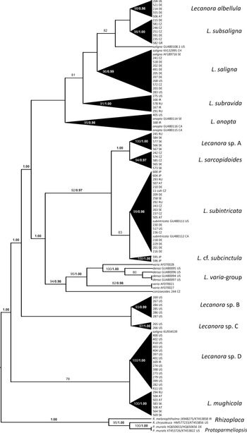
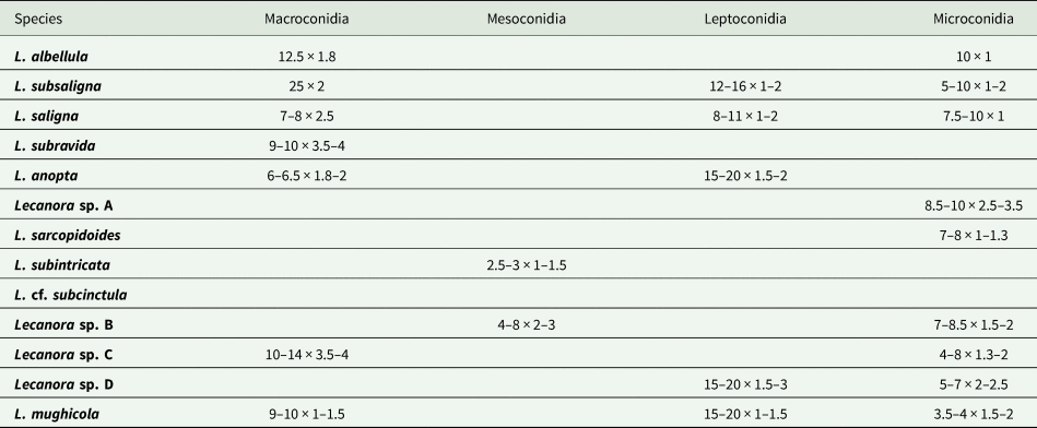
The Lecanora saligna-group appears to be paraphyletic, consisting of four separate clades (Fig. 1; Supplementary Material Fig. S2, available online) with the L. varia-group nested within. The ‘core’ clade was supported only in the ML analysis and contains what we currently recognize as L. albellula, L. subsaligna, L. saligna and L. subravida. Remarkably, all species known to produce macroconidia sensu van den Boom & Brand (Reference van den Boom and Brand2008) are found in this clade (Table 4; species order is according to their relatedness in Fig. 1). Lecanora anopta appears as sister to this ‘core’ clade but the joining node is not supported. Therefore, L. anopta belongs within the L. saligna-group but its true position and relationship within the group are as yet unclear. The sister group to these clades contains Lecanora sp. A, L. sarcopidoides, L. subintricata, L. cf. subcinctula and the L. varia-group. Two further clades appear paraphyletic at the base of these two groups, one comprising the undescribed North American taxa Lecanora sp. B and Lecanora sp. C, the other with Lecanora sp. D and L. mughicola. Pairwise distance values (Table 2) of the undescribed North American taxa showed low genetic distance intraspecifically, but distance values 10 times higher interspecifically. Rhizoplaca chrysoleuca, R. melanophthalma and Protoparmeliopsis muralis form the basal outgroup. The terminal clades are generally well supported but, with the exception of the L. subintricata clade and the L. varia-group, the internal groupings and the backbone of the tree do not receive high support. Most clades also seem to be geographically restricted, with a few exceptions of ‘alien’ specimens unexpectedly emerging within clades.
Maximum likelihood (ML) tree of the concatenated 2-loci data matrix for species of Lecanora. Infraspecific branches are collapsed so that black triangles represent ‘species’. Maximum likelihood bootstrap support (BS) and Bayesian posterior probabilities (PP) are given above the nodes. Support values < 75% (BS) or 0.95 (PP) were considered to be insufficient and are not displayed. At branch tips, specimens are represented by a 3 digit ‘Lecanomics ID’ (https://lecanomics.org) and a two letter country code (ISO 3166-1 alpha-2), or other identifying information (See Table 1).

Mean values of conidial measurements of Lecanora species. Conidia were not found associated with specimens of L. cf. subcinctula. Species are ordered in the table according to their relatedness in the phylogenetic tree (Fig. 1; Supplementary Material Fig. S2, available online).

The species
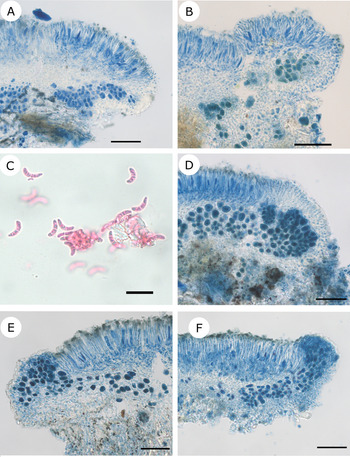
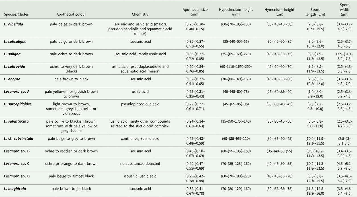
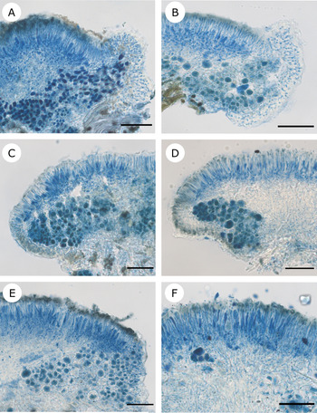
Lecanora albellula (Fig. 2A & B). Members of this clade are characterized by the small size of the apothecia, one of the smallest within the L. saligna-group (Table 5; species order is according to their relatedness in Fig. 1). The amphithecial cortex appears to be more or less uniform, commonly somewhat thinner laterally and becoming wider basally. All specimens contain isousnic acid. Specimens from Central Europe form a clade separate from North American samples, indicating that the name L. albellula was misapplied by Printzen (Reference Printzen2001). North American specimens identified as L. albellula were preliminarily named Lecanora sp. D (see Discussion below). The specimens studied here occurred on bark and wood, in mixed montane forests of Picea and Fagus from (sub)montane to subalpine habitats above 900 m a.s.l.
Lecanora albellula (A & B), L. subsaligna (C & D) and L. saligna (North American collections) (E & F). A, Germany, Bavaria, S. Kern TS3–14–5 (FR; L214), apothecial section in lactophenol cotton blue (LCB). B, Germany, Bavaria, L. Weber T3–27–10 (FR; L555), apothecial section in LCB. C, Czech Republic, Šumava Mts, Z. Palice 19576 (FR; L246), macroconidia in erythrosine. D, Czech Republic, Jizerské hory Mts, Z. Palice 21276 (PRA; L235), apothecial section in LCB under differential interference contrast (DIC). E, USA, California, K. Knudsen 13502, N. Pietrasiak & E. Hessom (NY–01220741; L283), apothecial section in LCB. F, USA, Oregon, B. McCune 34716 (hb. McCune; L268), apothecial section in LCB. Scales: A, B, D–F = 50 μm; C = 10 μm.

Summary of key traits used for the phenotypic characterization of the Lecanora saligna-group clades. Elevational range is based only on specimens used in this study. Measurement format is as follows: (lowest single measurement–) lowest mean value–highest mean value (–highest single measurement). Species are ordered in the table according to their relatedness in the phylogenetic tree (Fig. 1; Supplementary Material Fig. S2, available online).

Lecanora subsaligna (Fig. 2C & D). Recently described by van den Boom & Brand (Reference van den Boom and Brand2008), this species commonly has pale apothecial discs usually (but not always) lacking a pruina and having smooth apothecial margins. It has a thin amphithecial cortex, of uniform width. Lecanora subsaligna and L. albellula share several morphological characters and ecological preferences (often growing intermixed on twigs), rendering them very difficult to differentiate. Apothecia in L. albellula can have strongly convex discs, whereas in L. subsaligna they are only weakly convex. Specimens of L. subsaligna were the only ones with U-shaped macroconidia of c. 25 × 2 μm (Table 4). Sample L246 appeared to produce crescent-shaped, 1–3-septate falcate conidia, 8.2–9.5 × 2.5 μm (Fig. 2C) not previously reported from L. subsaligna, but also observed in one sample of L. albellula (L204). This either suggests that the length and shape of macroconidia of L. albellula are not useful diagnostic characters, or indicates the presence of a mixed sample and requires further study of material. Lecanora subsaligna produces only isousnic acid, whereas L. albellula can produce isousnic acid, usnic acid, and rarely traces of squamatic acid. Most of the specimens of L. subsaligna included in our data set originate from Central Europe (Bavaria and the Czech Republic), sharing a habitat preference for mixed forests at middle and montane elevations, and a substratum preference of wood and bark. However, one sample from Greece collected at sea level also nested within the L. subsaligna-clade.
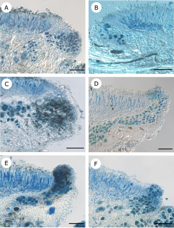
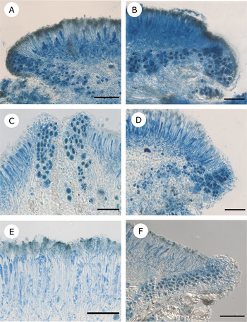
Lecanora saligna (Figs 2E & F, 3A & B). This species differs from L. albellula and L. subsaligna by having slightly larger, darker apothecial discs, a higher hypothecium (Table 5), an amphithecial cortex that gradually becomes thinner (sometimes almost disappearing) towards the hymenium and reniform, aseptate macroconidia, 7–8 × 2–5 μm (Table 4). Apothecia typically have ochre to reddish brown discs and a whitish, subrugose, sometimes verrucose margin. Most collections contained only isousnic acid but we examined one specimen from the Czech Republic (241) that may contain neousnic acid, a distinctive compound so far reported only for L. saligna and L. quercicola (van den Boom & Brand Reference van den Boom and Brand2008). This clade contains specimens mainly from Central Europe collected on bark and wood within mixed montane forests of Picea abies and Fagus sylvatica.
Lecanora saligna (European collections) (A & B), L. subravida (C–E) and L. anopta (F). A, Czech Republic, Šumava Mts, Z. Palice 21284 (PRA; L241), apothecial section in LCB. B, Germany, Bavaria, S. Kern TS3–17–4 (FR; L202), apothecial section in LCB (DIC). C, USA, California, K. Knudsen 16879 (NY–02203860; L275), apothecial section in LCB (DIC). D, Russia, Adygea, Z. Palice 22535 (PRA; L578), apothecial section in LCB (DIC). E, Iran, Mazandaran Province, M. Sohrabi & C. Printzen 14917 (FR; L166), apothecial section in LCB. F, USA, Utah, S. Leavitt 18.081 (BRY–C; L405), apothecial section in LCB. Scales: A–F = 50 μm.

Lecanora subravida (Fig. 3C–E) can easily be distinguished by its large, ochre to dark brown or black apothecial discs, a subrugose to verrucose, persistent pale margin, and a conspicuous algal layer surrounded by an amphithecial cortex that is extremely thin near the hymenium, becoming much wider at the base (in some sections it can resemble that of L. varia). Lecanora subravida also has a higher hymenium than L. albellula, as well as wider ascospores than L. albellula and L. subsaligna (Table 5). Its major secondary compound is usnic acid; isousnic, pseudoplacodiolic and squamatic acids were detected as minor/trace compounds. Our samples of L. subravida were collected on wood in montane and subalpine conifer and broad-leaved forests in the Russian Caucasus, the Alborz Mountains of northern Iran, and in a mixed conifer forest with Quercus in the San Bernardino Mountains of California, USA. It is newly reported here from Iran.
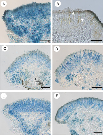
Lecanora anopta (Figs 3F, 4A & B) has several characters that distinguish the clade from the other species in the L. saligna ‘core’ group, mainly the presence of the pigment Cinereorufa-green (Meyer & Printzen Reference Meyer and Printzen2000) in the epihymenium, occasionally streaking into the hymenium, and orange-brown, sclerotized spores (or ‘guttulae’; Hedlund Reference Hedlund1892; Pérez-Ortega et al. Reference Pérez-Ortega, Spribille, Palice, Elix and Printzen2010) within the hymenium. Due to its often convex and subglobose apothecial discs with a biatorine appearance, the species may be confused with poorly developed or damaged L. saligna, L. sarcopidoides or L. cadubriae (see also the description of L. pseudosarcopidoides in van den Boom & Brand (Reference van den Boom and Brand2008)). Similar to other members of the L. saligna clade, we found macroconidia of (5.5–)6–6.5(–7.5) × (1.5–)1.8–2(–2.1) μm (Table 4) with a pseudoseptum (disappearing in KOH) formed in relatively large pycnidia sometimes exceeding 0.1 mm. These conidia differ slightly from the mesoconidia of 7–9 × 1 μm reported by Pérez-Ortega et al. (Reference Pérez-Ortega, Spribille, Palice, Elix and Printzen2010) but conform to the dimensions given by Hedlund (Reference Hedlund1892). In the literature, L. anopta has been reported from Fennoscandia, Central Europe, the USA and Canada; specimens used in this study come from a subalpine oak forest in the Alborz Mountains of northern Iran, and from an aspen/spruce forest on the south-eastern corner of Utah, USA, both growing on wood. This is the first report for Iran.
Lecanora anopta (A & B), Lecanora sp. A (C & D) and L. sarcopidoides (E & F). A, Iran, Golestan Province, M. Sohrabi & C. Printzen 15029 (FR; L168), apothecial section in LCB. B, same collection in water, note orange-brown patches in hymenium (arrows). C, Slovakia, Muránska planina, D. Blanár, A. Guttová, J. Halda & Z. Palice 27840 (PRA; L584), apothecial section in LCB. D, Slovakia, Muránska planina, D. Blanár, A. Guttová, J. Halda & Z. Palice 27765 (PRA; L566), apothecial section in LCB. E, Czech Republic, Šumava Mts, Z. Palice 27894 (PRA; L574), apothecial section in LCB. F, Slovakia, Muránska planina, A. Guttová, J. Halda & Z. Palice 27751 (PRA; L573), apothecial section in LCB. Scales: A–F = 50 μm.

Lecanora sp. A (Fig. 4C & D). Corticolous specimens of this clade from Slovakia were originally identified as L. cf. latens sensu Printzen (Reference Printzen2001) due to their small inconspicuous apothecia (Table 5). Better developed material from wood in the Caucasus was recently published as L. aff. anopta (Urbanavichus et al. Reference Urbanavichus, Vondrák, Urbanavichene, Palice and Malíček2020). This material is similar to pale morphs of L. anopta, including the presence of orange-brown guttulae (see above), but differs in having narrower ascospores and by containing usnic acid. Both corticolous and lignicolous material of this species have relatively pale, distinctly pruinose apothecia that become slightly convex and may appear biatorine, with a clearly delimited, narrow amphithecial cortex in sections. The thallus is more or less endosubstratal and usnic acid is present as a major metabolite. An unidentified trace compound, apparently a satellite of usnic acid, was detected in the Caucasian material. The corticolous specimens were collected in a relatively dry, well-lit forest of Quercus cerris in Muránska planina National Park in Slovakia, previously affected by fire (c. 600 m a.s.l.). Material from the Russian Caucasus grew on wood of a lying Abies trunk at c. 1800 m a.s.l. in a humid, upper montane forest dominated by Abies nordmanniana with intermingled Fagus orientalis. The samples are very similar to L. latens isotypes (FR) which, however, produce zeorin in addition to usnic acid and appear to have a very different ecology (the coast of California, USA).
The apothecia of L. sarcopidoides samples (Fig. 4E & F) used in this study have a characteristic colour ranging from bluish violet to (pale) pinkish brown (often a mixture of both), commonly with a coarse white pruina, and a very thin to almost disappearing margin, which is often concolorous with the thallus (if present). Its amphithecial cortex is remarkably thick but even, composed of hyphae surrounded by a coarse gelatinous layer. The species is also well characterized by the production of pseudoplacodiolic acid as the major compound. Usnic acid was reported as a minor compound by van den Boom & Brand (Reference van den Boom and Brand2008) but was not detected here. Instead, placodiolic acid was detected as a trace admixture in two samples from PRA (Herbarium of the Institute of Botany of the Czech Academy of Sciences) not included in our phylogeny. Lecanora sarcopidoides might be misidentified as L. subintricata, since both may grow intermingled on the same piece of substratum. Both can sometimes have a similarly coloured disc (pale brown to brown), especially in specimens devoid of obvious pruina. Both species also form a heavily gelatinized thalline cortex, made of anticlinally arranged hyphae with lumina of c. 1.5–2 μm. However, the apothecial discs of L. subintricata are more regularly rounded with a well-developed yellowish margin, whereas the margin of L. sarcopidoides tends to be white rather than yellowish and is often poorly developed or disappearing. Lecanora subintricata also has slightly smaller and more rounded ascospores. If not overmature, apothecia of L. sarcopidoides have a coarse whitish pruina whereas in L. subintricata the pruina is either finely granulose or entirely lacking. The specimens studied by us were collected on wood or bark in well-lit, boggy upper montane/boreal forests of Picea abies and Pinus sylvestris in the Czech Republic (Šumava Mts). Single specimens originate from the wood of a fence (Šumava Mts) and from close-to-native well-lit forest dominated by Quercus petraea agg. (Q. dalechampii/polycarpa) in Slovakia (western Carpathians).
Lecanora subintricata (Fig. 5A & B). Apothecia of this species are well rounded, with a flat to weakly convex dark brown, dark grey to yellowish grey disc, with a very conspicuous yellow margin, due to the presence of usnic acid. Extreme phenotypes of L. subintricata, however, may possess a thin and discontinuous margin, and a weakly convex ochre to brownish disc, which might lead to confusion with L. sarcopidoides (see above). Its amphithecial cortex is similar to that of L. sarcopidoides. Specimens of this clade were collected all over the Northern Hemisphere, in Central and Northern Europe, Russia, Japan and North America, on wood and bark.
Lecanora subintricata (A & B), L. cf. subcinctula (C & D) and Lecanora sp. B (E & F). A, Sweden, Dalarna, J. Hermansson 19606 (UPS–L–767688; L263), apothecial section in LCB. B, Austria, Salzburg, C. Ivanovich LUN19 & L. Weber (FR; L505), apothecial section in LCB. C, Japan, Tochigi Prefecture, C. Printzen 15192 (FR; L595), apothecial section in LCB. D, Japan, Tochigi Prefecture, C. Printzen 15286 (FR; L596), apothecial section in LCB. E, USA, Montana, B. McCune 37125 (hb. McCune; L267), apothecial section in LCB (DIC). F, USA, California, K. Knudsen 16266 & J. Kocourková (NY–02100405; L287), apothecial section in LCB, hymenium with pigmented paraphysis tips. Scales: A–F = 50 μm.

Lecanora cf. subcinctula (Fig. 5C & D) forms a sister clade to L. subintricata. The two samples have rounded pale beige to greyish to brown, epruinose apothecial discs with a sometimes weakly glossy surface. The apothecial margins are more or less concolorous with the disc but almost disappearing. Its amphithecial cortex is remarkably even and uniform throughout, showing brown pigmentation all the way to the base. We also observed this character in L. anopta, L. sarcopidoides and Lecanora sp. C. Although externally resembling L. subintricata, L. cf. subcinctula can be distinguished from its sister clade by its longer and narrower ascospores (Table 5) and the complete lack of pruina. The species assignment of our two samples remains somewhat uncertain because we have not yet seen type material to confirm their identity. The two specimens from Japan were collected on wood in disturbed, anthropogenic environments, at c. 1400–1600 m a.s.l.
Material of Lecanora sp. B (Fig. 5E & F) appears to conform to L. saligna sensu Printzen (Reference Printzen2001). Externally, specimens resemble the largely European L. saligna. However, Lecanora sp. B can be distinguished from L. saligna by its narrower ascospores (Table 5). Both species share isousnic acid as the major compound; however, neousnic acid is occasionally present in L. saligna but has never been detected in Lecanora sp. B. The samples of Lecanora sp. B included in this study originate from California, Montana and Oregon, USA, and were almost all collected on wood in forests of Pinus, Quercus and Juniperus. One sample occurred on a fence post in an open, heavily grazed pasture.
Lecanora sp. C (Fig. 6A & B). This clade is formed by only two samples identified initially as L. saligna (= Lecanora sp. B). These specimens show distinctly glossy apothecia not present in any other species studied here. Externally, they strongly resemble Lecanora sp. B but they possess wider ascospores and a thicker more conspicuous amphithecial cortex that tends to widen laterally. In contrast, Lecanora sp. B has a thin, barely visible amphithecial cortex (Fig. 5E & F). Secondary metabolites were not detected in our two samples of Lecanora sp. C. More material will be required to properly assess the phenotypic variability of this clade. These specimens were both collected on worked wood in an urban area of Oregon, USA.
Lecanora sp. C (A & B), Lecanora sp. D (C & D) and L. mughicola (E & F). A, USA, Oregon, B. McCune 29514 (hb. McCune; L266), apothecial section in LCB. B, USA, Oregon, B. McCune 35801 (hb. McCune; L265), apothecial section in LCB (DIC). C, USA, Utah, S. Leavitt 5702 (BRY–C; L410), apothecial section in LCB, margins of two densely packed apothecia. D, Iran, Golestan Province, M. Sohrabi & C. Printzen 15031 (FR; L169), apothecial section in LCB. E, Austria, Salzburg, C. Ivanovich LUN15 & L. Weber (FR; L504), apothecial section in LCB, hymenium with subterminally branched paraphyses. F, Slovakia, Muránska planina, D. Blanár, A. Guttová, J. Halda & Z. Palice 27767 (PRA; L569), apothecial section in LCB (DIC). Scales: A–F = 50 μm.

Lecanora sp. D (Fig. 6C & D) corresponds to L. albellula sensu Printzen (Reference Printzen2001). Specimens from this clade differ from specimens in the L. albellula clade by having slightly larger apothecial discs and ascospores, as well as a higher hymenium and hypothecium (Table 5). The apothecia usually densely cover the substratum, growing side by side or overlapping, with little to no thallus in between. The amphithecial cortex is thin laterally and gradually widens at the base. The major secondary metabolites are usually isousnic and usnic acids, rarely only isousnic acid or usnic acid. Found on either bark or wood, all but one of the samples originate from North American forests of Cercocarpus, Pinus and Quercus, above 2100 m. A single specimen from Iran (169) collected at 2430 m on Quercus in a subalpine forest dominated by Quercus macranthera, Acer and Zelkova is also included in this clade.
Lecanora mughicola (Fig. 6E & F) is a distinctive species. The apothecia are often more irregular (or polygonal) in outline than those of other members of the L. saligna group with discs that are mostly flat, typically dark brown to jet black and with margins level with the disc. The ascospores are the largest in the group (Table 5), and rarely 1-septate. Its amphithecial cortex is thick and usually even, but commonly somewhat thicker laterally. Our samples contained only isousnic acid. Lecanora mughicola may resemble underdeveloped Lecanora sp. D, another upland species with isousnic acid, but the apothecia are not as densely spaced or overlapping as they are in Lecanora sp. D. Most of the samples we studied were collected in Central Europe, with one collection from the Russian Caucasus. All specimens were collected on hard wood substrata in either open areas or well-lit mixed conifer forests, in montane to subalpine habitats.
Discussion
The Lecanora saligna-group (i.e. ‘group 3’ sensu Printzen (Reference Printzen2001)) as traditionally defined appears to be paraphyletic, with the L. varia-group being nested within it next to L. subintricata and related species. This relationship is, however, supported only in the Bayesian analysis. Pérez-Ortega et al. (Reference Pérez-Ortega, Spribille, Palice, Elix and Printzen2010) already indicated the paraphyletic nature of the group: two specimens of L. subintricata emerged as basal to the L. polytropa-group, whereas L. saligna and L. anopta appeared monophyletic next to Protoparmeliopsis and Myriolecis. Hence, none of the analyses so far (including the 2-locus phylogeny by Zhao et al. (Reference Zhao, Leavitt, Zhao, Zhang, Arup, Grube, Pérez-Ortega, Printzen, Śliwa and Kraichak2016)) have provided significant support for the polyphyly of the group. In order to resolve the taxonomic status of the L. saligna-group, a more comprehensive multi-loci approach is clearly necessary. Such an analysis should also include more samples of Myriolecis, Rhizoplaca, Protoparmeliopsis and the L. varia-group in order to better assess the relationships within the group, and those of the L. saligna-group with its sister genera.
Morphology, chemistry and ecology seem to offer useful traits for species delimitation within the L. saligna-group. However, considering the relatively small number of samples included in this study, our conclusions must be treated with caution. Characters supporting the phylogenetic delimitation of species include amphithecial cortex, hymenium, ascospore size, and the occurrence of secondary metabolites (Table 5). One particularly noteworthy feature of the clade comprising Lecanora sp. A, L. sarcopidoides, L. subintricata, L. cf. subcinctula, and the L. varia-group is the production of usnic or pseudoplacodiolic acid (L. sarcopidoides) instead of isousnic acid. The core group around L. saligna, also including L. albellula and L. subsaligna, as well as Lecanora sp. B, Lecanora sp. D and L. mughicola produce isousnic acid as a major compound, sometimes with additional usnic acid and traces of other substances. The exceptions are Lecanora sp. C, which according to our TLC results does not produce any secondary metabolites, and L. subravida, a chemically aberrant species with a complex chemistry which produces usnic acid as a major metabolite as well as pseudoplacodiolic acid as a trace/minor associate compound. Nevertheless, isousnic acid is probably regularly present as a secondary compound. The core group around L. saligna is further characterized by the occurrence of crescent-shaped macroconidia that have so far not been detected in any of the other species.
The two basal lineages display some interesting biogeographical patterns. Lecanora sp. B, Lecanora sp. C and Lecanora sp. D were collected mainly in the USA, indicating a geographical delimitation between L. saligna-group members from North America and the Old World. Genetic distances within and among the taxa comprising the clades of Lecanora sp. B, Lecanora sp. C and Lecanora sp. D, show small distance values intraspecifically (Table 2), but an order of magnitude higher when compared interspecifically, indicating that each clade is quite homogeneous but genetically circumscribed and distinct from the other unnamed clades.
Lecanora mughicola is widely distributed in Europe (van den Boom & Brand Reference van den Boom and Brand2008) but has also been reported from North America by Printzen (Reference Printzen2001) and Ryan et al. (Reference Ryan, Lumbsch, Messuti, Printzen, Śliwa, Nash, Nash, Gries and Bungartz2004). It remains to be seen whether North American material is conspecific with European collections, or whether it belongs to yet another undescribed lineage.
As discussed above, the two samples of L. cf. subcinctula were initially identified as extreme forms of L. subintricata. However, their phylogenetic segregation as a sister clade of L. subintricata, together with the morphological differences outlined above, called for a re-evaluation of these specimens. Nylander (Reference Nylander1890) described L. subcinctula as ‘Thallus albidus tenuissimus aut obsoletus. Apothecia testaceo-pallida planiuscula (latit. 0.5 mm vel minora), marginata et saepius margine (perithecio) subobscurato. Sporae 8nae oblongae, long 0.010–0.016, crass 0.0035 mm’ and placed it near L. subintricata. This description fits our specimens relatively well but study of the type material is necessary to confirm the assignment of our samples.
Lecanora sp. C was first distinguished by us because of its glossy apothecia. The two samples included here form a well-supported sister group to Lecanora sp. B, but also differ from the latter species by their wider ascospores (Table 5). The delimitation of these two species from each other, however, has no statistical support. More samples and further phylogenetic analyses based on more loci will be necessary to clarify the status of Lecanora sp. C and its phylogenetic affinities.
The small number of phenotypic characters available for species determination and their variability often make it difficult to assess the ‘cryptic’ diversity of lichens and correctly identify samples based solely on morphological characters. This has often led to the misapplication of European names for lichens found in North America. The clades here called Lecanora sp. B and Lecanora sp. D comprise almost exclusively samples from North America and correspond to L. saligna and L. albellula sensu Printzen (Reference Printzen2001). According to our results, these North American taxa are not even closely related to their European namesakes and most likely represent two undescribed species. In order to better assess the North American taxa of the group, it would be necessary to also study fresh material of other species related to the L. saligna-group described from North America, such as L. mughicola sensu Printzen (Reference Printzen2001) and L. coniferarum Printzen, in a future phylogenetic analysis.
Despite the phylogenetic independence of North American taxa, however, biogeographical patterns are apparently rather complex. One sample from Montana (L268) nested within European samples of L. saligna, whereas one specimen of the otherwise exclusively North American Lecanora sp. D was collected in Iran. Based on our limited sampling, we can only speculate whether these species are in fact more widely distributed and under-collected in parts of their range or whether there are underlying biological reasons for these disjunctions. Lecanora saligna from Montana was collected on an anthropogenic substratum, a fence pole in a parking lot. We regard it as possible that the species was introduced by man, as has been discussed for western North American occurrences of Xanthoria parietina (Lindblom Reference Lindblom1997; Fraser et al. Reference Fraser, Bowman, Gianopoulos and Newcombe2016). In the case of Lecanora sp. D, it is interesting to note that mountain ranges in western North America and south of the Caspian Sea share some ecological features. Both fall within the cold semi-arid climate (BSk) according to the Köppen-Geiger classification (Peel et al. Reference Peel, Finlayson and McMahon2007) and thus share abiotic and vegetational characteristics. For example, the higher elevations and southern slopes of the Alborz Mountains in Iran are, in places, dominated by Juniperus woodlands (Jafari & Akhani Reference Jafari and Akhani2008). By comparison, the majority of US samples of Lecanora sp. D were collected within the Great Basin National Park in Utah. According to the 2012 Great Basin National Park Vegetational Inventory Project, the park area is dominated by a wide range of conifer-based communities, where Juniperus is a dominant part of foothill biomes (Cogan et al. Reference Cogan, Taylor and Schulz2012). However, the material of Lecanora sp. D studied by Printzen (Reference Printzen2001) was collected mainly on Abies sp. and Pseudotsuga engelmannii, indicating that ecological factors and substratum preferences are still not well understood in this group.
Due to the often small, inconspicuous apothecia, together with the largely endosubstratal thallus of the specimens initially studied, Lecanora sp. A strongly resembles L. latens Printzen (Printzen Reference Printzen2001) in outer appearance. An isotype of L. latens from the Herbarium Senckenbergianum (FR) also matches anatomically with samples of Lecanora sp. A. However, L. latens is so far known only from California, where it occurs at elevations between 150 and 550 m, whereas our samples of Lecanora sp. A were collected in two dissimilar areas and habitats in continental Europe and the Caucasus (see above). Considering the geographical segregation between L. saligna and Lecanora sp. B, as well as L. albellula and Lecanora sp. D, and the apparent ecological differences, we are inclined to believe for now that Lecanora sp. A is not conspecific with L. latens. Sequencing of fresh North American material of L. latens will be required to confirm the identity of the European samples.
Acknowledgements
We dedicate this paper to Peter Crittenden for his outstanding services to the lichenological community as Editor of The Lichenologist. The collectors and curators of the herbaria mentioned in the text are gratefully acknowledged for putting lichen samples at our disposal. James Lendemer kindly read and commented on a previous version of the manuscript. This work benefited from the sharing of expertise within the DFG priority program SPP 1991 ‘Taxon-Omics’, and support from DFG grant PR 567/19–1 to CP. ZP was supported by the research development programme RVO n. 67985939.
Author ORCID
Cristóbal Ivanovich, 0000-0002-2810-0084.
Supplementary Material
To view Supplementary Material for this article, please visit https://doi.org/10.1017/S0024282921000074