Introduction
Globally, it is estimated that over 36 million people are infected with the human immunodeficiency virus (HIV). It is estimated that there are 27 million HIV positive people in Sub-Saharan Africa, 5.5 million of whom reside in South Africa.
The natural history of HIV infection without antiretroviral treatment is characterised by three phases. The initial acute phase lasts up to 12 weeks, and is followed by a chronic or latent phase, which may last over a decade without treatment; this is followed, finally, by the acquired immunodeficiency syndrome (AIDS) phase, marked by AIDS-defining illnesses and a reduced cluster of differentiation four glycoprotein (CD4) T cell count of <200 cells/mm3. Because of concerns regarding side effects of highly active antiretroviral therapy and viral resistance, treatment is generally only commenced when the CD4 count drops to below 200 cells/mm3.
Human immunodeficiency virus/AIDS is now probably the most common parotid pathology in Sub-Saharan Africa. In a Brazilian study, the parotid glands of 100 patients who had died of AIDS were histologically examined. None of the patients had suffered antemortem salivary symptoms. Fifty-one per cent of the parotid glands revealed significant histological changes: 29 had non-specific chronic sialadenitis; 22 had infectious pathologies including mycobacterioses, cytomegalovirus, cryptococcus and histoplasmosis; six had lymphoepithelial cysts; one had non-Hodgkin's lymphoma; and there was one case of Warthin's tumour.Reference Vargas, Mauad, Böhm, Saldiva and Almeida1 Despite the lack of a non-AIDS control group, this study does suggest that salivary pathology is very common in patients with HIV.
Human immunodeficiency virus related benign lymphoepithelial cysts are typically bilateral (in up to 80 per cent of patients) and multiple (in up to 90 per cent).Reference Dave, Pernas and Roy2 They generally present in the chronic or latent phase of HIV infection, when the CD4 count is between 300 and 600 cells/mm3. They occur in up to 10 per cent of children with HIV, and may be the first indication of HIV infection.Reference Dave, Pernas and Roy2
The differential diagnosis of parotid cysts includes: branchial cysts; salivary duct cysts; traumatic sialocoeles; Sjögren's syndrome; lymphangiomas; cryptococcus; polycystic parotid disease; cystic tumours such as Warthin's, mucoepidermoid carcinoma and cystadenocarcinoma; cystic metastases from cutaneous squamous cell carcinoma and melanoma; hydatid cysts; tuberculous abscesses; and even possible confusion with a lipoma.
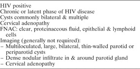
The key diagnostic points for HIV-related benign lymphoepithelial cysts are listed in Table I.
Key diagnostic points for HIV-related benign lymphoepithelial cysts

HIV = human immunodeficiency virus; FNAC = fine needle aspiration cytology
The pathophysiology of HIV-related lymphoepithelial cysts is unresolved. It has been suggested that cysts may arise from epithelial ductular inclusions in lymph nodes.Reference Soberman, Leonidas, Berdon, Bonagura, Haller and Posner3 It has also been proposed that, because of the favourable response to antiretrovirals, HIV-induced cytokines and lymphoid hyperplasia may stimulate ductal epithelium to produce secretions, which form cysts without communicating ducts.Reference Soberman, Leonidas, Berdon, Bonagura, Haller and Posner3
As regards clinical course, lymphoepithelial cysts increase in size, become disfiguring and mark the patients as being HIV positive. However, they do respond well to antiretrovirals, and do not appear to predispose to lymphoma.Reference Dave, Pernas and Roy2
The management of HIV-related benign lymphoepithelial cysts is as follows. Patients with CD4 counts below 200 cells/mm3 qualify for antiretroviral therapy, and the cysts usually resolve with this treatment, without further intervention.Reference Dave, Pernas and Roy2 Patients in the chronic or latent phases of HIV, with CD4 counts above 200 cells/mm3, present a management dilemma. They may be managed by simple observation, and some might receive antiretrovirals for other reasons. Simple, repeated aspiration is unhelpful. The multicentric nature of the disease makes it difficult to access and drain all the cysts. Low-dose radiation therapy has had some temporary success;Reference Kooper, Leemans, Hulshof, Claessen and Snow4 however, with HIV now a chronic, long-term illness, such treatment raises ethical concerns about radiation-induced malignancy, and may compromise radiation therapy for subsequent AIDS-related malignancy. Radiation therapy also has the potential to cause painful xerostomia.Reference Kooper, Leemans, Hulshof, Claessen and Snow4, Reference Suskind, Tavill and Handler5 Parotidectomy exposes medical staff to HIV for the sake of what is essentially a cosmetic problem.
Sclerotherapy has met with some success.Reference Suskind, Tavill and Handler5 However, the sclerosants used thus far have generally been unavailable in developing world countries, and injectable tetracycline has been discontinued. Therefore, we undertook a study of alcohol injection sclerotherapy.
Doxycycline has been proven as a successful sclerosing agent, both in children and adults. It has a low pH and is thought to induce an inflammatory response with secondary adhesion and obliteration of the cyst.Reference Suskind, Tavill and Handler5, Reference Lustig, Lee, Murr and Kingston6 However, this drug is no longer available in South Africa.
Another agent that has recently been used is sodium morrhuate, a detergent sclerosant which works by a mechanism of protein theft denaturation. The only reported complications with this agent were post-operative oedema and tenderness over the injected sight. However, all patients thus treated were taken to the operating theatre to perform the procedure. There was an average 91.5 per cent reduction in cyst size.Reference Marcus and Moore7
Pure ethanol has been used in the past as a sclerosing agent for renal, thyroid and hepatic cysts, with good success and no significant complications.Reference Mohsen and Gomha8–Reference Valcavi and Frasoldati10 It is extremely cheap, which makes it an ideal choice for use in the developing world.
Material and method
Human immunodeficiency virus positive, adult patients seen at Groote Schuur Hospital between July 2005 and September 2006 with a clinical diagnosis of benign lymphoepithelial cysts of the parotid glands were included in the study. Patients' CD4 counts were determined, and patient consent was obtained as per the study protocol, which was approved by the University of Cape Town ethics committee.
The equipment used for sclerotherapy was simple, and included local anaesthetic, a butterfly needle, a vial of 95 per cent alcohol (which costs only 10 American cents (99 South African cents)). The procedures were performed in the ENT out-patient department.
After infiltrating the skin with local anaesthetic, the butterfly needle was inserted into the cyst and secured to the skin with tape. The cyst was aspirated to dryness. A volume of alcohol, equal to 25 per cent of the volume aspirated, was then injected into the cyst and re-aspirated after 10 minutes. All clinically apparent cysts were injected, until no cysts were palpable. The aspirate was only sent for cytological analysis if the referring institution had not already done so.
Results
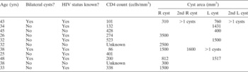
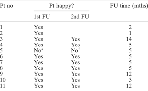
Eleven young adult patients were studied, 27 per cent of whom had not previously been aware of their HIV positive status (Table II). None were receiving antiretroviral medication. Nine patients had known CD4 counts, three of which were ≤200 cells/mm3. Cysts were bilateral in 27 per cent of patients. The cyst volumes initially aspirated varied between 3 and 35 mls. Injections were not painful, and there were no complications. The average follow-up time was 5.7 months (Table III). Three patients required a second injection sclerotherapy procedure. All but one patient were pleased with the cosmetic results (Table III).
Patient data

Yrs = years; HIV = human immunodeficiency virus; CD4 = cluster of differentiation four glycoprotein T cell; R = right; L = left
Follow-up statistics

* 10 ml aspirated; †nodes palpable, no cyst. Pt no = patient number; FU = follow up; mths = months
Discussion
Benign lymphoepithelial cysts can be very large and disfiguring for HIV patients, who often feel stigmatised by this visible manifestation of HIV infection. For patients who do not qualify for antiretroviral therapy, simple, repeated aspiration is unhelpful, low-dose radiation therapy raises ethical concerns, and parotidectomy is an invasive and time-consuming surgical procedure posing significant risk to the facial nerve.
Injection sclerotherapy has previously been reported, using a number of sclerosants such as a doxycycline and sodium morrhuate. Alcohol has been successfully used for thyroid, hepatic and renal cysts. Alcohol is very cheap and readily available, and is therefore an ideal agent especially in the developing world. Different studies of sclerotherapy for non-parotid cysts have varied both the volume of alcohol injected and the time it is left in situ. We arbitrarily selected a volume of 25 per cent of the cyst aspirate and an in situ time of 10 minutes. If the skin at the injection site was infiltrated with local anaesthesia, the procedure was pain-free. When local anaesthesia was omitted, there was a mild burning sensation which lasted a few seconds. The procedure is therefore well suited to an ambulatory care setting.
• Human immunodeficiency virus (HIV)/acquired immunodeficiency syndrome is now probably the most common parotid pathology in Sub-Saharan Africa
• The pathophysiology of lymphoepithelial cysts in HIV positive patients is unresolved. It has been suggested that cysts may arise from epithelial ductular inclusions in lymph nodes
• Benign lymphoepithelial cysts can be very large and disfiguring, and patients often feel stigmatised by this visible manifestation of HIV infection
• Alcohol sclerotherapy is a simple, safe, cheap, effective, ambulatory treatment for HIV-related benign lymphoepithelial cysts, particularly for patients who do not qualify for, or have no access to, antiretroviral therapy
Lustig et al. have reported that cysts injected with doxycycline may require re-injection, and that smaller cysts show a more favourable response. Larger cysts are replaced by a palpable, fibrotic mass.Reference Lustig, Lee, Murr, Deschler and Kingdom11 This is in agreement with our clinical experience.
Ultrasound studies in cases of HIV-related salivary gland disease show the presence of multiple parotid cysts with solid components.Reference Dave, Pernas and Roy2, Reference Martinoli, Pretolesi, Del Bono, Derchi, Mecca and Chiaramondia12 Some of our patients required re-injection. However, it was not possible to determine whether the cysts we ‘re-injected’ were the originally injected cyst(s) or completely new cysts. It is therefore possible that outcomes could be improved if multiple cysts were identified before aspiration and injected under ultrasound guidance. However, in the developing world, and especially in HIV clinics, reliance must be placed on clinical, rather than ultrasonographic, evaluation.
Conclusions
Alcohol sclerotherapy is a simple, safe, cheap, effective, ambulatory treatment for HIV-related benign lymphoepithelial cysts, particularly in patients who do not qualify for, or have no access to, antiretroviral therapy.
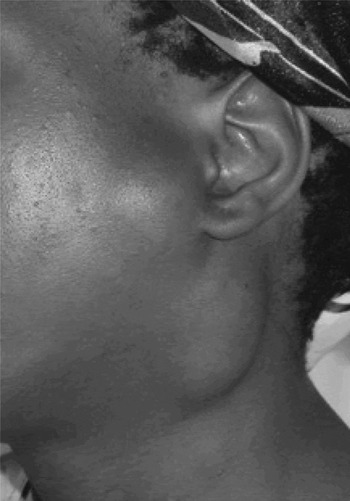
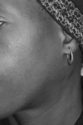
Pre-sclerotherapy human immunodeficiency virus related salivary gland disease.

Post-sclerotherapy result.