Introduction
The giant liver fluke (Fascioloides magna) is a liver parasite which infects a variety of wild and domestic ruminants in North America and Europe (Swales, Reference Swales1935; Erhardová-Kotrlá, Reference Erhardová-Kotrlá1971). On this last continent, fascioloidosis occurs currently in Austria, Croatia, the Czech Republic, Hungary, Italy, Serbia and Slovakia (Králová-Hromadová et al., Reference Králová-Hromadová, Bazsalovicsová, Štefka, Špakulová, Vávrová, Szemes, Tkach, Trudgett and Pybus2011). During recent decades the range of F. magna distribution has increased, and other European countries are clearly at risk of its introduction. Several lymnaeid snails are known to be susceptible to F. magna infection. In North America, five species: Fossaria bulimoides techella, F. modicella, F. parva, Stagnicola caperata and S. palustris nuttalliana have been reported as natural hosts, while another five lymnaeids have been successfully infected under laboratory conditions (Dunkel et al., Reference Dunkel, Rognlie, Johnson and Knapp1996). In Europe, Galba truncatula is the most common intermediate host of F. magna, both in the field and in the laboratory (Erhardová-Kotrlá, Reference Erhardová-Kotrlá1971; Hirtová et al., Reference Hirtová, Modrý and Faltýnková2003). Another three European species: Lymnaea palustris (Chroustová, Reference Chroustová1979), Omphiscola glabra (Rondelaud et al., Reference Rondelaud, Novobilský, Vignoles, Treuil, Koudela and Dreyfuss2006) and Radix peregra (Faltýnková et al., Reference Faltýnková, Horáčková, Hirtová, Novobilský, Modrý and Scholz2006) can also sustain larval development of this parasite in the laboratory. Furthermore, Foreyt & Todd (Reference Foreyt and Todd1974) obtained full larval development of the parasite in an Australian species, Austropeplea (Lymnaea) tomentosa, when exposed to a North American isolate of F. magna.
In view of these findings, one may wonder whether this digenean can infect additional lymnaeids in South American countries. As the importation of feral animals (mainly deer) for zoos or even for reproduction in hunting lodges exists in Argentina, the introduction of F. magna-infected animals and the infection of local snails are possible. For this reason, we have chosen two populations of South American lymnaeids, named Lymnaea viatrix prior to the present study, that are currently raised under laboratory conditions at CEDIVE (the Center for Veterinary Diagnostic and Investigations, Faculty of Veterinary Sciences, University of La Plata, Buenos Aires, Argentina) for production of F. hepatica metacercariae. These two populations were directly involved in the transmission of fasciolosis in their regions of origin (Argentina and Uruguay: Mera y Sierra et al., Reference Mera y Sierra, Agramunt, Cuervo and Mas-Coma2011) and their efficiency for F. hepatica transmission seemed to be two- or threefold greater than that of French G. truncatula (Sanabria et al., Reference Mera y Sierra, Agramunt, Cuervo and Mas-Coma2011). To verify the above hypothesis regarding F. magna, two experiments were carried out: (1) to determine the capacity of Lymnaea to sustain larval development of the parasite; and (2) to specify how many cercariae these snails produced when the method by Rondelaud et al. (Reference Rondelaud, Fousi, Vignoles, Moncef and Dreyfuss2007) for the breeding of amphibious snails is used. The results were compared to those noted in French G. truncatula infected and raised according to the same protocol (reference group). Simultaneously, another experiment was conducted to identify precisely the two populations of South American snails, using molecular biology, because the systematics of these lymnaeids is controversial and identification cannot be made with the sole use of morphological criteria (Duffy et al., Reference Duffy, Kleiman, Pietrokovsky, Issia, Schijman and Wisnivesky-Colli2009; Mera y Sierra et al., Reference Mera y Sierra, Artigas, Cuervo, Deis, Sidoti, Mas-Coma and Bargues2009).
Materials and methods
Collection of snails and adult worms of Fascioloides magna
The first South American population (strain U) of snails has been raised at CEDIVE since 1996 and originated from Paysandú, Uruguay (36°00′ S, 57°30′ W). The other population (strain SP) has been bred in the laboratory since 2008 and came from San Pedro, Buenos Aires, Argentina (33°40′ S, 59°39′ W). Two French populations of G. truncatula were also used. The first originated from a road ditch at Chézeau Chrétien, commune of Chitray, department of Indre (46°40′27″ N, 1°21′21″ E) and was used for molecular biology. The other population of G. truncatula was collected from a road ditch (45°55′33″ N, 2°2′33″ E) in the commune of Saint Michel de Veisse, department of Creuse, and was used for experimental infections. The habitat of this last population was located on siliceous soil so that the upper shell height of adults (8–9 mm) ranged in the same scale of values as that of South American adults. Snails of height 4 mm were collected from each population.
Eggs of F. magna were collected from adult flukes recovered from the livers of naturally infected red deer (Cervus elaphus) hunted in the district of Krivoklátsko (Central Bohemia, Czech Republic). Prior to hatching, the eggs were washed several times with spring water and were incubated for 12 days at 24°C in the dark (Ollerenshaw, Reference Ollerenshaw1971).
Molecular identification of South American lymnaeids by polymerase chain reaction–restriction fragment length polymorphism and sequencing
Five snails of each population (SP, U and G. truncatula) were used for molecular taxonomical determination. The entire foot of each fresh specimen was sectioned with a scalpel blade. Foot samples were processed using the DNeasy Blood and Tissue Kit (QIAGEN, Hilden, Germany). Lysis using proteinase K was performed overnight. Once extracted, the internal transcribed spacer (ITS)-1 segment of their nuclear ribosomal DNA was amplified using the following primers: ETTS2 (5′-TAACAAGGTTTCCGTAGGTGAA-3′) and ITS-1r (5′-CGAGCGAGTGATCCACCGC-3′), for forward and reverse senses, respectively (Carvalho et al., Reference Carvalho, Cardoso, Lira, Rumi, Roche, Berne, Müller and Caldeira2004).
The polymerase chain reaction (PCR) mix was prepared to have a final volume of 25 μl, containing 12.5 μl of Qiagen Multiplex, 1 μl of each primer, 5 μl of genomic rDNA and 5.5 μl of DNAse and RNAse-free water. The PCR reaction was performed using a Perkin-Elmer Thermo Cycler (Applied Biosystems, Warrington, Cheshire, UK) as follows: initial denaturation (95°C, 3 min); 35 cycles, each comprising denaturation (95°C, 45 s), annealing (55°C, 1 min) and extension (72°C, 2 min); final extension (72°C, 5 min).
Amplified products were digested with the endonuclease HpyF3I (DdeI) (Fermentas GmbH, St. Leon-Rot, Germany) which restricts the sites 5′…C ↓ TNAG…3′. This enzyme was chosen for the present study because it was previously reported as the most accurate for differentiating lymnaeid species (Carvalho et al., Reference Carvalho, Cardoso, Lira, Rumi, Roche, Berne, Müller and Caldeira2004). Incubation was performed at 37°C for 3.5 h into a final volume of 15 μl, containing 5 μl of DNA, 8.5 μl of DNAse and RNAse-free water, 1 μl of Tango buffer (Fermentas GmbH) and 0.5 μl of enzyme. All products were separated on 2% agarose gels using a 100 bp ladder and stained with ethidium bromide. They were visualized using a UV transilluminator coupled with the software GELSMART 7.0 (Clara Vision, Verrières le Buisson, France). Band size was measured with the online available software Gel Analyzer 2010a (http://www.gelanalyzer.com).
For each South American population (SP or U), two amplicons were chosen randomly and purified using a rapid PCR purification kit (Marligen Biosciences, Ijamsville, Maryland, USA) according to the manufacturer's instructions. Sequencing reactions were performed using the ABI Big Dye kits (Applied Biosystems) and the same forward and reverse primers as those employed in the PCR amplification of ITS-1. Both strands were sequenced utilizing the ABI Prism 3130xl genetic analyser (Applied Biosystems). The forward and reverse sequences obtained were aligned and analysed using the software FinchTV 1.4.0 (Geospiza Inc., Seattle, Washington, USA) and Clustal X 2.0.11 (EBI, Hinxton, Cambridgeshire, UK). Sequences were submitted to GenBank under the following accession numbers: JF960165 and JF960166 for population SP, and JF960167 and JF960168 for population U. A consensus sequence was then obtained for each population of South American snails and was compared to GenBank reported sequences using the online tool BLASTN (http://blast.ncbi.nlm.nih.gov).
Experimental infections of snails
Two experiments were carried out. In experiment A, the propensity of each snail species for F. magna infection was studied. One hundred and fifty snails, each measuring 4 mm in shell height, were randomly chosen from each population. Each snail was then exposed to two miracidia for 4 h at 20°C in 3.5 ml of spring water and then raised at the same temperature according to the method of Rondelaud et al. (Reference Rondelaud, Fousi, Vignoles, Moncef and Dreyfuss2007). Snails were finally raised in groups of ten individuals in 14-cm Petri dishes (volume of spring water, 60 ml) for 30 days. Food consisted of dried leaves of lettuce and dead leaves of grass (Molinia caerulea), while several stems of live Fontinalis sp. ensured oxygenation of the water layer. The dissolved calcium in spring water was 35 mg/l. The Petri dishes were placed in an air-conditioned room under the following conditions: a constant temperature of 20°C ( ± 1°C), natural photoperiod of 10 h light. At day 30 post-exposure (pe), each surviving snail was isolated in a 35-mm Petri dish with pieces of dead grass, lettuce and spring moss, and placed at 20°C. A daily surveillance was made, (1) to change spring water and food if necessary, and (2) to count the metacercariae in the dishes. When the first cercarial shedding occurred, the surviving snails were subjected to a thermal shock every 3 days by placing their Petri dishes at 10–13°C for 3 h (outdoors) to stimulate cercarial exit (this temperature was chosen according to our experience with cercarial shedding of snails infected with digeneans). At the death of each snail, the shell was measured using callipers.
Experiment B was carried out to determine cercarial production of F. magna in snails dissected at day 65 pe at 20°C. Each experimental group consisted of 100 snails. Snail exposure to miracidia and maintenance were similar to those of experiment A. At day 65, each surviving snail was dissected under a stereomicroscope to count free rediae, intraredial cercariae and free cercariae.
The two parameters calculated were snail survival at day 30 pe and the prevalence of F. magna infection (calculated in relation to the number of snails surviving at day 30 pe). Prevalence took into account the number of cercariae-shedding snails (CS snails) and the number of individuals containing cercariae but without shedding (NCS snails) in experiment A, and all snails carrying larval forms of F. magna in experiment B. For each parameter, the difference between the values recorded for the three snail groups was analysed using a χ2 test.
In experiment A, the other parameters calculated were the growth of CS snails during the experiment, the length of the prepatent period (time of cercarial differentiation inside rediae), the length of the patent period (time during which cercariae exit from the snail), the life period after the last exit of cercariae and the total number of metacercariae. In experiment B, the number of free rediae, the quantity of free cercariae and the quantity of intraredial cercariae were considered. Individual values recorded for these last eight parameters were averaged and their standard deviations were calculated considering snail groups. One-way analysis of variance was used to establish levels of statistical significance calculated using the Statview 5.0 software (Deltasoft Meylan, France).
Results
Identification of South American lymnaeids
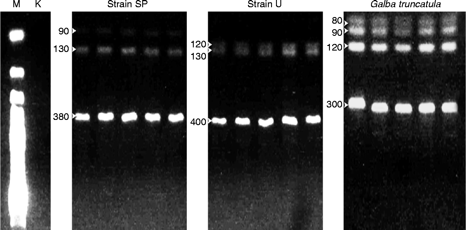
The approximate size of amplified bands for snail samples was 600 bp for population SP and G. truncatula, and 650 bp for population U. In South American lymnaeids, three bands (fig. 1) were noted in the patterns derived from the restriction fragments; 90, 130 and 380 bp for population SP and 120, 130 and 400 bp for population U. In contrast, four bands (80, 90, 130 and 300 bp) were noted for the French G. truncatula (fig. 1). The observed restriction patterns clearly allowed separation of population SP from population U.
Restriction fragments of rDNA ITS-1 segment from each snail population treated with endonuclease HpyF3I. Lanes: M, 100 bp marker; K, negative control; SP, Argentinean population of Lymnaea neotropica; U, Uruguayan population of Lymnaea viatrix; Galba truncatula, French G. truncatula. Band sizes are shown beside each band.

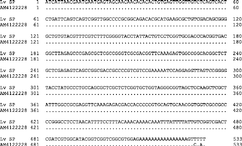
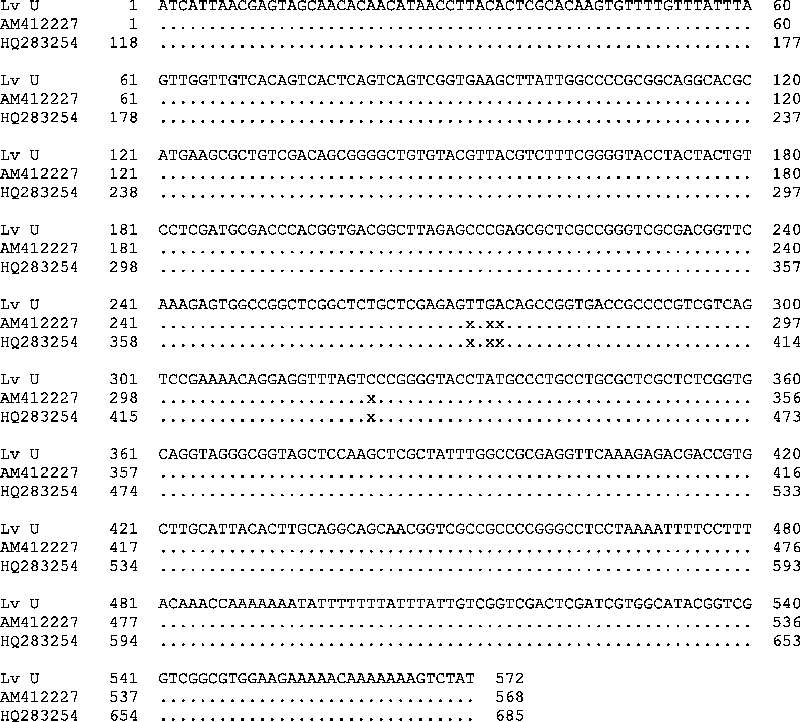
Sequence analysis of the ITS-1 amplicons showed differences among both populations of South American snails. Population SP (fig. 2) showed 99.6% identity with the previously reported sequence of Lymnaea neotropica (GenBank, accession number AM412228). On the other hand, population U (fig. 3) exhibited greater similarity to the sequences of L. viatrix var. ventricosa and L. viatrix (GenBank, accession numbers AM412227 and HQ283254, respectively) and was thus classified as L. v. ventricosa in the present study. This population differed from the GenBank sequences by the addition of four bases at positions 272, 274, 275 and 322.
Comparison of ITS-1 sequences for population SP (GenBank, accession numbers JF960165 and JF960166) and Lymnaea neotropica (GenBank, accession number AM4122228).
Comparison of ITS-1 sequences for population U (GenBank, accession numbers JF960167 and JF960168), L. viatrix var. ventricosa (GenBank, accession number AM412227) and L. viatrix (GenBank, accession number HQ283254). X indicates a lack of a base in the previously reported sequences.
Lymnaeid species and snail infection
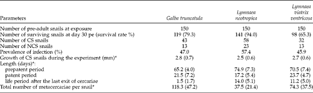
The results of experiment A are shown in table 1. Compared to the reference G. truncatula, the survival rate of snails at day 30 pe (table 1) was significantly greater for L. neotropica (χ2= 13.96, P< 0.001) and significantly lower for L. v. ventricosa (χ2= 7.34, P< 0.01). In both groups of South American snails, the prepatent period was significantly longer (F= 3.58, P< 0.05) than that of the reference group, whereas the number of shed cercariae was significantly lower (F= 6.70, P< 0.01). The prevalence of infection, the snail growth during the experiment, and the patent periods were not significantly different between the snail groups tested. In contrast, the South American snails survived on average 11.2–14.0 days after the last exit of their cercariae and these periods were significantly longer (F= 7.84, P< 0.01) than in the G. truncatula group.
Main characteristics of Fascioloides magna infection in three species of lymnaeids (experiment A).
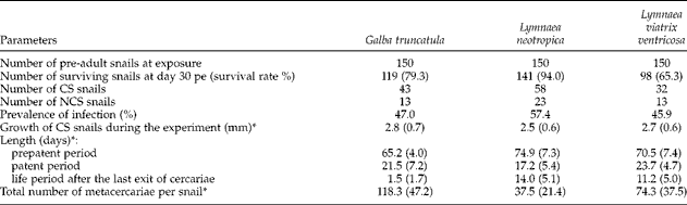
pe, post-exposure; CS snails, cercariae-shedding snails; NCS, snails containing cercariae but without shedding.
* Mean value (SD).
In experiment B, a significant difference (χ2= 10.97, P< 0.001) between survival rates of G. truncatula and L. neotropica was noted (table 2), whereas there was no difference between G. truncatula and L. v. ventricosa. Infection rates were not significantly different between the three snail groups. Compared to the reference group, the number of free rediae and that of free cercariae were significantly lower in the tested South American lymnaeids (free rediae: F= 3.29, P< 0.05; free cercariae: F= 16.58, P< 0.01). At day 65, the number of cercariae remaining in the body of rediae ranged within the same scale of values and so no significant difference was recorded.
The number and prevalence of larval stages of Fascioloides magna in three species of lymnaeids on day 65 post-exposure (pe) at 20°C (experiment B).
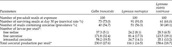
* Mean value (SD).
If the number of shed cercariae (table 1) is compared to the total cercarial production within the snail's body (table 2), the percentage of larvae that exited from the snail was 51.3% for G. truncatula and 46.8% for L. v. ventricosa, while it was only 32.2% for L. neotropica. In this respect, L. neotropica seemed to be a less suitable intermediate host of F. magna than L. v. ventricosa.
Discussion
The presence of L. neotropica has been reported at Mendoza in western Argentina (Mera y Sierra et al., Reference Mera y Sierra, Artigas, Cuervo, Deis, Sidoti, Mas-Coma and Bargues2009) and is mentioned here for the first time in the eastern part of this country (Buenos Aires). In the same way, our results on L. v. ventricosa confirm the report by Carvalho et al. (Reference Carvalho, Cardoso, Lira, Rumi, Roche, Berne, Müller and Caldeira2004) indicating the presence of this snail in Uruguay (unspecified locality). Compared to sequences of L. viatrix (AM412227 and HQ283254) the sequence of L. v. ventricosa showed four additional bases. As these differences were a constant in both forward and reverse strands for all sequences, it may be concluded that these characteristics represent a local genotype. Similar differences have already been reported, for example, in allopatric samples of Lymnaea stagnalis (Remigio & Blair, Reference Remigio and Blair1997). In view of this finding, it is necessary to take into account the fact that ITS-1 segments might show population variations, as demonstrated by Almeyda Artigas et al. (Reference Almeyda Artigas, Bargues and Mas-Coma2000). Even if the use of ITS-1 sequence is sufficient for species discrimination of both South American lymnaeids, snail identification could be improved in further studies by additional molecular markers such as mitochondrial DNA.
The present study demonstrates that Lymnaea neotropica and L. v. ventricosa can be included as new intermediate hosts for F. magna. The longer prepatent periods and lower numbers of cercariae noted for these two species (in the context of cercarial shedding, table 1) might represent the case of an incomplete adaptation process of the snail–parasite system. This interpretation is supported by the larger values of standard deviations noted for these parameters (table 1).
After the exit of the last cercariae, L. neotropica and L. v. ventricosa can survive on average for 14.0 and 11.2 days, respectively, before their death, whereas these snails still contained free cercariae in their bodies during this period. This finding is difficult to comment on because the ‘post-shedding period’ was not recorded in our experimental infections of L. neotropica and L. v. ventricosa, nor in the French G. truncatula with a French isolate of F. hepatica. Two complementary hypotheses may be proposed. First, the two South American species could be more able to sustain larval development of F. magna than the French population of G. truncatula. Second, the life period after the last released cercariae might also be subjected to unknown characteristics of F. magna larval development. Under constant conditions in the laboratory, there was little cercarial shedding of F. magna from European populations of G. truncatula (Erhardová-Kotrlá, Reference Erhardová-Kotrlá1971; Vignoles et al., Reference Vignoles, Novobilský, Rondelaud, Bellet, Treuil, Koudela and Dreyfuss2006; Rondelaud et al., Reference Rondelaud, Fousi, Vignoles, Moncef and Dreyfuss2007) and also from O. glabra (Rondelaud et al., Reference Rondelaud, Novobilský, Vignoles, Treuil, Koudela and Dreyfuss2006). Owing to the thermal shock used to stimulate cercarial shedding, the presence of metacercariae (table 1) was noted for a mean of 17.2 days (L. neotropica) and 23.7 days (L. v. ventricosa). The interruption of this process in the following days may be the consequence of too few free cercariae remaining in the snail body to cause another cercarial shedding.
In conclusion, both South American lymnaeids, like A. tomentosa in Australia (Foreyt & Todd, Reference Foreyt and Todd1974) and O. glabra in France (Rondelaud et al., Reference Rondelaud, Novobilský, Vignoles, Treuil, Koudela and Dreyfuss2006), can be potential intermediate hosts for larval development of F. magna, whereas fascioloidosis has not been recorded in these countries. These findings demonstrate the high potential of the parasite to spread in new areas by adapting to local snail species. The facility of adaptation of F. magna to new intermediate hosts enhances the risk of parasite transmission in these areas, mainly as a result of local deer migrations and/or importation of infected ruminants.
Acknowledgements
This study was partly supported by the Swedish Foundation for Agricultural Research (contract no. H1050003), the Czech Science Foundation (grant no. P502/10/P248), the Czech Ministry of Education (grant nos. MSM LC06009 and MSM 0021620828) and by the University Research Centre (UNCE) of Charles University, Prague (grant no. 204017). The authors thank Dr David A. Morrison for revising the English text.