Introduction
The vast majority of animal experiments with gastrointestinal (GI) nematodes in rodents are still carried out with single-pulse infections, in which animals receive a single, relatively large dose of infective larvae at one point in time. However, in nature animals are more likely to be exposed to infection repeatedly with smaller doses of larvae, and perhaps just the occasional larger dose (Beaver, Reference Beaver1953; Udonsi et al., Reference Udonsi, Nwosu and Anya1980; Hominick et al., Reference Hominick, Dean and Schad1987). Two similar but distinct protocols have been used to mimic this latter situation experimentally. First, so-called trickle infection protocols entail administration of only a few larvae daily (often restricted to weekdays) and this protocol has been used, for example, in experiments with Nippostrongylus brasiliensis and Trichuris muris (Jenkins & Phillipson, Reference Jenkins and Phillipson1971; Behnke & Wakelin, Reference Behnke and Wakelin1973; Wakelin, Reference Wakelin1973; Bancroft et al., Reference Bancroft, Else, Humphreys and Grencis2001). An intermediate protocol, the so-called repeated infection protocol, based on giving animals doses of larvae once a week has also been used in rodent model systems (Brailsford & Behnke, Reference Brailsford and Behnke1992a; Behnke et al., Reference Behnke, Lowe, Clifford and Wakelin2003), but both protocols are still rarely exploited in experiments in rodent-GI nematode model systems. In contrast, protocols of infection based on daily or weekly infections with GI nematodes have been adopted frequently in work with parasites of ruminants, where this type of infection protocol has become more or less standard practice (Jackson et al., Reference Jackson, Angus and Coop1983; Chiejina et al., Reference Chiejina, Behnke, Musongong, Nnadia and Ngongeha2010).
Repeated/trickle infection protocols create a highly dynamic situation in the intestine with a range of possible outcomes for the parasite. In some species, worms accumulate until expulsion occurs, in others there may be worm turnover, and in others still, acquired immunity is elicited rapidly and, apart from the very first batches of larvae, subsequent batches fail to establish (Michel, Reference Michel1970; Donald & Waller, Reference Donald, Waller, Symons, Domald and Dineen1982; Courtney et al., Reference Courtney, Parker, McClure and Herd1983; Jackson et al., Reference Jackson, Angus and Coop1983; Barger et al., Reference Barger, Le Jambre, Georgi and Davies1985). Quite clearly the characteristics of immune responses under all these conditions may be distinct from those observed following single-pulse inocula, as experiments with T. muris have shown, where in contrast to single-pulse low-intensity infections that typically induce type 1 responses and result in worms surviving, repeated low-level infections are more likely to stimulate type 2 responses with resultant worm expulsion, once a threshold worm burden has been exceeded (Bancroft et al., Reference Bancroft, Else, Humphreys and Grencis2001).
Hamsters tolerate very long chronic primary infections with A. ceylanicum after a single-pulse primary infection, lasting over 200 days, although worm burdens are reduced slowly over this period of time (Behnke, Reference Behnke, Schad and Warren1990). When adult worms have been removed by chemotherapy after 4 weeks or so of infection, hamsters acquire resistance to challenge rapidly and secondary infections are mostly resisted in the first few days after challenge, with expulsion of the majority of L4 stages before they moult to the adult stage (Behnke et al., Reference Behnke, Guest and Rose1997). Nevertheless, some larvae negotiate this hazardous phase of infection successfully and manage to moult, and the resulting adult worms then survive for many months. When hamsters are repeatedly infected with smaller doses they control worm burdens rapidly (Brailsford & Behnke, Reference Brailsford and Behnke1992b), and there is little evidence of the sort of build-up of worms that occurs, for example, in poor-responder mouse strains subjected to repeated infections with Heligmosomoides bakeri (Behnke et al., Reference Behnke, Lowe, Clifford and Wakelin2003) or in sheep infected frequently with Trichostrongylus vitrinus (Jackson et al., Reference Jackson, Angus and Coop1983).
The mucosal changes accompanying primary infections with A. ceylanicum have been documented in detail with respect to the mucosal architecture and the dynamics of the changing cell populations involved in resistance (Alkazmi et al., Reference Alkazmi, Dehlawi and Behnke2006, Reference Alkazmi, Dehlawi and Behnke2008). Villous height fell dramatically within 2 weeks of infection, while the crypts of Lieberkühn became deeper, and the degree of change in both cases was dependent on the numbers of worms that had settled in the intestine. However, after removal of the worms, recovery of the mucosa was rapid, indicating that the persistence of these changes was dependent on the presence of worms, and data showed that as long as adult worms were still present in the intestine the gut did not return to normal for over 10 weeks (Alkazmi & Behnke, Reference Alkazmi and Behnke2010). By 2–3 weeks after initial infection there was a marked increase in goblet cells, mast cells and eosinophils in villi, but Paneth cell numbers in the bases of the crypts typically fell after infection with A. ceylanicum (Alkazmi et al., Reference Alkazmi, Dehlawi and Behnke2006, Reference Alkazmi, Dehlawi and Behnke2008).
The aim of the current experiment was to document the mucosal changes in animals subjected to repeated infections, during which it is known that immunity is generated very rapidly (Brailsford & Behnke, Reference Brailsford and Behnke1992b), and to compare these to those taking place in animals given a conventional single-pulse infection, with chronic survival of a stable worm burden, in order to identify changes that may be linked to protective immunity in the former case and to lack of it in the latter.
Materials and methods
Parasites
Hamsters were infected with a strain of A. ceylanicum that has been maintained at the University of Nottingham since 1984, and was originally obtained from Dr Rajasekariah of Hindustan CIBA-Geigy Ltd., Bombay, India. This strain is believed to be of dog origin. The methods employed for maintenance of the parasite, for worm recovery and faecal egg counts have all been described previously in full (Garside & Behnke, Reference Garside and Behnke1989; Behnke et al., Reference Behnke, Guest and Rose1997).
Hamsters
The Golden hamsters (DSN strain) used in this study were originally obtained from Harlan Olac in 1983 and since then they have been maintained in the animal house of the School of Biology as a closed colony. Only female hamsters were used in this experiment and these were 3–4 months old on the day of the first infection. Animals were kept under conventional animal house conditions, with pelleted food and tap water supplied ad libitum. Cages were cleaned twice weekly to prevent re-infection. Animals were first weighed 2 weeks before infection and thereafter twice weekly until the completion of the experiment.
Since the colony was maintained under conventional animal house conditions, the animals were exposed to various micro-organisms present in the environment. In order to prepare hamsters for infection and reduce other competing intestinal micro-organisms, all animals were pre-treated for 1 week with Emtryl (May & Baker, dimetridazole at a concentration of 1 g/l in drinking water), then for another week with Terramycin (Pfizer, oxytetracycline hydrochloride, 3 g/l in drinking water), and were returned to normal drinking water for 1 week prior to infection.
Measurement of mucosal architecture and the cellular mucosal responses to infection
The methods used to measure the height of villi, the depth of the crypts of Lieberkühn and mitotic activity have been described comprehensively in earlier publications (Alkazmi et al., Reference Alkazmi, Dehlawi and Behnke2006). Methods for assessing the mast cell, goblet cell, eosinophil and Paneth cell responses have also been reported in full earlier (Alkazmi et al., Reference Alkazmi, Dehlawi and Behnke2008). In all the histological observations reported here, except for Paneth cell densities, we counted cells/mm2 of mucosal tissue on appropriately stained sections, using the Weible 2 graticule as described by Kermanizadeh et al. (Reference Kermanizadeh, Crompton and Hagan1997). Hence, counts are expressed as cells/mm2 of mucosal tissue rather than as cells/villus crypt unit (VCU) as in some other studies. Further details of the methodology, including illustrations of typical staining can be found in Alkazmi (Reference Alkazmi2004).
Experimental design
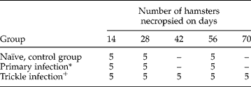
Fifty-five hamsters were allocated randomly to three experimental treatment groups, as shown in table 1, and, with few exceptions, values for the parameters of interest (see legends to the figures for exact details) were mostly based on five hamsters from relevant groups at each time point. Fifteen naïve control hamsters were left untreated throughout the period of the experiment. A further 15 hamsters were infected orally with 50 L3 on day 0 and were necropsied in groups of five animals on the days shown in table 1. Twenty-five hamsters were exposed to weekly infections of 30 L3. In total they received 10 infections of 30 larvae each. At each of these infections a further two naïve hamsters were also infected and these latter animals were killed 14 days after infection to assess the infectivity of the inocula used for the repeated infection protocol throughout. Worm recovery from these animals showed that between 4 and 10 worms had established in these tracer animals on each occasion, and using the means for each time point, the animals in the repeatedly infected treatment group had received on average 62 larvae capable of developing into adult worms in naïve hosts.
Experimental design.
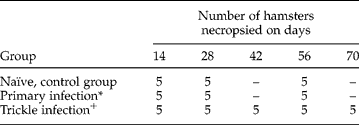
* All the animals in this group were given 50 L3 on day 0.
+ All the animals in this group were given 30 L3 on days 0, 7, 14, 21, 28, 35, 42, 49, 56 and 63.
Statistical analysis
Where relevant, data are presented as group mean values ± standard error of the mean (SEM). Because the worm burden data did not meet the assumptions of normality, the two infected treatment groups were compared by the non-parametric two-way analysis of variance (ANOVA) (Barnard et al., Reference Barnard, Gilbert and McGregor2007). Correlations between variables, usually values of the target variables (cell types, villus height, etc.) with time, were analysed by Spearman's non-parametric correlation test, and where a priori predictions were possible we implemented one-tailed tests and corresponding probabilities. To compare responses between the two infection protocols (single-pulse primary infection versus repeated infections) we restricted analysis to these two groups (excluding the naïve control group), and confined it to days 14, 28 and 56, when animals from both treatments were necropsied. Values in naïve control groups were mostly markedly different from those among infected animals, and were included to provide a baseline for the quantified parameters but mostly did not require inclusion in statistical analysis. In all cases we checked R 2 values and the distribution of residuals for conformity to a normal distribution. Where the assumptions of normality were not met, the data were log transformed for analysis or analysed by non-parametric tests. In some cases we conducted a posteriori tests on small groups of animals necropsied on a specific day (hence normality could not be assumed), and in these cases we used the Mann–Whitney U-test for two group comparisons. With the exception of non-parametric ANOVA, which was carried out using bespoke software based on Barnard et al. (Reference Barnard, Gilbert and McGregor2007), all other tests and calculations were conducted in SPSS for Windows (Release 16.0.0; SPSS Inc., Chicago, Illinois, USA), and we employed P= 0.05 as our cut-off for statistical significance.
Results
Worm burdens
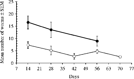
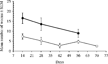
The mean worm burdens recovered at necropsy from each of the two treatment groups exposed to infection are illustrated in fig. 1. In both groups mean worm burdens fell significantly with time, as expected (Garside & Behnke, Reference Garside and Behnke1989; Behnke, Reference Behnke, Schad and Warren1990; Brailsford & Behnke, Reference Brailsford and Behnke1992b) and hence we used one-tailed tests (for primary infection, r s= − 0.493, n= 15, P= 0.031; and for the repeated infection, r s= − 0.442, n= 25, P= 0.013). Analysis of the worm burdens at shared time points (days 14, 28 and 42) by a non-parametric two-way ANOVA (treatment and time), gave H1= 9.87, P= 0.017 for the effect of treatment, and it is evident (fig. 1) that despite the greater numbers of larvae given to the repeatedly infected animals, those carrying the single-pulse primary infection had higher worm burdens throughout. There was no significant interaction between treatment and time.
Changes in worm burdens during the course of the experiment in hamsters given the single-pulse primary infection (filled-in circles) and those treated with the repeated infection protocol (open circles). For statistical analysis, see the text.
Small intestinal villi
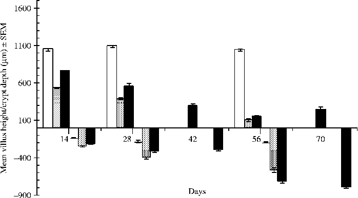
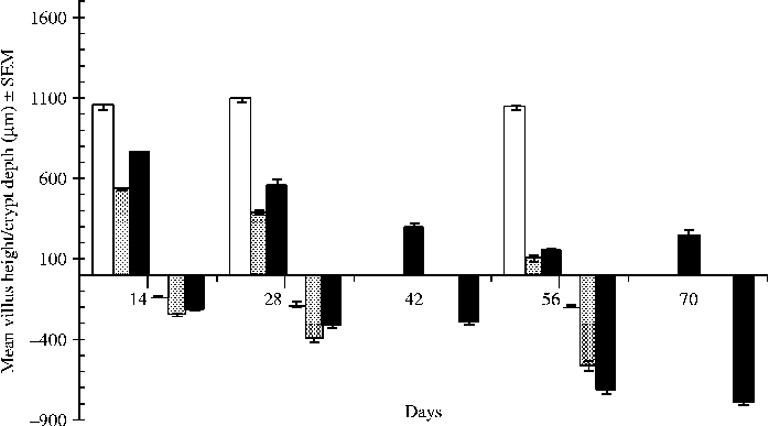
Changes in the height of the villi are summarized in fig. 2. There was no significant difference in the height of the villi in control animals during the period of the experiment, the mean values of 1049.5 μm (day 14), 1091.25 μm (day 28) and 1044.25 μm (day 56) recorded in this group being consistent with, and well within, the mid-range for uninfected hamsters published earlier (Alkazmi et al., Reference Alkazmi, Dehlawi and Behnke2006). Very clearly, villous height fell markedly in both of the infected groups, but there was a significant difference between groups (two-way general linear model (GLM) with time and treatment as factors, confined to the two infected groups on days 14, 28 and 56, F 1,24= 54.1, P< 0.001, adjusted model R 2= 94.9%), initially somewhat faster in the group given the single-pulse primary infection, which had a higher adult worm burden than the animals subjected to repeated infections, but then by day 56 values converging with time (interaction between time and treatment F 2,24= 6.9, P= 0.004). By day 56 villus height in the animals given the single-pulse primary infection had dropped by 89.6% and in the repeatedly infected animals by 85.2%.
Changes in the height of villi (upper panel) and in the depth of the crypts of Lieberkühn (lower panel) in naïve control hamsters (open columns), and in hamsters given the single-pulse primary infection (stippled columns) and those treated with the repeated infection protocol (filled-in columns). For statistical analysis, see the text.
Crypts of Lieberkühn
In contrast to the villi, the crypts of Lieberkühn became deeper with time post-infection (fig. 2) in both infected groups (correlation with time as one variable, primary infection group r s= − 0.945, n= 15, P< 0.001; repeated infection group r s= − 0.902, n= 25, P< 0.001). In this case, when the analysis was confined to the two infected groups, there was no significant difference between them, but there was a significant interaction (F 2,24= 14.9, P< 0.001), arising mainly because initially (days 14 and 28) the crypts were deeper in the hamsters given the single-pulse primary infection, but by day 56 the reverse was true. Crypts in the naïve control group were much shorter throughout the experiment but, even so, there was a significant increase in their depth between days 14 and 56 (correlation with time, r s= − 0.76, n= 15, P= 0.001 and depth increasing by 46.7%). Nevertheless, even on day 56, the depth of crypts in control naïve hamsters was just 185.26 μm, compared to 697.25 μm in the animals subjected to repeated infections, and even the extreme values for the control group were well within the normal range for uninfected hamsters (Alkazmi et al., Reference Alkazmi, Dehlawi and Behnke2008).
Mitotic figures per crypt
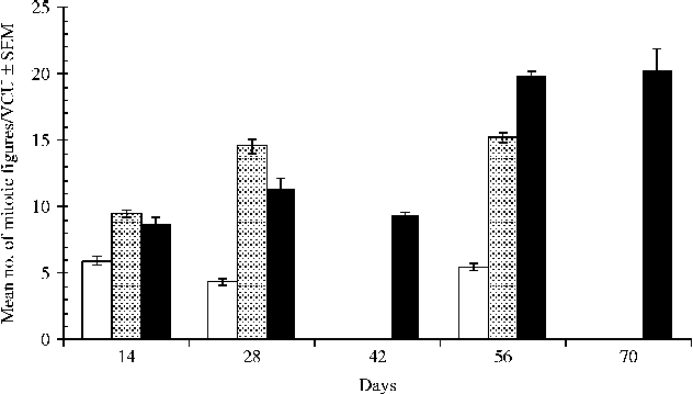
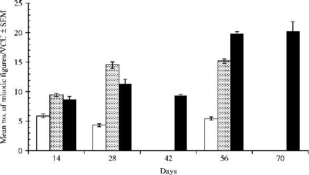
The mean values for mitotic figures are illustrated in fig. 3. There was little change among the naïve control hamsters, with mean values ranging from 4.23 (day 28) to 5.7 (day 14), well within the normal range of uninfected hamsters (Alkazmi et al., Reference Alkazmi, Dehlawi and Behnke2006), and there was no significant change with time (correlation with time, r s= − 0.21, n= 15, P= NS). In comparison to the control group, the numbers of mitotic figures were markedly elevated in both infected groups (e.g. on day 56, × 2.84 and × 3.72 in hamsters given primary and repeated infections, respectively), and in both groups they increased significantly with time (for the primary infection group r s= 0.85, n= 15, P< 0.001; for the repeated infection group r s= 0.78, n= 25, P< 0.001). Comparing the two infected groups on days 14, 28 and 42, there was no significant main effect of treatment, but there was a significant interaction (two-way GLM with time and treatment as factors, F 2,24= 27.9, P< 0.001, adjusted model R 2= 91.1%). As can be seen from fig. 3, initially (days 14 and 28) values were higher in the animals given the single-pulse primary infection but by day 56, there were more mitotic figures in the repeatedly infected group.
Changes in the mitotic figures of crypts in naïve control hamsters (open columns), and in hamsters given the single-pulse primary infection (stippled columns) and those treated with the repeated infection protocol (filled-in columns). VCU refers to villous crypt units. For statistical analysis, see the text.
Mast cells
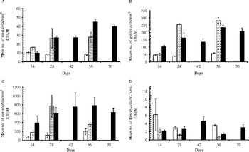
In naïve control hamsters the concentration of mast cells in mucosal tissues appeared to drop (fig. 4A) but this reduction over time was not significant (correlation with time as one variable, two-tailed test on the control group r s= − 0.492, n= 15, P= 0.063). We expected to see mast cell numbers increasing with time in animals given the single-pulse primary infection (Alkazmi et al., Reference Alkazmi, Dehlawi and Behnke2008) and this was borne out (one-tailed test, r s= 0.491, n= 15, P= 0.032). Repeated infections had not been studied in this context earlier, but even with a two-tailed test the increase in mast cell numbers was highly significant (fig. 4A and r s= 0.859, n= 25, P< 0.001). Confining further analysis to the two infected groups and restricting it to data collected on days 14, 28 and 56, when tissue samples were taken from both groups, showed that there was no significant difference in values between the two infected groups and no interaction. Although the figure shows higher values in the repeatedly infected hamsters on day 56, the number of mast cells in hamsters sustaining a prolonged primary infection is known to remain elevated above background naïve control levels for over 90 days after infection (Alkazmi & Behnke, Reference Alkazmi and Behnke2010).
Changes in cellular compartments of the intestinal mucosa in naïve control hamsters (open columns) and in hamsters exposed to either a single-pulse primary infection (stippled columns) or to repeated infections (filled-in columns). (A) Mast cells; (B) goblet cells; (C) eosinophils; (D) Paneth cells. VC, villous crypt. For statistical analysis, see the text.

Goblet cells
As expected (Alkazmi et al., Reference Alkazmi, Dehlawi and Behnke2008), there was a rapid increase in goblet cells in hamsters exposed to the single primary infection but a similar response was also observed in those given the repeated infections (fig. 4B). Among the infected hamsters necropsied on days 14, 28 and 56, the main effect of treatment was not significant (F 1,24= 3.01, P= 0.095). However, there was a significant interaction (F 2,24= 7.9, P= 0.002, adjusted model R 2= 79.5%), because values were higher initially (day 14) in the repeatedly infected animals, but subsequently (day 28 and day 56) rose higher in those given the primary infection. At the height of the response (day 56), the concentration of goblet cells in the primary infection group was 4.4 × greater compared with the naïve controls. At the same time, values were 4.0 × higher in repeatedly infected animals. In the naïve control group, the mean values ranged from 38.6 to 58.3 goblet cells/mm2 of intestinal mucosa, which is towards the bottom end of the normal range for uninfected hamsters of this strain (Alkazmi et al., Reference Alkazmi, Dehlawi and Behnke2008).
Eosinophils
Eosinophil cell counts in the mucosa were perhaps the most variable of all the four cell types examined in this experiment. Very clearly, eosinophil numbers were much higher in both infected groups compared to the naïve controls (e.g. for means on day 28, × 6.9 in the single-pulse infected hamsters and × 5.4 in those repeatedly infected), but there was no significant difference between the two groups of infected hamsters whether analysis was on raw or log transformed data. Figure 4C shows that in the repeatedly infected animals, the elevated numbers were sustained throughout to day 70, but suggests a drop in the animals given the single-pulse primary infection by day 56. Post hoc comparison of the values in the primary infection and repeatedly infected groups on day 56 indicated a significant difference (Mann–Whitney U-test, z= 2.61, P= 0.009).
Paneth cells
In contrast to the other cell types, Paneth cell counts have been shown to decline following single-pulse primary infections (Alkazmi et al., Reference Alkazmi, Dehlawi and Behnke2008), and this trend was quite evident here also (fig. 4D). Mean Paneth cells counts on day 56 were only 17.5% of the counts in the naïve controls on that day (correlation with time r s= − 0.78, n= 15, P= 0.001). Note that the relatively high mean Paneth cell count (and SEM) on day 14 in naïve control hamsters arose through one of the animals on that day showing an unusually high count (21.75) compared to the others, whose values were in the normal range for uninfected hamsters, varying only from 1.05 to 3.05. Interestingly, no fall in Paneth cells was observed in the repeatedly infected hamsters and the difference between the two infected groups of hamsters was significant (two-way GLM with treatment and time as factors, restricted to days 14, 28 and 56, main effect of treatment F 1,24= 5.66, P= 0.026, adjusted model R 2= 32.4%) but there was no significant interaction because in both groups lower counts were evident on day 56. Nevertheless, overall, taken across the five time points of the experiment and despite the dip in counts on day 56, Paneth cell numbers in the repeatedly infected animals were much like those of the naïve control animals and did not vary significantly with time (correlation with time, r s= 0.027, n= 25, P= NS).
Discussion
As reported by Brailsford & Behnke (Reference Brailsford and Behnke1992b), hamsters given weekly repeated infections with a few larvae of A. ceylanicum did not accumulate increasing worm burdens, despite in this experiment receiving an estimated 62 larvae capable of developing into adults (based on worm burdens from tracer hamsters) over the whole of the experimental period. In fact, few adult worms were recovered even at the first time point on day 14 after just two doses of 30 L3 each (on days 0 and 7). The most parsimonious explanation of these results, therefore, is that after establishment of the very first batches of incoming larvae, mainly from the day 0, day 7 and day 14 inocula, subsequent larvae mostly failed to establish and to grow into adult worms, although we cannot eliminate the possibility of some worm turnover, as suggested by Brailsford & Behnke (Reference Brailsford and Behnke1992b). Either way, the hamsters quickly acquired strong immunity to challenge, while continuing to harbour some adult worms throughout the course of the experiment. Clearly, an active host protective immune response preventing further colonization of the gut and accumulation of adult worms must have been operating throughout the latter 7 weeks of the experiment in hamsters subjected to repeated infections.
Interestingly, this scenario contrasts with the result of a recent experiment in which a challenge infection was superimposed on an established adult worm infection, and establishment of challenge infection larvae was higher in the presence of persisting adult worms, compared with animals where the immunizing infection adult worms were first cleared by treatment with anthelmintic (Alkazmi & Behnke, Reference Alkazmi and Behnke2010). One explanation may be that while the presence of long-persisting adult worms facilitates the establishment of a second batch of larvae, the multiple delivery of larvae prior to maturation of the adult worms (in the current experiment hamsters would have received two separate infections, on day 0 and day 7, before patency, which is usually on day 14 in DSN hamsters (Garside & Behnke, Reference Garside and Behnke1989)), may induce a higher state of acquired immunity capable of preventing further establishment of larvae. It may be pertinent that Bancroft et al. (Reference Bancroft, Else, Humphreys and Grencis2001) found that repeated low-level dosing with T. muris encouraged a type 2 response that is typically driven by the cytokines interleukin (IL)-4 and IL-13 and associated with protective immunity to GI nematodes (Else & Finkelman, Reference Else and Finkelman1998; Nair et al., Reference Nair, Guild and Artis2006), rather than the non-protective type 1 response (associated with interferon (INF)-γ) seen in chronically infected mice harbouring low-intensity primary infections. In the current experiment, host-protective immunity in the repeatedly infected group developed rapidly, within 2 weeks of the first inoculum being administered, and it is pertinent therefore to examine closely the mucosal changes that occurred for evidence of correlates of immunity that may shed some light on the mechanisms involved.
Although we did not assess any correlates of type 1 immune responses in this experiment, our results do not concur entirely with Bancroft et al.'s (Reference Bancroft, Else, Humphreys and Grencis2001) findings in at least one crucial respect, because it is clearly evident that the type 2 response, as reflected in marked mast cell, eosinophil and goblet cell responses, was well developed in the chronically infected primary-infection-harbouring hamsters. As with the single-pulse primary infections, the mucosal changes in the intestines of repeatedly infected hamsters, even though they carried fewer adult worms at any point in the experiment, were intense and mostly long-lasting. The erosion of villous height and the time course for the deepening of the crypts were similar, and where a difference was observed in the early stages of the experiment it was most likely attributable to the difference in worm burdens between the groups. For example, on days 14 and 28 villi were taller in the repeatedly infected hamsters compared with those given the single-pulse infection, when worm burdens were heavier in the latter treatment group. Similarly, while there was no overall difference in mitotic figures in villi, the single-pulse primary infection group had higher values in the early stages of the experiment (see fig. 3). Neither mast cell nor goblet cell responses differed significantly between the two infected groups, despite the marked increases in both cell types as the experiment progressed, although there were significant interactions indicating a switch between the protocols in generating a higher response at specific times.
Consideration of two responses, however, may be more instructive. First, in hamsters given the single-pulse primary infection the intense eosinophil infiltration of the mucosa observed by day 28 appeared to fall with time. This concurs with the results of a challenge experiment in which much higher eosinophil levels were recorded on days 10 and 31 after infection, reaching in excess of 500 eosinophils/mm2, compared with days 73 and 94, by which time the eosinophil response had declined to levels of between 200 and 300 cells/mm2 (Alkazmi & Behnke, Reference Alkazmi and Behnke2010), a pattern that is not dissimilar to that observed here. A very similar pattern was reported also by Alkazmi & Behnke (Reference Alkazmi and Behnke2011) in another challenge experiment, so quite clearly the intense levels of eosinophil infiltration observed early during primary exposure, reaching in excess of 600 cells/mm2, fall with time in these chronic infections. The marked contrast, then, with repeatedly infected animals, where eosinophil levels stayed elevated, at over 700 cells/mm2 even 10 weeks after the first dose of larvae, may reflect a response to the continued stimulation of the mucosa by the arrival of fresh larvae. Eosinophilia in humans has been regarded as the hallmark of helminth infections for decades (Behm & Ovington, Reference Behm and Ovington2000; Cadman & Lawrence, Reference Cadman and Lawrence2009) and eosinophils are known to be capable of adhering to and destroying helminths (Kazura & Aikawa, Reference Kazura and Aikawa1980; Meeusen & Balic, Reference Meeusen and Balic2000; Klion & Nutman, Reference Klion and Nutman2004), including hookworm larvae (Desakorn et al., Reference Desakorn, Suntharasamai, Pukrittayakamee, Migasena and Bunnag1987). It would not be inconsistent, therefore, to suggest that eosinophils play some role in preventing hookworm larvae from invading the gut mucosa in hamsters subjected to repeated infections.
Second, a consistent finding of our experiments with A. ceylanicum in hamsters has been the decline in Paneth cells after single-pulse infections (Elphick & Mahida, 2005; Alkazmi et al., Reference Alkazmi, Dehlawi and Behnke2008; Alkazmi & Behnke, Reference Alkazmi and Behnke2010, Reference Alkazmi and Behnke2011). We found that after clearance of worms, and also after subsequent challenge infection, there was a marked rebound effect, with Paneth cell numbers reaching much higher levels than in naïve controls, and this coincided with loss of incoming larvae in the first 2 weeks of the challenge infection (Alkazmi et al., Reference Alkazmi, Dehlawi and Behnke2008; Alkazmi & Behnke, Reference Alkazmi and Behnke2010). We suggested that the decline in Paneth cell numbers in single-pulse primary infections may have been a consequence of immunomodulation by the adult worms, a possibility that is entirely consistent with the growing evidence that hookworms are immunomodulatory (Fujiwara et al., Reference Fujiwara, Cancado, Freitas, Santiago, Massara, Santos Carvalho, Correa-Oliveira, Geiger and Bethony2009), and that in immune animals the failure of hookworms to control Paneth cell numbers may be responsible for worm loss. Here, in the current experiment, the drop in Paneth cell numbers after a single-pulse primary infection was once again clearly apparent, and consistent with our earlier suggestion. In contrast, the repeatedly infected animals, which resisted incoming larvae from the first few weeks of the experiment onwards, maintained Paneth cells in the mucosa in numbers similar to those seen in control naïve animals.
The precise roles of eosinophils and Paneth cells in host-protective immunity to hookworm infections, or its lack, in this model system are far from resolved as yet, but further progress in dissecting their individual contributions is not going to be straightforward because of the lack of suitable reagents and genetically manipulated hamsters. Hookworms are clearly fascinating organisms against which immune responses are, at best, only mildly effective, at least in humans, even after repeated infections, resulting in chronic infections that last for many years (Behnke, Reference Behnke1987). In recent years a lot has been learned about the battery of secretory products of hookworms and how these manipulate components of the host immune system to potentially block host-protective immunity (Hotez et al., Reference Hotez, Hawdon, Capello, Jones and Pritchard1995; Hsieh et al., Reference Hsieh, Loukas, Wahl, Bhatia, Wang, Williamson, Kehn, Maruyama, Hotez, Leitenberg, Bethony and Constant2004; Loukas et al., Reference Loukas, Constant and Bethony2005; Teixeira-Carvalho et al., Reference Teixeira-Carvalho, Fujiwara, Stemmy, Olive, Damsker, Loukas, Corrêa-Oliveira, Constant and Bethony2008). In the years ahead, further investment in developing animal model systems for experimental approaches, in conjunction with studies in infected people, will help to complete the picture of exactly how these organisms have evolved such effective strategies against the sophisticated mammalian adaptive immune system, and this should enable development of appropriate and effective counter-strategies in the form of therapeutic treatments such as vaccines (Bethony et al., Reference Bethony, Cole, Guo, Kamhawi, Lightowlers, Loukas, Petri, Reed, Valenzuela and Hotez2011).
Acknowledgements
We thank the Ministry of High Education of the Kingdom of Saudi Arabia for the provision of a postgraduate studentship for L.M.M.A. We are also grateful to Dr Catherine Lawrence and Professor P. Garside for training provided for L.M.M.A. in the initial stages of this project.