Implications
Differences in epimural microbiota and immunity parameters at ileal level together with reduced feed efficiency of fattening lambs are described as a consequence of early feed restriction during the suckling period. This information may allow implementing different strategies to increase feed efficiency of early feed restricted animals (udder problems, lack of lamb vitality or low milk production), thus reducing the units of feedstuff consumed by the animal during the fattening period to produce a kilogram of meat and increasing the profitability for the farm.
Introduction
The World Organization for Animal Health has estimated that ~20% of animal production is lost due to unhealthy animals. This is due to the fact that immune defenses are energetically very expensive, so nutritional resources directed towards an immune response are removed from other functions such as growth (Rauw, Reference Rauw2012). Therefore, in order to increase animal performance, is fundamental to understand the different factors modulating the immune response (Animal Task Force, 2013).
One of these factors might be related to early feed restriction of animals (Greenwood and Cafe, Reference Greenwood and Cafe2007) during the postnatal phases, when the colonization of mucosal epithelium of the gastrointestinal tract by commensal bacteria takes place (Taschuk and Griebel, Reference Taschuk and Griebel2012). Interestingly, once established the epimural microbiota seems to be less influenced by the diet than microbiota associated to digesta contents (Petri et al., Reference Petri, Schwaiger, Penner, Beauchemin, Forster, McKinnon and McAllister2013; Yáñez-Ruiz et al., Reference Yáñez-Ruiz, Abecia and Newbold2015), so any factor (e.g. early feed restriction) modifying the composition of the microbiota firmly attached to these cells may modulate not only the nutrient processing (Santos et al., Reference Santos, Giráldez, Trevisi, Lucini, Frutos and Andrés2018a and Reference Santos, Valdés, Giráldez, López, France, Frutos, Fernández and Andrés2018b) but also the gut-associated lymphoid tissue, thus exerting nutritional programming of immune health and hence long-term effects on lifetime feed efficiency.
However, even though several studies have described the composition of microbiota in the rumen, jejunum, colon and caecum contents of steers or lambs differing in feed efficiency (Myer et al., Reference Myer, Smith, Wells, Kuehn and Freetly2015a, Reference Myer, Wells, Smith, Kuehn and Freetly2015b, Reference Myer, Wells, Smith, Kuehn and Freetly2015c and Reference Myer, Wells, Smith, Kuehn and Freetly2016; Santos et al., Reference Santos, Valdés, Giráldez, López, France, Frutos, Fernández and Andrés2018b), there are scarce studies focused on ileal epimural microbiota of ruminants (Mao et al., Reference Mao, Zhang, Liu and Zhu2015). This is surprising, as ileum is involved not only in nutrient absorption but also in the development of immune response (ileal Peyer’s patches, iPP) during early life. Moreover, ileum is a far smaller, physiologically and anatomically homogenous region of the small intestine (Verdonk et al., Reference Verdonk, Spreeuwenberg, Bakker and Verstegen2001). Consequently, ileum was chosen as the target region of the small intestine in order to study if both ileal epimural microbiota and some parameters related to the mucosal associated lymphoid tissue are altered by early feed restriction, thus promoting long-term effects on feed efficiency traits of Merino lambs during the fattening period. Our working hypothesis was that early feed restriction modifies both ileal epimural microbiota and immunity parameters at this level, thus adversely impacting feed efficiency traits during the fattening period of Merino lambs.
Material and methods
Animals and diets
All handling practices followed the recommendations of the Directive 2010/63/EU of the European Parliament and of the Council on the protection of animals used for scientific purposes and the IGM-CSIC Animal Experimentation Committee (protocol number 2015-04).
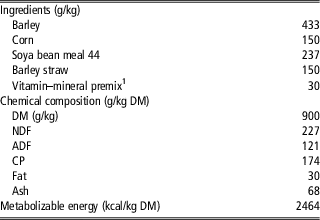
A total of 24 male Merino lambs, penned individually with their corresponding ewe during the suckling period, were used in this experiment. The lambs were stratified on the basis of live BW at birth (average BW 4.81±0.256 kg), treated with a subcutaneous injection of vitamin E and Se (1 ml per animal of Selevit Complex, Syva) to prevent white muscle disease, and then assigned randomly to one of two experimental treatments (n=12 per dietary treatment) during the suckling period. The first group of lambs (ad libitum, ADL) was kept permanently with the sheep whereas the other group (restricted, RES) was separated from the dams from 0900 to 1800 h and milk restricted. Dams of the RES group were injected with oxytocin to remove alveolar milk and then milked at 1700 h before the reintroduction of lambs. Lambs were weighed twice a week throughout the experiment and vaccinated against enterotoxaemia and pasteurella (Heptavac P Plus, MSD Animal Health) at 4 weeks of age. When each lamb reached 13.5 kg of BW it was weaned progressively (free access to a complete pelleted diet and alfalfa, whereas it was allowed only 2 h with the dam) until it weighed 15 kg. Then, all the animals were penned individually, had free access to fresh drinking water and were offered the same complete pelleted diet at the same level (35 g/kg BW per day) to avoid differences in DM intake during the fattening period as explained elsewhere (Santos et al., Reference Santos, Valdés, Giráldez, López, France, Frutos, Fernández and Andrés2018b). The average daily gain was calculated by linear regression between the lamb BW and the weighing day. All the lambs received the complete pelleted diet once a day at 0900 h, with the amount of feed offered being adjusted twice a week on the basis of the BW. Ingredients and chemical composition of the complete pelleted diet are summarized in Table 1.
Ingredients and chemical composition of the complete pelleted diet fed during the fattening period

1 10 g/kg CaCO3, 5 g/kg NaCl, 7 g/kg NaHCO3, 4 g/kg CaHPO4, 2 g/kg ClNH4, 2 g/kg premix [manufactured by DMS Nutritional Products, S.A. and containing (per kg) 2.25 g vitamin A, 0.02 g vitamin D3, 10 g vitamin E, 0.25 g vitamin K3, 0.50 g vitamin B1, 0.50 g vitamin B2, 2.50 g vitamin B5, 0.50 g vitamin B6, 5 g vitamin B12, 5 g vitamin B3, 50 g choline chloride, 17.50 g Fe, 15 g Zn, 2.5 g Cu, 20 g Mn, 0.05 g Co, 0.25 g I, 0.10 g Se, 259 g Ca and 50.5 g Mg].
Sampling and storage
All the animals were slaughtered after a fattening period of at least 50 days when they reached the target BW of 27 kg. Feed was withdrawn 6 h before slaughtering; then lambs were weighed, stunned, slaughtered by exsanguination from the jugular vein, eviscerated and skinned according to the Council Regulation (EC) No. 1099/2009 on the protection of animals at the time of killing. A 10 cm segment of ileum was collected proximal to the ileocecal valve immediately after slaughter. Ileal samples were rinsed three times with sterile phosphate-buffered saline solution to remove the digesta and divided into several portions. Tissue samples were preserved in different ways for analysis of microbial composition (stored at −80°C during 48 h, then freeze-dried), flow cytometry (sterile phosphate-buffered saline solution), histological or immunohistochemical examination (fixed by immersion in 10% buffered formalin for 1 week), immunoglobulin A (IgA) quantification (stored at −20°C), and gene expression (RNAlater Invitrogen, Lithuania; stored at −80°C).
DNA extraction and quantitative real-time polymerase chain reaction of bacteria attached to the ileal mucosa
The luminal part of the freeze-dried ileal mucosa (15 mg) was scraped with a scalpel and subsequent microbial DNA purification with the Ultra-Deep Microbiome Prep kit (Molzym, Bremen, Germany) was performed according to manufacturer’s instructions. The concentration of DNA was measured in each sample with a NanoDrop UV-Vis Spectrophotometer (NanoDrop Technologies, Wilmington, DE, USA), and then quantitative real-time PCR was used to quantify total bacteria attached to the ileal mucosa and the relative abundance of some genus very related to the health status of the host (e.g. Prevotella spp., Lactobacillus spp. and Bifidobacterium spp.) according to the procedures described by Andrés et al. (Reference Andrés, Bodas, Tejido, Giráldez, Valdés and López2016), Vargas et al. (Reference Vargas, Andrés, Snelling, López-Ferreras, Yáñez-Ruiz, García-Estrada and López2017) and Delroisse et al. (Reference Delroisse, Boulvin, Parmentier, Dauphin, Vandenbol and Portetelle2008), respectively. Finally, quantitative real-time PCR data were normalized according to the previous concentration of DNA, and results were expressed as fold change (log2 of ‘number of copies’ per milligram of genomic DNA, RES/ADL) of overrepresented species in ileal epimural microbiota (positive value if RES/ADL>1; negative value if ADL/RES>1).
Mechanical disruption of ileal tissue and flow cytometry
In order to quantify leucocyte population in the ileum, a single colour flow cytometry analysis was performed. Mucosal leucocytes were labelled with the leucocyte common antibody CD45 (Charavaryamath et al., Reference Charavaryamath, Fries, Gomis, Bell, Doig, Guan, Potter, Napper and Griebel2011). The mucosa of ileal samples was scraped off with a scalpel; the homogenates were mixed with 8 ml of cold phosphate-buffered saline solution (pH 7.4) and digested in a masticator paddle blender (IUL Instruments, Barcelona, Spain). Contents were filtered, added carefully to 8 ml of Lymphoprep (Axis-Shield PocAS, Oslo, Norway) and centrifuged at 1000×g and 21°C for 30 min. Layer of leucocytes was extracted, moved to a falcon tube with 20 ml of Hanks’ Balanced Salt Solution at 4°C and centrifuged at 270×g and 4°C for 10 min. Then, the pellet was washed twice by centrifugation at 270×g and 4°C for 10 min in RPMI 1640 medium with 10% foetal bovine serum (Lonza, Basilea, Switzerland) and 1% antibiotic antimycotic solution. The pellet was re-suspended in 5 ml cold of RPMI–foetal bovine serum. Cells were counted using Bio-Rad TC10 (Bio-Rad Laboratories, Hercules, CA, USA), and adjusted with RPMI–foetal bovine serum to a final concentration of 2×10⁶ cells/ml. For antibody labelling, briefly, 50 μl of suspension were incubated for 60 min at 4°C with a specific primary monoclonal antibody against CD45 antigen (Corpa et al., Reference Corpa, Juste, Marín, Reyes, González and Pérez2001) diluted 1 : 5 in phosphate-buffered saline solution, centrifuged at 142×g and 4°C for 3 min, washed twice by centrifugation at 270×g and 4°C, then re-incubated for 30 min at 4°C in the dark with 50 μl of fluorescein isothiocyanate conjugated rabbit antimouse immunoglobulin (Dako, Glostrup, Denmark) diluted 1 : 50. After two further washes, cells were kept at 4°C until analysis on a FACScan flow cytometer (BD, San Jose, CA, USA) equipped with CellQuest software (BD). Results were expressed as the percentage of positive stained cells in sample populations of 10 000 individual cells.
Histological and morphometric analysis
Formalin-fixed samples from ileum were trimmed and processed for paraffin embedding and histological examination (haematoxylin–eosin staining). Slides were examined with a Leica DM2000 LED microscope and digital pictures were taken at 4× magnification. Thickness of mucosa, submucosa and tunica muscularis was measured at 10 different sites in each picture using the image processing and analysis software ImageJ v1.6.0_14 (National Institutes of Health (NIH), Bethesda, MD, USA).
Immunohistochemical analysis
Immunohistochemical analysis was performed according to the procedure described by Arranz-Solís et al. (Reference Arranz-Solís, Benavides, Regidor-Cerrillo, Horcajo, Castaño, Ferreras, Jiménez-Pelayo, Collantes-Fernández, Ferre, Hemphill, Pérez and Ortega-Mora2016). Briefly, cross sections were cut from the ileum wall samples and placed onto poly-l-lysine-coated slides for immunohistochemical labelling of T (CD3 antigen), B (CD20 antigen) and M-cells (cytokeratin 18 antigen). Endogenous peroxidase activity was blocked in deparaffinized sections by immersion in 3% hydrogen peroxide in methanol for 30 min in darkness at room temperature and rehydrated slides were rinsed The antigen retrieval was performed using heat-based methods (Table 2). After washing, sections were incubated with 100 µl of the primary antibodies diluted in phosphate-buffered saline solution overnight at 4°C in a humidified chamber.
Primary antibodies used in immunohistochemistry of ileal samples from fattening lambs

CD=cluster of differentiation.
After washing, sections were incubated for 40 min at room temperature with 100 µl of EnVision+/HRP solution (Dako, Glostrup, Denmark). After washing, antibody localization was determined using 100 µl of 3.3-diaminobenzidine (Sigma-Aldrich Corp., Saint Louis, MO, USA) as chromogenic substrate for peroxidase. Sections were counterstained with haematoxylin for 30 s and mounted. Quantification of labelled cells was performed under a light microscope with a 40× objective. The number of T and B cells was counted in ten random fields within the lamina propria of the ileum.
Quantification of M-cells labelling intensity was performed measuring the % of positive labelled area in the total area of epithelial cells in iPP dome with the plugin IHC Profiler (Varghese et al., Reference Varghese, Bukhari, Malhotra and De2014) of ImageJ. Domes were photographed under a 20× objective, and epithelial cells of domes were delimited as regions of interest. Positive labelling was automatically measured by the software.
Ileal mucosa antibody recovery and immunoglobulin A quantification
Immunoglobulin A was quantified according to the procedure described by Ahmed et al. (Reference Ahmed, Sebastiano, Sweeney, Hanrahan, Glynn, Keane, Mukhopadhya, Thornton and Good2015). Briefly, ileal samples were unfrozen overnight at 4°C; then the surface was scraped with a scalpel and 2 g were collected in a falcon tube with 6 ml of ice cold phosphate-buffered saline solution supplemented with protease inhibitors (Sigma-Aldrich Corp.). Samples were vortexed, incubated overnight at 4°C and centrifuged at 3000×g for 30 min at 4°C. Protein concentration of each supernatant was measured with the Pierce BCA protein assay kit (Thermo Fisher Scientific, Waltham, MA, USA), and adjusted to 500 µg/ml using phosphate-buffered saline solution supplemented with protease inhibitors. The amount of IgA was measured in ileal mucosa using a Genorise ELISA IgA kit following manufacturer’s instructions (Genorise Scientific, Devon-Berwyn, PA, USA).
RNA extraction and real-time reverse transcription polymerase chain reaction
Total RNA was extracted from 100 mg of ileal samples (including iPP) according to the procedure described by Abecia et al. (Reference Abecia, Jiménez, Martínez-Fernández, Martín-García, Ramos-Morales, Pinloche, Denman, Newbold and Yáñez-Ruiz2017). Briefly, samples were homogenized with 0.9 mm stainless steel bead and 1 ml of TRIzol Reagent (Invitrogen, Carlsbad, CA, USA), using a Bullet Blender homogenizer (Next Advance, Troy, NY, USA). The homogenate was incubated for 5 min at room temperature before adding 200 μl chloroform/ml of TRIzol-containing tissue homogenate. The aqueous phase was precipitated with 500 μl isopropanol/ml of TRIzol-containing tissue homogenate, and applied to an RNeasy Mini-column (Qiagen, Hilden, Germany). RNA bound to the column was DNase treated using RNase-Free DNase Set (Qiagen) and collected in 50 μl volume eluent. The RNA quantity was measured using ND 1000 spectrophotometer (NanoDrop Technologies, Wilmington, DE, USA) and RNA integrity number was evaluated using Bioanalyzer 2100 (Agilent Technologies, Santa Clara, CA, USA).
Total RNA (1 µg) was reversed transcribed using QuantiTect Reverse Transcription Kit (Qiagen) according to manufacturer’s instructions. RNA was used as template for real-time reverse transcription PCR analysis to evaluate the expression of genes encoding 10 toll-like receptors (TLRs) in the ileal epithelium using the gene-specific primer pairs described in Malmuthuge et al. (Reference Malmuthuge, Li, Fries, Griebel and Guan2012) relative to β-actin expression whereas for cytokines the amplification conditions and primers were those described by Martínez-Pérez et al. (Reference Martínez-Pérez, Robles-Pérez, Rojo-Vázquez and Martínez-Valladares2014). For each PCR reaction, 25 ng of cDNA were amplified with each primer set using the parameters described by Abecia et al. (Reference Abecia, Jiménez, Martínez-Fernández, Martín-García, Ramos-Morales, Pinloche, Denman, Newbold and Yáñez-Ruiz2017) on the Bio-Rad iCycler (Bio-Rad Laboratories Ltd., Mississauga, ON, Canada). Amplification data were expressed as change in quantification cycle (ΔC q value) and calculated as follows: ΔC q=C q (cytokines or TLRs)−C q (β-actin). A smaller ΔC q value equates to more abundant transcript.
Statistical analysis
Growth performance data together with those of quantification of microbiota, flow cytometry, morphometric analysis, immunohistochemistry, IgA quantification and gene expression were analysed by one-way ANOVA using the GLM procedure of SAS (SAS Institute Inc., Cary, NC, USA) with the suckling regime (ad libitum or restricted) as the only source of variation. In all cases, the individual lamb was considered as the experimental unit. Significance was declared at P<0.05.
Results
Animal performance
Average daily gain during the suckling phase was reduced for RES lambs (267 v. 191 g/day; P<0.001) and the age at weaning (BW of 15 kg) increased (42 v. 55 days; P<0.001). Furthermore, significant differences in gain to feed ratio (average daily gain/DM intake) were observed during the fattening phase (0.320 v. 0.261 for the ADL and RES groups, respectively; P<0.001) despite the similar DM intake during this period of time. Thus, RES animals required more days (74 v. 62 days; P<0.001) to reach the intended BW at slaughter (27 kg).
Ileal epimural microbiota
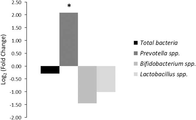
No differences were observed for total bacteria, Lactobacillus spp. or Bifidobacterium spp. between experimental groups (ADL v. RES lambs, Figure 1). However, a significant increase of the relative abundance of Prevotella spp. in the ileal epimural microbiota of early feed restricted lambs was detected (Figure 1; log2FC=2.09).
(colour online) Relative quantitation compared with ad libitum lambs (ADL) of 16S rRNA copy numbers of microbial groups attached to the ileal mucosa after early feed restriction (RES) during the suckling period. Fold-changes for specific amplicon groups were calculated as log2 ratio of normalized abundances. *Significant difference in copy number (P<0.05) was calculated using Tukey’s method (SAS).

Immunity parameters
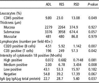
The results regarding the counts of CD45+ cells (flow cytometry), thickness of ileal layers (histologic examination), M-cells labelling intensity, B or T infiltrating lymphocytes counts (immunohistochemistry), and IgA concentration in ileal mucosa (ELISA) are summarized in Table 3.
Leucocyte counts measured by flow cytometry in the ileum, thickness of ileal layers, infiltrating lymphocyte counts in ileal lamina propria, intensity of staining of M-cells in ileal mucosa and ileal immunoglobulin A concentration of fattening lambs being fed ad libitum (ADL) or restricted (RES) during the suckling period

RSD=residual standard deviation; CD=cluster of differentiation; IgA=immunoglobulin A.
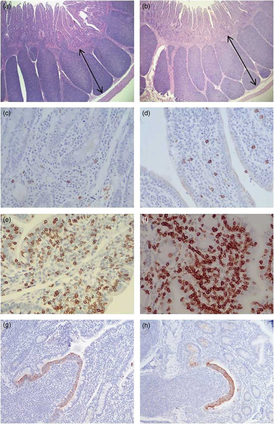
Greater percentages for CD45+ were detected in the ileum of the RES group when compared with the ADL lambs (P=0.049). Moreover, morphometric analysis revealed a trend towards a greater thickness of ileal submucosa layer in the RES group (P=0.057) where the iPP are located (Figure 2). Likewise, a higher infiltration of B and T cells in the ileal lamina propria was observed in the RES lambs (P=0.007 and 0.042, respectively; Table 3; Figure 2). Also, significantly higher percentages of the epithelium region in the iPP domes were labelled with high (P=0.068) or medium intensity (P=0.008) when M-cell marker was assessed in the RES group (Table 3; Figure 2), whereas those negatively labelled showed a trend towards being significantly higher (P=0.067) in the ADL group. Similarly, secretory IgA at ileal mucosa level was greater for the early feed restricted lambs (P=0.037).
(colour online) Thickness of submucosa layer (arrows) in ileal segments of ad libitum (ADL) (a) and early feed restricted (RES) lambs (b). B cells labelled in the lamina propria of ADL (c) and RES lambs (d). T cells labelled in the lamina propria of ADL (e) and RES animals (f). Cytokeratin 18 labelling (M-cells) in Peyer’s patches domes of ADL (g) and RES lambs (h). Magnification was 4× for pictures A and B; 40× for pictures (c, d, e and f); and 20× for pictures (g) and (h).

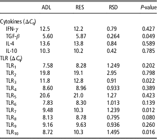
Cytokines and TLRs gene expression at the ileal epithelium (including iPP) is shown in Table 4. No significant differences in mRNA expression of interferon gamma (IFN-γ), interleukin 4 (IL-4) and interleukin 10 (IL-10) were observed. However, transforming growth factor beta (TGF-β) was down-expressed in the RES group (P=0.043). In addition, gene expression of several genes was significantly downregulated in the early feed restricted group (e.g. TLR3, P=0.022; TLR7, P=0.012; and TLR10, P=0.016).
Cytokines and toll-like receptors (TLRs) mRNA expression in the ileal mucosa of fattening lambs being fed ad libitum (ADL) or restricted (RES) during the suckling period

C q=quantification cycle. ΔCq=C q(cytokines or TLRs)–C q(β-actin). Lower C q represents higher RNA abundance level; IFN-γ=interferon gamma; TGF-β=transforming growth factor beta; IL-4=interleukin 4; IL-10=interleukin 10; RSD=residual standard deviation.
Discussion
Early feed restriction may impact colonization patterns of gastrointestinal mucosa during early postnatal period and immunity parameters at this level, thus promoting signalling events along the lifespan of the animal and adversely impacting feed efficiency during post-weaning phases (Greenwood and Cafe, Reference Greenwood and Cafe2007).
In the present study, the number of total bacteria attached to enterocytes was similar in both groups; nevertheless a higher presence of Prevotella spp. in the ileal epimural microbiota of the RES group was observed. Prevotella spp. strains are Gram-negative, anaerobic bacteria showing multiple virulence factors such as lipopolysaccharide, which facilitate the adhesion and invasion of host cells and chronic inflammatory conditions in the gut (Lukens et al., Reference Lukens, Gurung, Vogel, Johnson, Carter, McGoldrick, Bandi, Calabrese, Vande Walle, Lamkanfi and Kanneganti2014; Ley, Reference Ley2016). It must be remarked that these studies are relative to humans, whereas there is scarce information about the role of Prevotella spp. in the intestine of ruminants (Mao et al., Reference Mao, Zhang, Liu and Zhu2015). However, it is tempting to speculate that the adhesion of Prevotella spp. to the ileal mucosa might have promoted pro-inflammatory conditions at this level and, consequently, lower feed efficiency traits for this group of animals as will be explained below. In any case, in accordance with our results, the relative abundance of Prevotella spp. has been shown to be increased in the ruminal liquid of inefficient (high residual feed intake) bulls (McCann et al., Reference McCann, Wiley, Forbes, Rouquette and Tedeschi2014) and in the colon from steers showing low feed efficiency traits (Myer et al., Reference Myer, Wells, Smith, Kuehn and Freetly2015c). Regrettably, the reasons behind different efficiency traits in these studies were not ascertained.
The outgrowth of Prevotella spp. has been described to occur with a concomitant reduction in the relative abundance of lactic acid bacteria such as Lactobacillus spp. in the intestinal microbiome, especially under autoinflammatory disease conditions (Lukens et al., Reference Lukens, Gurung, Vogel, Johnson, Carter, McGoldrick, Bandi, Calabrese, Vande Walle, Lamkanfi and Kanneganti2014). This is important, as Lactobacillus acidophilus and Bifidobacterium spp. are beneficial organisms due in part to their ability to attach to enterocytes, thus inhibiting the binding of enteric pathogens by competitive exclusion. Lactic acid bacteria also generate signals for the production of cytokines modulating immune response, and decreasing inflammatory-intestinal damage (Villena et al., Reference Villena, Aso and Kitazawa2014; Lim et al., Reference Lim, Jeong, Jang, Han and Kim2016). However, in the present study, although numerically lower values were observed for these bacterial groups in the RES lambs, the differences in the abundance of Lactobacillus spp. and Bifidobacterium spp. did not reach the significance level, probably due to the low levels of these bacterial groups detected in both groups of lambs.
In addition, a clear influence of early feed restriction in several parameters related to the immune response was observed according to the results of the present study. In this sense it was enticing to speculate, and warrants further investigation, that the differences in the colonization of gastrointestinal mucosa promoted by early feed restriction during the suckling period of these lambs might have triggered those changes found in the immunological parameters evaluated. In fact, it has been suggested that a different exposure to pathogenic bacteria might alter the ontology of M-cells, which are essentially epithelial cells of the gastrointestinal mucosa specialized in sampling and translocating luminal antigens to the underlying lymphoid tissue (Prims et al., Reference Prims, Pintens, Vergauwen, Van Cruchten, Van Ginneken and Casteleyn2017). Therefore, a plausible hypothesis explaining these variations would be that the increased relative abundance of Prevotella spp. in the RES group might have contributed to enhance the expression of cytokeratin 18 antigen in the surface of M-cells, as suggested by the higher staining intensity of M-cells in the iPP domes of these animals. This is important, as the expression of cytokeratin 18 has been related to the M-cell function (e.g. antigen uptake; Gebert et al., Reference Gebert, Rothkotter and Pabst1994; Prims et al., Reference Prims, Pintens, Vergauwen, Van Cruchten, Van Ginneken and Casteleyn2017). Thus, this circumstance might explain, at least partially, the higher levels of CD45+ (flow cytometry), the higher infiltrations of B and T cells in the lamina propria, and the trend towards a greater thickness of the ileal submucosa layer (where iPP are located) in the RES lambs. In any case, the analysis of a more diverse immune cell population and in vitro functional studies are necessary to adequately evaluate this hypothesis.
Moreover, there were also significant differences between both groups of lambs as far as the amount of IgA in the ileal mucosa is concerned. IgA works across the gastrointestinal tract as an inhibitor of bacterial/viral adherence and penetration into the underlying epithelium, providing mucosal immune protection (Cerutti and Rescigno, Reference Cerutti and Rescigno2008). Therefore, it might be feasible to have a higher concentration of ileal IgA in the RES group produced to neutralize microbial products such as lipopolysaccharide in intestinal epithelial cells.
Finally, no differences in the level of expression of several cytokines were observed excepting for TGF-β, which was reduced in the RES lambs. According to Letterio and Roberts (Reference Letterio and Roberts1998) the downregulation of this cytokine in the RES group might be indicative of the existence of a negative regulatory mechanism to reduce lymphocytes infiltration in the lamina propria and the inflammatory response. In agreement with this hypothesis, the downregulation of some TLRs (pattern-recognition receptors of pathogen-associated molecular patterns located in several cell types such us macrophages, T and B cells or non-immune cells) observed in the ileal mucosa of RES lambs might have been implemented in an attempt to keep the homeostasis at intestinal level (Zhang and Ghosh, Reference Zhang and Ghosh2002; Malmuthuge et al., Reference Malmuthuge, Li, Fries, Griebel and Guan2012; Villena et al., Reference Villena, Aso and Kitazawa2014). However, as stated beforehand, further functional studies are necessary to adequately evaluate this hypothesis.
Conclusions
Early feed restriction during the suckling period of Merino lambs modifies both bacterial microbiota attached to the ileum and immunity parameters at this level. These changes might be related to a decreased amount of nutrients towards productive processes (e.g. growth or fattening) during the fattening period. Actions to improve gut microbiome towards a healthier profile (e.g. prebiotics, probiotics, and nutritional management to decrease Prevotella spp. abundance) should be tested during the early life of lambs as a feasible way to minimize the negative effects on feed efficiency of those animals exposed to feed restriction during the suckling period.
Acknowledgements
This work was funded by the Spanish Ministry of Economy, Industry and Competitiveness (MINECO, AGL2014-54124R). The authors would also like to thank MINECO (BFU2014-57964-R) for TLR quantification and CSIC (Proyecto Intramural Especial; Project 201540E084) for supporting J. Frutos with a training contract. Alba Santos gratefully acknowledges receipt of a pre-doctoral grant from the Spanish Ministry of Education, Culture and Sport (MECD, FPU15/01630). María Martínez-Valladares was funded by the Spanish ‘Ramón y Cajal’ Programme of the Ministry of Economy and Competitiveness (RYC-2015-18368).
Declaration of interest
None.
Ethics statement
All handling practices followed the recommendations of the Directive 2010/63/EU of the European Parliament and of the Council on the protection of animals used for scientific purposes and the IGM-CSIC Animal Experimentation Committee (protocol number 2015-04).
Software and data repository resources
None.