Tricuspid valve atresia is a rare CHD consisting of a plate-like shelf in the tricuspid valve position resulting in the obligatory right-to-left shunt, at the atrial level and a variably hypoplastic right ventricle. We report an interesting haemodynamic phenomenon of “functional” or “pseudo” tricuspid atresia leading to an echocardiographic finding mimicking tricuspid atresia in a neonate with Barth syndrome and severe cardiomyopathy.
Case report
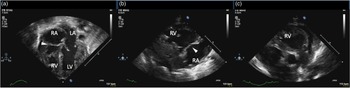
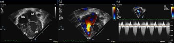
A 39-week-gestation male was born via vaginal delivery at an outside hospital. Mother received prenatal care and an antenatal ultrasound at 27 weeks of pregnancy which was unremarkable in terms of cardiac anatomy, ventricular function, or rhythm. Family history was significant for Barth syndrome in the mother’s previous child who had presented with dilated cardiomyopathy during late infancy. At birth, APGAR scores at 1 minute and 5 minutes were 1 and 1, respectively, and he required resuscitation with chest compressions. The patient required intubation, and once stabilised, the maximum SpO2 was about 80% even with FiO2 100%. There were no significant differences in the pre- and post-ductal oxygen saturations which were close to 80%. A chest X-ray showed significant cardiomegaly, which prompted for an echocardiogram. Initial echocardiogram revealed moderate biventricular hypertrophy and severe biventricular systolic dysfunction. In addition to hypertrophy and dysfunction, the left ventricle had a non-compacted appearance. The tricuspid valve appeared as a plate-like structure with a complete absence of forward flow across it (Fig 1, Supplemental video 1). However, mild-to-moderate regurgitation was seen across this plate-like structure implying patency of the tricuspid valve (Supplemental video 2). There was a moderate degree of pulmonary valve regurgitation with the minimal forward flow (Supplemental video 3). As expected, a large right-to-left shunt was seen across the atrial septum (Supplemental video 4) and a large left-to-right shunt was seen through the ductus arteriosus (Supplemental video 5). The estimated right ventricle fraction area change was 12% and tricuspid annular plane systolic excursion was 2.0 mm. The left ventricle ejection fraction measured was 20% (Simpson method). These measures suggested severely decreased biventricular function. There was no pericardial effusion. Due to the absence of forward flow across the tricuspid valve, prostaglandin infusion was initiated to maintain a steady source of pulmonary blood flow. Inotropic support with dobutamine, milrinone, and epinephrine was provided for ventricular dysfunction and inhaled nitric oxide was started at 20 ppm to decrease the pulmonary vascular resistance. The patient’s oxygen demand gradually decreased, and FiO2 was weaned down from 100% to 30% over the next 3 days. The inhaled nitric oxide and prostaglandin were also weaned off on days 3 and 4 of age, respectively. A repeat echocardiogram at day 3 of age demonstrated forward flow across the tricuspid valve with some leaflet excursion and moderate tricuspid valve regurgitation (Fig 2). There was a mild subjective improvement in right ventricle systolic function (the estimated right ventricle fraction area change; 19%) while there was no significant improvement in the left ventricle function. As the forward flow across the tricuspid valve improved and with improvement in the right ventricular function, there was an improvement in forward flow across the pulmonary valve. There was no significant change in pulmonary regurgitation compared with echocardiogram at day 1 of age. There was no significant change in the patent ductus arteriosus (Supplemental video 6). Persistent lactic acidosis and poor cardiac output led to multi-organ failure and life support was withdrawn at 6 weeks of age.
Transthoracic echocardiogram on day 1 of age. The tricuspid valve leaflets were not moving (arrowhead). ( a ) Apical four-chamber view. ( b ) Parasternal long-axis view. ( c ) Pulmonary valve was normal with open valve leaflets in parasternal long-axis view.

Transthoracic echocardiogram on day 3 of age. The tricuspid valve leaflets started to move. ( a ) Apical four-chamber view. Antegrade flow was started to be seen on colour Doppler ( b ) and continuous mode Doppler shows the flow pattern across the tricuspid valve ( c ).

On molecular testing, he was hemizygous for the familial pathogenic variant identified in TAZ (c.109 + 5G > A (intronic) confirming Barth syndrome. It is likely that the aetiology of his fulminant cardiomyopathy was a combination of Barth syndrome and neonatal birth asphyxia.
Discussion
We describe a very unusual neonatal presentation of Barth syndrome with unique echocardiographic findings resulting from the deranged/altered haemodynamic status. Cardiac findings included severe cardiomyopathy including a combination of dilated, hypertrophic cardiomyopathy and left ventricle non-compaction. Echocardiographic findings mimicking tricuspid atresia or “pseudo tricuspid atresia” where the tricuspid valve leaflet did not move at all and there was obligatory right-to-left atrial shunt. Despite no tricuspid valve leaflet excursion and no forward flow in diastole, there was significant regurgitation across this atretic appearing tricuspid valve.
The possible mechanisms for no forward flow across the tricuspid valve and no valve leaflet excursion could be (1) a stiff and tense right ventricle with moderate hypertrophy and severely decreased systolic/diastolic function, (2) persistently elevated pulmonary resistance, or (3) ventricular interaction due to severe left and right ventricle dysfunction. In our case, as the pulmonary vascular resistance decreased with the use of inhaled nitric oxide and milrinone; and with the improvement of right ventricular systolic function, there was an improvement in the forward flow across the tricuspid valve. Prostaglandin was weaned off on day 4 of age to let the ductus arteriosus close spontaneously. However, though the ductus became smaller in size, ductus arteriosus continued to be patent until her death. Wald et al summarised the clinical course of 28 patients with neonatal Ebstein’s anomaly with anatomical and functional pulmonary valve obstruction. Amongst 24 patients who received prostaglandin, 17 patients discontinued prostaglandin after the relief of the pulmonary obstruction, which resulted in uneventful ductal constrictions. They concluded the early ductal closure in the absence of outflow tract obstruction and early limitation of ductal patency after relief of the obstruction may contribute to improved survival in neonates with Ebstein’s anomaly.Reference Wald, Adatia, Van Arsdell and Hornberger1
On a detailed review of the literature, we found only one case reported by Patel and colleagues of functional tricuspid atresia in a neonate with cardiomyopathy who was diagnosed prenatally. This neonate had a normal initial foetal echocardiogram with a strong family history of cardiomyopathy and a follow-up echocardiogram at 30 weeks gestation showed functional tricuspid and pulmonary atresia with biventricular systolic dysfunction and left ventricle non-compaction. The echocardiographic findings were similar to our case, including the mixed type of cardiomyopathy and no forward flow across the tricuspid valve but regurgitation. This supports that a similar ha emodynamic derangement was playing a role for the lack of tricuspid valve leaflet excursion and forward flow resulting in “pseudo tricuspid atresia.” The baby was managed similarly with prostaglandin drip, inotropic, and ventilator support. The authors report improved antegrade tricuspid and pulmonary blood flow by 2 weeks of age and off prostaglandin by 3 weeks of age. In their case, the underlying cause of cardiomyopathy was CAP2 gene mutation, which is implicated in the regulation of actin dynamics, sarcomere organisation in striated muscles, and possibly cardiac conduction disorders.Reference Patel and Peterson2 Even though the clinical presentation was strikingly similar, our patient had a lethal outcome, unlike this patient. On molecular testing, our patient was found to have a mutation in the TAZ gene in the distal region of chromosome Xq28 as seen in Barth syndrome. This mutation affects the production of tafazzin, a protein located in mitochondria that alters cardiolipin, which plays a critical role in the mitochondrial inner membrane. This may affect adversely multiple systems in the body, especially those with high energy demands like the heart and skeletal muscles.Reference Sabbah3,Reference Dudek and Maack4 Clinical presentation can be highly variable, and cardiomyopathy is the major clinical feature especially dilated cardiomyopathy. The causes of neonatal cardiomyopathy include neonatal asphyxia, myocarditis, arrhythmia, neuromuscular diseases, and other genetic disorders. Identification of the exact cause is usually difficult, and possible in only one-third of the patients. Genetic form of dilated cardiomyopathy occurs in 25–50% of patients.Reference Soares, Rocha and Pissarra5 It is likely that neonatal asphyxia contributed to the clinical picture and to the severity and early presentation of cardiomyopathy in our patient.
Our case highlights an interesting haemodynamic phenomenon of “pseudo tricuspid atresia” seen echocardiographically in patients with significant haemodynamic derangement resulting from cardiomyopathy. Regurgitation may be the only “telltale sign” for the patency of the valve in this situation. As cardiac functioning improves, forward flow across the valve is established with judicious use of inotropes, afterload reducing agent, and or pulmonary vasodilator.
Supplementary material
For supplementary material accompanying this paper visit https://doi.org/10.1017/S104795112100322X
Acknowledgments
None.
Financial support
This research received no specific grant from any funding agency, commercial, or not-for-profit sectors.
Conflicts of interest
None.
Author contributions
D.T.: writing the manuscript; all authors: critically reviewing the manuscript.