Mycetoma
A paranasal sinus fungus ball or sinus mycetoma is a non-invasive fungal infection seen in immunocompetent persons. Aspergillus fumigatus is the most frequently isolated organism.
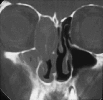
Affected people often present with long-standing symptoms of nasal obstruction, a unilateral, purulent nasal discharge, cacosmia and occasionally proptosis (Figure 1). In the majority of patients, only one sinus is affected.Reference Klossek, Serrano, Peloquin, Percodani, Fontanel and Pessey1 The maxillary sinus is most commonly involved, with partial or complete opacification and bone thickening; sclerosis or bone destruction can also occur.
Coronal computed tomography scan showing mycetoma affecting the right ethmoid sinuses. The patient presented with mild proptosis.

Computed tomography (CT) shows opacification of the involved sinus, often associated with flocculent calcifications. Histopathological investigation should reveal that this material is composed of a dense, matted conglomeration of fungal hyphae, separate from the mucosa of the sinus; it should also demonstrate no evidence of allergic mucin in the sinus, nor granulomatous reaction in the mucosa.
Surgical removal of the fungus ball is the treatment of choice. If the person then becomes immunocompromised, invasive fungal sinusitis may develop.Reference Ferguson2
Allergic aspergillosis
Allergic fungal sinusitis is a non-invasive disorder seen in immunocompetent individuals.
The criteria for diagnosis of this condition have been revised several times. However, most authors agree on the following criteria: presence in patients with chronic rhinosinusitis (confirmed by CT scan), nasal polyposis (unilateral or bilateral), and characteristic allergic mucin containing clusters of eosinophils and Charcot–Leyden crystals; presence of fungal organisms within that mucin (detectable on staining or culture); and presence of type one (immunoglobulin (Ig) E mediated) hypersensitivity to fungi.Reference Kuhn and Swain3
Aspergillus species are believed to be the predominant cause of allergic fungal sinusitis. More recent series suggest that various dematiaceous (brown-pigmented) environmental moulds, including alternaria, bipolaris, cladosporium, curvularia and drechslera species, can also be responsible.Reference Manning and Holman4
Allergic aspergillosis occurs in young, immunocompetent adults with chronic, relapsing rhinosinusitis which is unresponsive to antibiotics, antihistamines or corticosteroids. Patients do not have underlying immunodeficiencies, and 50–70 per cent are atopic. There is no male or female predominance. Cases of allergic fungal sinusitis have been described from different parts of the world, but the condition appears to be most prevalent in warm, humid areas such as the Indian subcontinent, Australasia and the southern United States (where it accounts for about 7 per cent of all sinus surgery).Reference Cody, Neel, Ferreiro and Roberts5
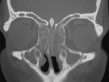
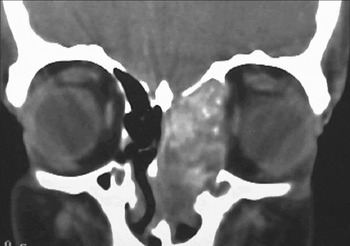
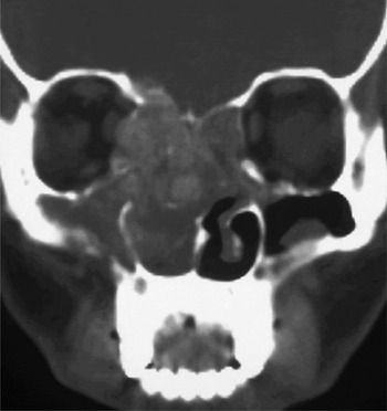
There are no unique, pathognomonic symptoms. Patients often present with unilateral nasal polyposis and thick, yellow-green nasal or sinus mucus. Nasal polyposis can be unilateral or bilateral (Figure 2) and may form an expansive mass that causes bone necrosis (Figure 3). Should the lamina papyracea of the ethmoid bone be affected, the condition can expand this area and cause proptosis. Polypoid material can also push the nasal septum into the contralateral airway. Computed tomography scans often reveal a characteristic serpiginous opacification of more than one sinus, mucosal thickening and erosion of bone; however, there is no tissue invasion (Figure 4).
Coronal computed tomography scan showing bilateral allergic fungal sinusitis.

Coronal computed tomography scan showing allergic aspergillosis eroding the surrounding bone.

Coronal computed tomography scan showing allergic aspergillosis expanding and eroding the surrounding bone, giving the impression of invasion. However, no mucosal invasion was present, and aspergillus precipitin levels were very elevated.

The treatment of allergic fungal sinusitis includes surgical debridement to remove polyps and the allergic mucin. Adjunctive medical management is beneficial because not all the fungal elements can always be removed. In studies, post-operative systemic corticosteroids reduced recurrence of disease, but there was a high recurrence rate.Reference Kupferberg, Bent and Kuhn6, Reference Kuhn and Javer7
Allergic aspergillosis has been likened to allergic bronchopulmonary aspergillosis; in other words, it is a systemic reaction to an allergen in the respiratory tract.Reference Schubert8 Oral itraconazole has been studied in a randomised, controlled trial researching the pulmonary form of allergic fungal sinusitis, allergic bronchopulmonary aspergillosis, and has been shown to be effective.Reference Stevens, Schwartz, Lee, Moskovitz, Jerome and Catanzaro9 In allergic fungal sinusitis, there have been anecdotal reports of the use of post-operative itraconazole.Reference Jonathon, Lund and Milroy10, Reference Rains and Mineck11 Rains et al. reported retrospectively on 139 patients with allergic fungal sinusitis treated with steroids and post-operative itraconazole; they found that this regime may reduce the need for revision surgery.Reference Rains and Mineck11
Our experience, along with that of others (K R Meganadh, personal communication), is that the use of pre-operative itraconazole for four weeks markedly reduces the extent of surgery required. When given together with a six-week post-operative course, itraconazole reduces recurrence rates.Reference Andes, Proctor and Bush12 Long-term, repeated courses of oral corticosteroids reduce symptomatic recurrence; however, such treatment must be avoided in patients with diabetes, blood dyscrasias, immunodeficiency, glaucoma, osteoporosis and hepatitis, amongst a range of contraindications.Reference Kuhn and Javer7 Topical steroids also help reduce symptomatic recurrence. Total serum IgE and endoscopic examination have been used for early monitoring and detection of recurrent disease.
There is no published evidence that topical antifungal treatment is of benefit. Immunotherapy has been advocatedReference Mabry, Marple and Folker13 but has yet to be proven by a controlled study.
Eosinophilic mucin rhinosinusitis
This condition has been described by Ferguson.Reference Ferguson14 In contrast to allergic aspergillosis, in which approximately 40 per cent of patients have asthma, over 90 per cent of eosinophilic mucin rhinosinusitis patients have asthma. Eosinophilic mucin rhinosinusitis occurs bilaterally, whereas allergic fungal sinusitis can be unilateral. There is no evidence of aspergillus infection in these patients, but the eosinophilic mucus and mucosal eosinophilia are similar to those seen in allergic fungal sinusitis. It has been proposed that eosinophilic mucin rhinosinusitis is similar to allergic fungal sinusitis but is driven by a different mechanism.Reference Ferguson15
Chronic invasive fungal sinusitis
Chronic invasive fungal sinusitis is a slowly progressive disease that is seen in both immunocompromised and immunocompetent individuals. It is usually caused by aspergillus, but can also be caused by alternaria, bipolaris, curvularia and exserohilum. Many of these organisms are ubiquitous in the environment, being found in the air and soil and on decomposing organic matter; others are plant pathogens.
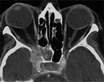
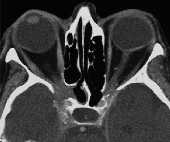
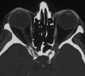
Granulomatous invasive fungal sinusitis often presents with long-standing symptoms of nasal obstruction, unilateral facial discomfort and/or enlarging mass, or with a silent proptosis.Reference de Carpentier, Ramamurthy, Denning and Taylor16, Reference Clancy and Nguyen17 This condition may begin as a paranasal sinus fungus ball and then become invasive, perhaps as a result of the immunosuppression associated with diabetes mellitus or corticosteroid treatment. If left untreated, the infection can spread to invade adjacent structures, including the orbit and brain (Figures 5, 6 and 7).
Axial computed tomography scan showing chronic, invasive fungal sinusitis invading the cavernous sinus.

Axial computed tomography scan of same patient as in Figure 5, three months after itraconazole commencement.

Axial computed tomography scan of same patient as in Figure 5, two years after itraconazole commencement and one year after treatment cessation.

In patients with chronic invasive sinusitis, non-contrast CT scans will reveal a hyperdense mass within the involved sinus, with associated erosion of the sinus walls. Histological analysis reveals profuse fungal growth with localised tissue invasion and non-caseating granulomas with giant cells. The granulomatous response is often intense enough to cause pressure necrosis of bone and can cause proptosis.
Unless removed, the fungus can spread into the orbit and brain. It is important to distinguish this condition from mycetoma (a chronic, expanding fungal disease) and from allergic fungal sinusitis (which can erode bone and expand into neighboring areas). Chronic invasive aspergillosis not only causes bone loss visible on CT scanning but also, most importantly, causes soft tissue invasion visible on histological analysis.
The orthodox treatment of life-threatening fungal infections is considered to be surgical debridement followed by amphotericin B.Reference Romett and Newman18–Reference Stringer and Ryan20 However, the use of this drug is complicated by the frequently associated infusion-related side effects and nephrotoxicity, and it is unsuitable for maintenance therapy outside a hospital setting. Furthermore, it has been shown not to be consistently effective. Denning and Steven showed that amphotericin B resulted in an overall response rate of only 55 per cent in patients with invasive aspergillosis, with a generally poorer outcome in severely immunocompromised patients.Reference Denning and Steven21
The advent of the azoles (itraconazole fluconazole and voriconazole) represented a major advance in the management of fungal infections, particularly in immunocompromised patients. The advantages of itraconazole include its anti-aspergillus activity, ease of administration, and limited toxicity compared with amphotericin B. In invasive pulmonary aspergillosis, response rates for itraconazole have been shown to be similar to those for amphotericin B.Reference Denning, Lee, Hostetler, Pappas, Kauffman and Dewsnup22, Reference Steven and Lee23 This suggests that oral itraconazole provides a suitable alternative to amphotericin B in the treatment of invasive aspergillosis, although long courses may be required.Reference Stevens, Kan, Judson, Morrison, Dummer and Denning24
Until relatively recently, the use of itraconazole for chronic invasive fungal sinusitis has been confined to a small number of cases in which attempts to control the disease by surgical debridement, with or without intravenous amphotericin B, have failed.Reference Rowe-Jones and Freedman25–Reference Dhiwaker, Thaker and Bahadur27 Based on evidence from a unpublished series (K R Meganadh, personal communication), and from our own experience of oral itraconazole as primary therapy for sinonasal aspergillosis,Reference Browning, Sim, Timms, Vernon, McConachie, Allibone and Jones28 we believe that itraconazole has a role in the primary treatment of patients with chronic (granulomatous) invasive fungal sinusitis.
Chronic invasive fungal sinusitis is often advanced by the time of diagnosis, with erosion of the skull base and possibly even involvement of the cavernous sinus. Treatment consists of itraconazole, 100 mg twice daily, unless the disease is of the acute fulminant type with blood vessel invasion, in which case aggressive surgery and intravenous amphotericin is needed. It is important to monitor liver function during and one month after treatment, and monthly thereafter. Side effects are rare, but caution is required in patients with cardiac disease. The duration of treatment is not established, but 12 months appears to be adequate.
Fulminant aspergillosis
Acute fulminant (invasive) fungal sinusitis is a rapidly progressive disease most commonly seen in immunocompromised individuals or poorly controlled diabetics. Immunocompetent individuals are seldom affected. In the absence of treatment, the disease is rapidly fatal in 50–80 per cent of patients.Reference Gillespie, O'Malley and Francis29
The commonest causes of acute fulminant fungal sinusitis are moulds of the mucorales order, including the rhizopus and rhizomucor species. The mucorales can be distinguished from other moulds, such as aspergillus species, by their characteristic broad, non-septate hyphae with right-angled branching. It is not possible, however, to differentiate aspergillus species from fusarium species, Scedosporium apiospermum or other non-pigmented moulds, on the basis of their microscopic appearance in tissue. All produce branching, septate, non-pigmented hyphae. Isolation of the aetiological agent in culture is essential in order to identify the fungal species. Other, less frequent causes of fulminant sinusitis include aspergillus species, particularly A flavus and A fumigatus.
The infection can spread rapidly from the nasal mucosa and sinus into the orbit and brain.Reference Radner, Witt and Edwards30 The aetiological agents have a predilection for vascular invasion, causing thrombosis, infarction and ischaemic necrosis of tissues. Prolonged neutropenia and metabolic acidosis are well recognised as important risk factors for rhinocerebral mucormycosis and fulminant aspergillus sinusitis among patients with haematological malignancies, haematopoietic stem cell transplant recipients and individuals with diabetes mellitus.Reference deShazo19 Other contributing factors include corticosteroid use, des ferrioxamine treatment and human immunodeficiency virus infection. In immunocompromised persons, acute invasive fungal sinusitis presents with fever, unilateral facial swelling, unilateral headache, nasal obstruction or pain, and a serosanguinous nasal discharge. Black, necrotic lesions on the hard palate or nasal turbinates are a characteristic diagnostic sign. The middle turbinate is the most commonly affected site.Reference Gillespie, O'Malley and Francis29 As the infection spreads into the orbit, periorbital or perinasal swelling occurs and progresses to disfiguring destruction of facial tissue. Ptosis, proptosis, ophthalmoplegia and loss of vision can occur.
The mainstay of treatment of acute, fulminant invasive fungal rhinosinusitis continues to be a combination of aggressive surgical debridement and antifungal therapy. The drug of choice in the immunocompromised or diabetic patient is amphotericin B (1.0–1.5 mg/kg per day). If the disease fails to respond to the conventional formulation of amphotericin B, treatment should be changed to one of the lipid-based formulations of the drug at dosages of 3–5 mg/kg or higher. This should be continued until the patient recovers, or for at least two weeks before reverting to conventional amphotericin B. Administration of lipid-based amphotericin B is also recommended for patients in whom the conventional formulation is contraindicated because of renal impairment, or for those who develop sideeffects which would otherwise necessitate discontinuation of the drug.
Conclusion
Fungal infections of the nose and paranasal sinuses need to be recognised in order to avoid significant mortality and morbidity. Suspicion should be aroused in cases of purulent rhinosinusitis which do not respond to two or more courses of antibiotics, and on the basis of radiological features. The diagnosis is based on culture, skin prick tests, CT scans and aspergillus precipitin titres. Treatment depends on whether the disease is allergic, invasive or non-invasive, as well as on the organism. Any predisposing factors also need treating. The use of oral itraconazole significantly reduces morbidity in both allergic and chronic invasive aspergillosis.