

Introduction
Agenesis of the corpus callosum (ACC) is an abnormality of brain structure, present at birth, characterised by partial or complete absence of the corpus callosum. Corpus callosum axons normally connect homologous regions in the left and right hemispheres of the cortex. Dysgenesis of the corpus callosum often results in anatomical defects in cell proliferation and migration, in glial and axon development and guidance, and functionally in severe intellectual disability (ID) (Kamnasaran, Reference Kamnasaran2005; Paul et al., Reference Paul, Brown, Adolphs, Tyszka, Richards, Mukherjee and Sherr2007).
From the olfactory point of view, ACC, often related to olfactory bulb agenesis (Engle, Reference Engle2010), is sometimes similar to Kallmann's syndrome, a pathological condition characterised by congenital anosmia and hypogonadotropic hypogonadism. In Kallmann's syndrome, the hypothalamus fails to release the gonadotropin-releasing hormone (Hanna et al., Reference Hanna, Marsh, Swistun, Al-Gazali, Zaki, Abdel-Salam and Gleeson2011; Seminara, Hayes, & Crowley, Reference Seminara, Hayes and Crowley1998). Gonadotropin-releasing hormone normally arises from the olfactory placode in the developing nose, migrating into the olfactory bulb and hypothalamus.
Olfaction is based on a few hundred olfactory receptor classes, expressed by millions of olfactory neurons organised in the olfactory epithelium at the interface with the external environment. Each olfactory neuron seems to express only one type of receptor and each glomerulus, located in the olfactory bulb, seems to receive signals from olfactory neurons carrying the same type of receptor (Imai, Suzuki, & Sakano, Reference Imai, Suzuki and Sakano2006).
The olfactory system is extremely complex, having a rich cortical representation, including the entorhinal and the orbitofrontal cortices. These two components are able to transform the volatile molecules into their respective odour perception, thus they largely contribute to odour perception and olfactory memory.
Odour perception is disturbed by a number of neurological conditions, including Alzheimer's and Parkinson's diseases, schizophrenia, depression, autism and early life exposure to toxins. In such cases, olfactory evaluation can be extremely useful, being the only sensory system negatively affected by such events (Wilson et al., Reference Wilson, Xu, Sadrian, Courtiol, Cohen and Barnes2014). Moreover, odours are considered a trigger for brain areas involved in stress states, such as the limbic system, in people with a normal olfactory function (Avilov & Sudakov, Reference Avilov and Sudakov2008), but the literature on ‘olfactory stress’ in people with hyposmia and/or anosmia is relatively limited. Several methods are currently available for olfactory evaluation, including the Sniffin’ Sticks Identification Test, employed in this study and described later.
The association between decreased odour perception and neurological disorders also seems to occur in pathologies affecting children but, to the best of our knowledge, findings are currently limited and mostly debatable. For this reason, it may be interesting to evaluate the possibility of assessing olfaction in children with ACC who present with ID and suspected hyposmia or anosmia. Psychophysical evaluation may also be useful to measure objectively the olfactory response in children with complex neurodevelopmental disorders and who have difficulty complying with requests.
Therefore, the aims of this study were: (1) to evaluate whether this validated method to assess olfaction in typically developing children may provide clinical information in a child with severe hypoplasia of corpus callosum, ID and suspected anosmia; and (2) to verify the stress parameters as a further method to recognise anosmia in children with neurodevelopmental disorders who cannot participate in ordinary olfactory testing.
Methods
Participants
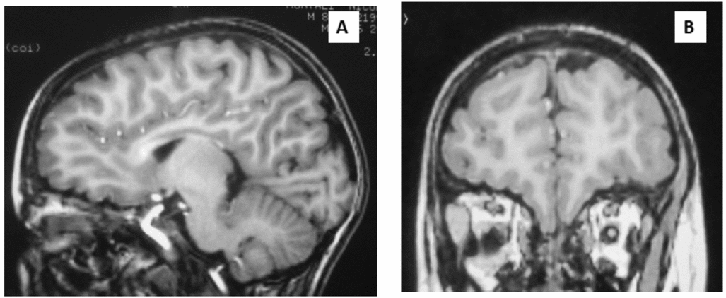
A 13-year-old boy with moderate ID (mental age of 5 years) was tested with the Sniffin’ Sticks Odor Identification Test (Hummel, Kobal, Gudziol, & Mackay-Sim, 2007). The child was diagnosed as affected by a chromosomopathy (unbalanced translocation 17p13.3–17p13.1 detected at array comparative genomic hybridisation (aCGH) analysis), severe hypoplasia of the corpus callosum and of the olfactory bulbs (Figure 1). The patient also evidenced partially empty sella turcica at brain magnetic resonance imaging (MRI), and suspected anosmia, with absence of mutation of gene KAL1 and no hypogonadotropic hypogonadism. Cognitive function was assessed by Wechsler Intelligence Scale for Children – III, which showed a moderate ID, more pronounced in the performance domain.
Sagittal (A) and coronal (B) T1-weighted magnetic resonance image (MRI) of the studied patient, showing severe hypoplasia of the corpus callosum and olfactory bulbs.

Three control participants, one with ID and two typically developing controls, were also evaluated. The child affected by ID presented with the same chronological and the same mental age of the patient, but a normal brain MRI scan. The two typically developing (TD) control participants were aged 13 and 5, equal to the chronological and mental age of the patient.
Olfactory Testing
‘Sniffin’ Sticks’ is a test of nasal chemosensory performance based on pen-like odour-dispensing devices and consisting of three tests of olfactory function, namely tests for odour threshold, discrimination and identification (Hummel, Sekinger, Wolf, Pauli, & Kobal, Reference Hummel, Sekinger, Wolf, Pauli and Kobal1997; Kobal et al., Reference Kobal, Hummel, Sekinger, Barz, Roscher and Wolf1996). In particular, the odour identification test seems to be the easiest sub-test to be employed with children affected by ID, although there is no definitive evidence on large populations and further evaluation is required. Previous research has established the test–retest reliability of this kit, and its validity in comparison to recognised measures of olfactory sensitivity (Cain, Gent, Goodspeed, & Leonard, Reference Cain, Gent, Goodspeed and Leonard1988).
The test has already been employed in several published studies involving healthy adults (Tonacci, Borghini, Mercuri, Pioggia, & Andreassi, Reference Tonacci, Borghini, Mercuri, Pioggia and Andreassi2013), elderly people affected by neurodegeneration (Förster et al., Reference Förster, Vaitl, Teipel, Yakushev, Mustafa, Fougère and Steinbach2010; Hummel et al., Reference Hummel, Fliessbach, Abele, Okulla, Reden, Reichmann and Haehner2010), as well as people exposed to environmental toxins (Tonacci et al., Reference Tonacci, Baldus, Corda, Piccaluga, Andreassi, Cremonesi and Picano2014), demonstrating its applicability in a number of different conditions.
This particular test was chosen in preference to other commercially available tests because it is extremely easy to administer, even in children with ID who could have issues in performing other tests, such as the NIH Toolbox Odor Identification Test (Dalton et al., Reference Dalton, Doty, Murphy, Frank, Hoffmann, Maute and Slotkin2013). This latter test was specifically designed for children, with the odours microencapsulated into scratch-and-sniff cards, but they are not particularly suitable for children with particular neurodevelopmental disorders.
In the identification task, involving 16 odorised sticks, the patient was asked to identify the odour using a display of four visual descriptors, representing photos of the correct and three distractor odour sources. The images (e.g., orange, blueberry, strawberry, pineapple) were chosen according to the suggestions of experienced neuropsychiatrists. The patient was also asked to identify verbally the photos corresponding to the odours prior to commencement of the olfactory testing, to ensure that he recognised what they represented. The interval between odour presentations was 20–30 s. The score range is from 0 to 16. The same task was also performed by the three control participants.
Psychophysical Tests
During the olfactory assessment, psychophysical tests were performed to assess the stress state of the subject under study. In particular, skin conductance, measured by galvanic skin response, related to sympathetic tone, an indirect measure of emotion, and heart rate and heart rate variability, which reflect the sympathovagal balance, were monitored continuously. Skin conductance was checked using electrodes stuck to the palmar surface of the non-dominant hand, while heart rate was detected using a chest belt developed by our research team, which showed performances comparable to those of commercial tools (Billeci et al., Reference Billeci, Brunori, Crifaci, Tartarisco, Scardigli, Pioggia and Morales2012a, Reference Billeci, Pioggia, Brunori, Crifaci, Tartarisco, Balocchi and Morales2012b). The protocol consisted of 90 s of basal acquisition, 400 s of acquisition during the task and 40 s of acquisition after the task. A control test recall was also administered to the patient after 3 weeks to assess whether his anxiety was related to the olfactory task or to a generic clinical task (a sort of ‘white coat syndrome’). More specifically, the control test consisted of a similar acquisition compared with the standard protocol, employing blank felt-tip pens instead of odorants to check eventual differences between the olfactory protocol and the control test in terms of psychophysical response to odorants.
According to the study hypothesis, due to the non-invasive and well-accepted nature of the olfactory testing, there should have been a different response depending on the different odours in the participants with normal olfaction, while we expected a less evident trend in those with an olfactory deficit, with a response more related to the test administration than to the nature and hedonic tone of the odour presented.
Data Analysis
Statistical analysis was divided into two main parts. For the olfactory testing, we applied a descriptive statistical analysis (Z-score), calculated on the basis of the normative data for the age that corresponded to the mental age of the subjects. For the psychophysical test analysis, we employed the non-parametric Wilcoxon Rank Sum Test. A p value < .05 was considered significant.
Results
Olfactory testing
The patient scored 4/16 in the olfactory identification task. Comparing the score with the normative data for age- and gender-matched children (Hummel et al., Reference Hummel, Kobal, Gudziol and Mackay-Sim2007), this result was significantly lower (Z-score of –4.7) than the ‘normal’ values, the latter being in the range 8–16 (mean 12.41, SD 1.77). The deficit was confirmed by comparison with control participants. The patient control child with similar ID scored 12 (Z-score of –0.23), while the TD control participants scored 12/16 (for the 5-year-old TD participant; Z-score of –0.23) and 14/16 (for the 13-year-old TD participant; Z-score +0.90). These results showed that our patient had a clear hyposmia compared with a chronological age-, gender- and mental age-matched patient control participant, as well as with TD control participants.
Psychophysical Tests
Skin conductance was monitored during three phases, ‘baseline’ (before the olfactory task), ‘task’ (during the task) and ‘recovery’ (after the olfactory task), filtered and analysed. In the ‘baseline’ phase, the skin conductance signal of the patient had a value (mean ± SD) of 1.73 ± 1.06 mS; it increased to 3.17 ± 1.65 mS during the ‘task’ and to 4.54 ± 0.59 mS during the ‘recovery’, this last phase having a slope of –0.04, suggesting a tendency towards a relaxation of the subject after the task. The trend during the ‘task’ suggested a complete independence from the odorous stimuli, but rather an effect of the task itself on the stress state of the patient.
Similar findings were obtained with heart rate and heart rate variability analysis, both in the time and frequency domains. Heart rate increased from 98.1 ± 8.9 bpm during the ‘baseline’ phase, to 102.1 ± 7.5 bpm during the ‘task’. Inter-beat interval was significantly different in the two phases, with a lower value during the ‘baseline’ compared with ‘task’. In the frequency domain, the ratio between low frequency and high frequency components of the electrocardiograph signal (low frequency:high frequency ratio), which is an index of balance between sympathetic and parasympathetic systems, yielded a value of 2.94 during the ‘baseline’ phase, and 3.52 during the ‘task’. This was a significant difference, suggesting an increased sympathetic drive during the task.
During the control test recall, both the skin conductance and heart rate/heart rate variability analysis reported similar results, suggesting no influence of the olfactory task itself, but rather an anxiety due to the development of a generic clinical task.
In the patient control child with ID, a tendency towards a slight increase of the skin conductance value during the ‘task’ was observed, without any statistical significance (p > .05); also, the heart rate/heart rate variability signal showed a trend similar to that of skin conductance.
In contrast, both in the 5- and the 13-year-old TD controls, the skin conductance increased significantly during the task, with a following decrease during the ‘recovery’ phase. However, the trend during the ‘task’ was related to stimuli presentations, suggesting that different odours contributed differently to the subject's stress state. The same findings were noted in heart rate/heart rate variability analysis for both participants.
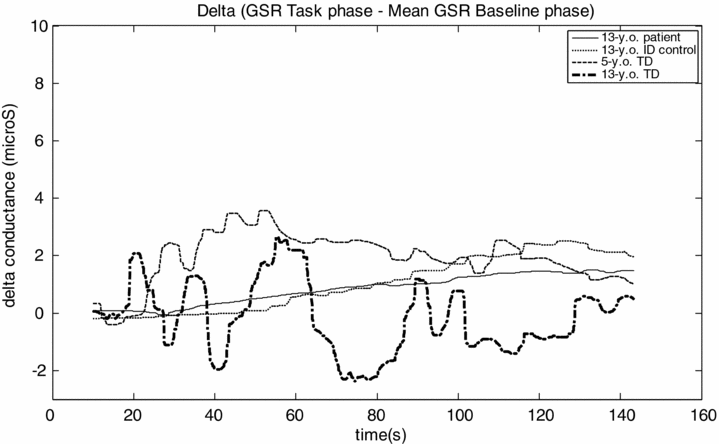
In the skin conductance evaluation, the ‘delta’ between ‘task’ and ‘baseline’ mean signals was calculated and values are displayed in Figure 2, showing a different trend between the patient with ACC and bulb hypoplasia and the two TD control participants.
Difference between filtered galvanic skin response (GSR) signal during the olfactory task phase and the mean GSR signal during the baseline phase, for the 13-year-old patient (solid line), the 13-year-old control with intellectual disability (ID) (dotted line), the 5-year-old typically developing control (TD) (dashed line) and the 13-year-old TD (dash-dotted line).
The patient showed a clear increase of the delta value independently from the quality of odour presented, while the two TD participants showed an increase or decrease of the delta depending on the pleasantness or unpleasantness of the odour proposed. The 13-year-old patient control with ID showed a similar trend when compared with the patient.
Discussion
The Sniffin’ Sticks Odor Identification Test documented a clear olfactory deficit in a 13-year-old boy with severe hypoplasia of the corpus callosum, partially empty sella turcica, hypoplasia of olfactory bulbs and intellectual disability. The identification score of 4/16 was significantly lower than normal values for mental age range and gender (Hummel et al., Reference Hummel, Kobal, Gudziol and Mackay-Sim2007) and also lower than those obtained from our patient control with ID and in typically developing control participants. Moreover, the score obtained was significantly lower if compared with the values obtained by adults exposed to environmental factors, such as ionising radiation (Tonacci et al., Reference Tonacci, Baldus, Corda, Piccaluga, Andreassi, Cremonesi and Picano2014).
During the olfactory testing, a psychophysical evaluation was also conducted. It is well known that psychophysical reactions to stimuli of varying nature could be different between people with ID and typically developing participants (Chaney, Reference Chaney1996; Ghanizadeh, Bahrani, Miri, & Sahraian, Reference Ghanizadeh, Bahrani, Miri and Sahraian2012; Legiša, Messinger, Kermol, & Marlier, Reference Legiša, Messinger, Kermol and Marlier2013; Sandberg et al., Reference Sandberg, Paton, Ahola, McCann, McGuinness, Hillary and Oja2000), providing useful information about the anxiety state of the evaluated subject.
The combination of olfactory evaluation and psychophysical testing could be particularly indicated in children with ACC, and/or signs of empty sella turcica on MRI scan, who have ID. In these conditions it may be difficult to verify whether the neuroradiological conditions are associated with anosmia by simply employing a traditional olfactory test, and the use of psychophysical evaluation could rectify this possible drawback of the olfactory testing.
The psychophysical analyses performed here, focused on skin conductance and heart rate variability, suggested a state of anxiety and stress of the patient, followed by a decrease of stress at the end of the olfactory task. No particular differences were found between odorants, suggesting that the stress state was related more to the task than to the odour presentation. A similar result was found during the control test in which pens without odour were presented to the patient with ACC. This effect was likely due to the fact that olfactory stimulation had no influence on these parameters, but a state of anxiety was present, possibly related to the generic task.
Conversely, both of the studied, typically developing, control participants showed a tendency towards an increase of the anxiety state, with a clear influence of the hedonic tone of the odour presented to them. In particular, some odours, with a more marked unpleasantness (e.g., garlic, fish), seemed to increase the anxiety of the children more than other, more pleasant ones (e.g., mint, orange). No particular influence of the pleasantness/unpleasantness of the odour presented was found in our patient, nor in the patient control with ID. This suggests that the employment of this tool could be useful in evaluating the olfactory psychophysical response in anosmic individuals, although its use does not seem to be specific enough in the presence of neurodevelopmental disorders.
The Sniffin’ Sticks Identification Test for the olfactory assessment seems feasible in children with complex neurodevelopmental disabilities, providing the caregiver with a functional evaluation and integration of the neuroradiological diagnosis of such conditions with an objective and repeatable test. Although the studied patient with ACC showed an increase of the stress state during this task, related more to the task development than to the odour presentation, with a similar trend in the patient control with ID, children without ID and with a normal olfactory function seemed to be more stressed when particular odorants were presented. Since response to the Sniffin’ Sticks is highly subjective, these results should be confirmed on larger numbers; however, it is important to note that stress situations can be monitored easily by using low-cost, portable devices, which can also be used with children with psychopathological behaviour.
These findings are also of interest in relation to recent indications for specific alterations to olfactory function in children and adolescents with many neurodevelopmental disabilities and psychiatric disorders, such as autism, anorexia nervosa or obsessive-compulsive disorder (Dudova & Hrdlicka, Reference Dudova and Hrdlicka2013; Dudova et al., Reference Dudova, Vodicka, Havlovicova, Sedlacek, Urbanek and Hrdlicka2011; Schecklmann et al., Reference Schecklmann, Pfannstiel, Fallgatter, Warnke, Gerlach and Romanos2012, Reference Schecklmann, Schwenck, Taurines, Freitag, Warnke, Gerlach and Romanos2013). Future studies are warranted to investigate the olfaction of children with brain malformation such as ACC, and various types of neurodevelopmental disabilities who are at risk for olfactory dysfunction, to better understand the processing of the odorous stimuli in their clinical condition.
Acknowledgements
This work was supported partially by RC 2012 of the Italian Ministry of Health.
Conflict of interest
None.