I. INTRODUCTION
Magnesium ferrite (MgFe2O4) is an important ferrite. It has a cubic structure of normal spinel type and is a soft magnetic n-type semiconducting material, which finds applications in memory and switching circuits and microwave devices, heterogeneous catalysis, adsorption, humidity sensors, magnetic technologies, and, more recently, in achieving local hyperthermia.Reference Willey, Noirclerc and Busca1–Reference Maehera, Konishi, Kamimori, Aono, Naohara, Kikkawa, Watanable and Kawachi4 Various methods have been used to prepare nanocrystalline ferrites. Conventional ceramic processing of ferrites requires a number of stages, including homogenization of the powder precursors, compaction of the reactants, and finally prolonged heat treatment at considerably elevated temperatures. However, the traditional methods of synthesis are not able to produce uniform and reproducible particles in the nanometer dimensions. One goal of modern ferrite research and development has been to identify simpler processing schemes that do not rely upon high temperature treatments for inducing solid-state reactions. One such scheme is the coprecipitation technique. The cubic spinel structure presents two types of interstitial sites, namely tetrahedral (A) and octahedral (B), surrounded by four and six oxygen atoms, respectively. A remarkable effect in spinel ferrites is the strong dependence of physical properties on cation composition and structural ordering, in particular the dependence on the metal distribution among the two available interstitial sites.
Ferrites possess high values of magnetization because of imbalanced site magnetic moments. Ferrites also have high values of resistivity, low dielectric loss, and high Neel temperatures. These properties have made them versatile materials for various technological applications. The crystallographic, electrical, and magnetic properties of ferrites depend strongly upon stoichiometry as well as processing parameters such as temperature, atmosphere, and pressure, which affect the distribution of cations among the available tetrahedral (A) and octahedral (B) sites in the spinel lattice. Control over cation distribution and the oxygen parameter provide a means of developing the desired physical properties for their proper use in industry.
According to literature, MgFe2O4 is a partially inverse spinel, and it can be considered as a collinear ferrimagnet whose degree of inversion depends upon the thermal history of the sample.Reference Knock and Dannheim5, Reference De Grave, Dauve, Govarert and Siller6 It has been reported that MgAl2O4 (mineral spinel) is a partially inverted spinel in which the ratio of Al3+ ions in the tetrahedral (A) and octahedral (B) sites is about 1:6.Reference Schmocaer, Boesch and Waidner7 Doping with trivalent ions like Al3+ for Fe3+ in MgFe2O4 influences the structural, electrical, and magnetic properties of the system in which ratio of Al3+ ions in the tetrahedral (A) and octahedral (B) sites is about 2:3.Reference Bara, Pedziwiatr, Standnik, Szytwa, Todorovic, Tomokowicz and Zarek8 The purpose of the present paper was to study the effect of Al3+ substitution on the physical properties of the MgAlxFe2−xO4 ferrites at different molar ratios (x = 0.0, 0.4, 0.8, and 1.0) using a variety of experimental techniques like x-ray powder diffractometer, infrared (IR) spectrometer, transmission electron microscope, and vibrating sample magnetometer (VSM).
II. EXPERIMENTAL TECHNIQUE
Nanoparticles of aluminum-substituted magnesium ferrites MgAlxFe2−xO4 (with x = 0.0, 0.4, 0.8, and 1.0) were prepared using coprecipitation technique. In the present study, all reagents were analytically pure and used without further purification. Stoichiometric compositions of magnesium chloride (MgCl2·6H2O), aluminum chloride (AlCl3·6H2O), ferric chloride (FeCl3), and sodium hydroxide (NaOH) were dissolved in 100 mL of distilled water. The solution was kept at 70 °C under vigorous stirring. After reacting for 60 min, the precipitated product was subjected to filtering, washing, and drying at 90 °C for 3 h. Finally, the prepared powders were sintered at 900 °C for 5 h to ensure the formation of a single ferrite phase.
Characterizations of the prepared compositions were made by XRD, DTA, transmission electron microscopy (TEM), and IR absorption spectroscopy. The structure and particle size of the prepared ferrites were determined using Philips X’pert powder diffractometer (Almelo, The Netherlands) supplied with Cu x-ray tube (40 kV, 30 mA), nickel filter, divergence slit 1°, receiving slit 0.1 mm, crystal monochromator, and proportional gas detector. Angular scans between 20° and 70° (2θ) were performed on the investigated samples with scanning speed of 0.05° and measuring time of 5 s per step. DTA measurements were performed using Shimadzu simultaneous differential thermal analyzer DTA-TG-60 (Kyoto, Japan) with accuracy of ±0.1 K and sensitively of ±10 μV under high purity nitrogen gas at a flow rate of 40 mL/min. TEM measurements to determine morphology and particles sizes of all compositions were made on TECNAI G2 S-T (Eindhoven, The Netherlands) with operating voltage 200 kV.
The IR spectrum of the prepared compositions was studied, in the range from 350 to 4000 cm−1, using Perkin-Elmer Spectrophotometer model 1430 (Norwalk, CT). Magnetization measurements were performed on a VSM (Lake Shore 7400 series, Westerville, OH) of 10−7emu sensitivity in the magnetic field range of −20 to 20 kG.
III. RESULTS AND DISCUSSIONS
A. Crystal structure analysis
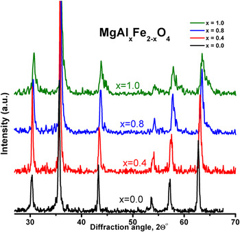
In mixed spinel ferrite MgAlxFe2−xO4 (with x = 0.0, 0.4, 0.8, and 1.0), one of the constituents, i.e., Mg2+ was fixed, while Fe3+ was replaced by Al3+. In Fig. 1, the recorded XRD patterns for all the prepared samples of Mg–Al ferrite show the well-known diffraction lines of the cubic spinel structure. The observed peaks were indexed in the light of the natural spinel structure MgAl2O4in which the more intense line of the pattern corresponds to the (311) plane. All patterns exhibit major peaks of spinel crystal structure. The diffraction from the crystal planes (220), (311), (400), (422), (511), and (440) appears for all the compositions, which confirmed that the crystal structure of the samples is a single cubic spinel ferrite phase.
X-ray diffraction patterns of MgAlxFe2−xO4 (x = 0.0, 0.4, 0.8, 1.0) ferrites.

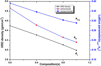
The interplanner distance d (Å) was calculated using Bragg’s law, and the values of the lattice constant were determined with an accuracy of ±0.003 Å using the external standard method.Reference Razik9 Variation of the lattice constant a (Å) is depicted in Fig. 2, which exhibits an obvious decrease with compositions (x). The reported value of the lattice constant of the magnesium ferrite MgFe2O4 is 8.376 Å as given by the JCPDS Card (No. 01-073-2410). The values of lattice constant of the present doped samples lie in the range of 8.39–8.29 Å. The overall decrease in the lattice constant is reasonable because the larger cation (Fe3+) with radius 0.64 Å is replaced by the smaller cation (Al3+) having radius of 0.51 Å in the ferrite system.
Variation of lattice parameters (a exp, a the), XRD density d x with composition (x).

The broad XRD lines indicate that the ferrite particles of the investigated samples are nanosized. The average particle size for each sample was calculated from the line broadening of the (311) peak using Scherrer equation:
 $$t = {{{{\rm{0}}{\rm{.9\lambda }}} \over {B{\rm{cos\theta }}_B
}}}\quad,$$
where t is the size of the particle, λ is the wave length of the x-ray radiation (Cukα1, 1.54056 Å), B is the full width at half maxima (FWHM) of the diffraction peak in radian, and θB is its angle. It was found that the average particle size for each sample lies in nanosize range (<100 nm) and decreases slightly with increasing of Al3+concentration.
The x-ray density (d x) for each sample was calculated by using the relation:
 $${d_{\rm{x}} = {{ZM} \over {NV}}}\quad,$$
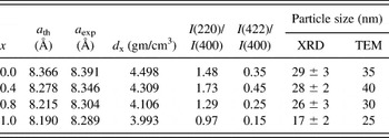
where Z is the number of molecules per unit cell (Z = 8), M is the molecular weight, N is Avogadro’s number, and V is the unit cell volume. The compositional dependence of x-ray density is obvious from the tabulated results in Table I. The calculated values of the x-ray (d x) densities decrease with increasing aluminum concentration (x), and this can be attributed to the decrease in the lattice constant and hence to the volume of the unit cell.
Structural parameters of MgAlxFe2−xO4 (x = 0.0, 0.4, 0.8, 1.0) ferrites obtained from XRD and TEM data.

According to Ohnishi and Teranishi,Reference Ohnishi and Teranishi10 the intensity ratios I(hkl)/I(400) are considered to be sensitive to the cation distribution parameter (x). The cation distribution of the studied ferrite was deduced through XRD pattern intensity analysis, IR spectrum, and magnetization data. Accordingly, the proposed cation distribution will be:
 $$\!\eqalign{ & {\rm{(Mg}}_{{\rm{0}}{\rm{.1}}}^{{\rm{2
+ }}} \,\,{\rm{ Fe}}_{{\rm{0}}{\rm{.8}}}^{{\rm{3 + }}}
{\rm{)[Mg}}_{{\rm{0}}{\rm{.9}}}^{{\rm{2 + }}} \,\,{\rm{
Fe}}_{{\rm{0}}{\rm{.2}}}^{{\rm{2 + }}} {\rm{
\,\,Fe}}_{{\rm{1}}{\rm{.0}}}^{{\rm{3 + }}} {\rm{],
}}\,\,\,\,\,\,\,\,\,\,\,\,\,\,\,\,\,\,\,\,\,\,\,\,\,\,\,\,\,\,\,\,\,\,x =
0.0,\cr & {\rm{(Mg}}_{{\rm{0}}{\rm{.1}}}^{{\rm{2 + }}}
\,{\rm{ Al}}_{{\rm{0}}{\rm{.15}}}^{{\rm{3 + }}} \,{\rm{
Fe}}_{{\rm{0}}{\rm{.55}}}^{{\rm{3 + }}}
{\rm{)[Mg}}_{{\rm{0}}{\rm{.9}}}^{{\rm{2 + }}} \,\,{\rm{
Al}}_{{\rm{0}}{\rm{.25}}}^{{\rm{3 + }}} \,{\rm{
Fe}}_{{\rm{0}}{\rm{.1}}}^{{\rm{2 + }}} \,{\rm{
Fe}}_{{\rm{0}}{\rm{.95}}}^{{\rm{3 + }}} {\rm{], }}\,\,x = 0.4, \cr
& {\rm{(Mg}}_{{\rm{0}}{\rm{.1}}}^{{\rm{2 + }}} \,\,{\rm{
Al}}_{{\rm{0}}{\rm{.3}}}^{{\rm{3 + }}} \,\,{\rm{
Fe}}_{{\rm{0}}{\rm{.5}}}^{{\rm{3 + }}}
{\rm{)[Mg}}_{{\rm{0}}{\rm{.9}}}^{{\rm{2 + }}} \,\,{\rm{
Al}}_{{\rm{0}}{\rm{.5}}}^{{\rm{3 + }}} \,\,{\rm{
Fe}}_{{\rm{0}}{\rm{.7}}}^{{\rm{3 + }}} {\rm{],
}}\,\,\,\,\,\,\,\,\,\,\,\,\,\,\,\,\,\,\,\,x = 0.8,\cr &
{\rm{(Mg}}_{{\rm{0}}{\rm{.1}}}^{{\rm{2 + }}} \,\,{\rm{
Al}}_{{\rm{0}}{\rm{.4}}}^{{\rm{3 + }}} \,\,{\rm{
Fe}}_{{\rm{0}}{\rm{.45}}}^{{\rm{3 + }}}
{\rm{)[Mg}}_{{\rm{0}}{\rm{.9}}}^{{\rm{2 + }}} \,\,{\rm{
Al}}_{{\rm{0}}{\rm{.6}}}^{{\rm{3 + }}} \,\,{\rm{
Fe}}_{{\rm{0}}{\rm{.55}}}^{{\rm{3 + }}} {\rm{],
}}\,\,\,\,\,\,\,\,\,\,\,\,\,\,\,\,x = 1.0\quad. \cr} $$
On the basis of the proposed cation distribution, the value of theoretical lattice constant a th for all the investigated samples has been determined using the relationReference Zaki and Mansour11:
 $$a_{{\rm{th}}} = {8 \over {3\sqrt 3 }}[(r_{\rm{A}} + R_{\rm{O}} ) +
\sqrt 3 (r_{\rm{B}} + R_{\rm{O}} )]\quad,$$
where R O is the radius of oxygen ion, r A the ionic radius of A-site, and r B the ionic radius for B-site. The obtained values of the theoretical lattice constants (a th) are listed in Table I. The difference between the theoretical and experimental values of the lattice constants may be attributed to the existence of porosity in the samples during the sintering process.
The morphology of the samples was investigated using TEM and is shown in Fig. 3. The average particle size obtained by this technique is around ∼33 nm for MgFe2O4 ferrite (x = 0.0), which is consistent with the value calculated by the XRD for the same sample (∼30 nm). Inspection of the particle size for all samples as a function of concentration showed that the average value for each composition lies in the nanoscale and it decreases with increasing Al3+ concentration as shown in Table I.
TEM micrographs of MgAlxFe2−xO4 [x = 0.0, 0.4, 0.8, 1.0, given as (a), (b), (c), and (d), respectively] ferrites. (color online)


Nanocrystal aggregation phenomenon is observed in the prepared samples. This phenomenon could be attributed to the nucleolus coalescing growth, which occurs due to the tendency that small (nanometrics) particles have to aggregate to achieve a lower free energy state reducing the specific superficial area by reducing the interfaces with the other particles.
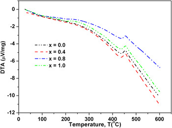
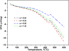
The spinel phase transition temperature found using the differential thermal analysis is shown in Fig. 4. The transition causes very small changes to the heat capacity of the sample, and the resulting thermal effect is barely distinguishable from the background. Candeia et al.Reference Candeia, Souza, Bernardi, Maestrelli, Santos, Souza and Longo12 synthesized the MgFe2O4 ferrite by polymeric precursor method and stated that the peak around 460 °C is related to the decomposition and combustion of the organic material. Hankare et al.Reference Hankare, Vader, Patil, Jadhav, Sankpala, Kadam, Chougule and Gajbhiye13 studied Mg–Cr ferrite and found that the spinel phase is formed at a temperature around 500 °C. Also, previous worksReference Bratton14, Reference Shiono, Shiono, Miyamoto and Pezzotti15 demonstrated that the magnesium aluminum ferrite decomposed to crystalline spinel and periclase at temperatures as low as 400–500 °C.
Thermograms (DTA) of MgAlxFe2−xO4 ferrite.

In addition, Ma et al.Reference Ma, Yue, Hua and Gao16 reported that the exothermic peak observed at 640 °C is assigned to the formation of MgFe2O4 spinel via the solid-state reaction of the mixed oxides prepared by coprecipitation, and the exothermic peak was observed around 300 °C by using citrate gel method. The substitution of trivalent Al ions instead of Fe ions into the lattice of MgFe2O4 ferrite would result in the decrease of Fe3+ ions in the octahedral site. Therefore, decrease in the population of Fe3+ with the increase of Al3+ ions could reduce the magnetization in all samples containing Al. Consequently, the transition temperature from ferromagnetic to paramagnetic should be reduced. This expectations disagree with the positions of the observed exothermic peaks in Fig. 4 for all compositions, which do not show a significant change and make it doubtful to be assigned to Curie temperature. However, there is a possibility to attribute the observation of the exothermic peaks around 450 °C to the phase transformation of the prepared samples from amorphous to crystalline state during the formation of the spinel structure. Moreover, it has been reported that the transition temperature from ferrimagnetic to paramagnetic for magnesium ferrite was found as 430 °CReference Purushotham, Reddy, Sagar, Kishan and Venugopal Reddy17 and by other workers as 400 °C.Reference Ladgaonkar, Vasambekar and Vaingankar18
B. IR spectral analysis
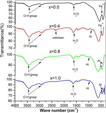
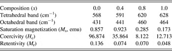
The IR spectra can give information on the valence states and the different variable modes in the crystal lattice. The room temperature IR spectra for MgAlxFe2−xO4 system with x = 0.0, 0.4, 0.8, and 1.0 are shown in Fig. 5. Many absorption bands were observed in the studied spinel ferrites in the range 350–4000 cm−1. The two main common bands of the tetrahedral and octahedral sites, which characterized the formation of spinel ferrites, are observed in the range (568–628 cm−1) and (431–460 cm−1), respectively.Reference Waldron19 These two bands are attributed to the intrinsic stretching vibrations due to the interaction of oxygen atom and cations in the tetrahedral and octahedral complexes, respectively. The band positions for all compositions are given in Table II. The shifting of bands toward high frequency is attributed to the decrease in the unit cell dimension as confirmed from x-ray diffraction pattern calculations and substitution of lighter cations Al3+ for Fe3+ in the system. The observed shift in band positions as a function of concentration (x) is summarized in Table II. The change in the band position is expected because of the difference in Fe3+–O2− distances in octahedral and tetrahedral complexes and also due to the more covalent bonding nature of Fe3+ ions at the A-sites than B-sites. Small absorption bands exist at 480 cm−1 for x = 0.0 and 490 cm−1 for x = 0.4, which indicate the presence of vacancies in B-sites due to the presence and oxidation of Fe2+ ions. Splitting occurred in the octahedral band beginning at concentration x = 0.4 and increased with increasing Al3+ content. This behavior can be associated with the migration of Al3+ ions from tetrahedral to octahedral sites and confirms the significant contribution of octahedral Al3+ cation to this mode of vibrations. Moreover, the recorded two shoulders in the IR spectra around 360 and 390 cm−1 for the high concentration of Al3+ samples (x = 0.8 and 10) confirm the increase of Al3+ concentration in B-sites as x increases. The bands around 800 cm−1 (which do not exist for x = 0.0) indicated the presence of Al3+ in the tetrahedral sites. The observed decrease in intensity of these absorption bands due to the increase of Al3+ content indicates the migration of some Al3+ ions from this site to B-site. These weak bands must be related to O2−–Al3+–O2− bonding mode, which is expected to lie in this region due to the increased Al3+ concentration.Reference Gillot, Bouton and Ferriot20 Moreover, this is a good indication for the transition of the studied samples from inverse spinel (MgFe2O4) to normal spinel (MgAl2O4). The absorbed water is featured by bands around 3424, 2923, and 1624 cm−1, which are assigned to O–H stretching and H–O–H bending modes of vibrations.Reference Giri, Sriharsha, Asthana, Gundu Rao, Nigam and Bahadur21 The unknown peak found at 2344 cm−1 only for the sample with x = 0.4 may be assigned to the fundamental stretching mode of the diatomic molecules polarized by the electric field created by Mg2+ ions, and this may enhance the magnetization for this sample (x = 0.4). The same band has been observed for Cu–Mg ferrite.Reference Deraz22
The IR spectra of MgAlxFe2−xO4 (x = 0.0, 0.4, 0.8, 1.0) ferrites.

Primary IR absorption bands and magnetic properties for Mg–Al ferrite.

C. Magnetic properties
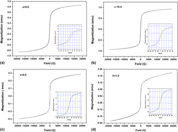
The magnetic parameters such as saturation magnetization (M S), coercivity (H C), and retentivity are extracted from MH loop for Mg–Al ferrites are shown in Fig. 6 and listed in Table II. The saturation magnetization is increased at x = 0.4 and then decreases. This increase can be attributed to the incorporation of aluminum ions into the crystal lattice of the MgFe2O4 ferrite and to the redistribution of cations on A- and B-sites. In spinel ferrite MgFe2O4, most of Mg2+ ions reside on B-sites and small fractions of these ions may goes to the A-sites.Reference Jani, Trivedi, Joshiand and Kulkarni23 The ratio of Al3+ ions in the tetrahedral (A) and octahedral (B) sites is about 2:3,Reference Chhaya, Trivedi and Kulkarni24 and this is realized in the proposed cation distribution. When Al3+ ions are introduced into the tetrahedral sites, this will encourage the migration of the Fe3+ ions to the octahedral sites, which increases the magnetization of the B-sublattices and gives rise to antiparallel spin coupling and spin canting resulting in weak A–B exchange interaction and increasing the saturation magnetization. The decrease in saturation magnetization for further substitution of Al3+ ions (x > 0.4) is owing to the magnetic disorder on B-sites caused by the paramagnetic aluminum ions on B-sites. Moreover, the number of Fe3+ in B-sites decreases due to an increase of Al3+ ions, which leads to the reduction of magnetization. The general trend of the intensities ratio estimated from XRD agrees well with magnetic saturation and confirms the proposed cation distribution. Moreover, the general trend of coercivity and retentivity values for the MgAlxFe2−xO4 system were found to decrease by increasing of Al3+ ions concentration as given in Table II. Magnetic device based on ferrite is expected to perform some kind of work by enhancing the magnetic field as an electrical current is passing through it. Such devices include the cores of electromagnets, motors, transformers, and generators, using AC currents. As the magnet cycled through the hysteresis loop continually and rapidly, the energy product of the loop should be as low as possible. Hence, a small coercivity and remanence are desirable. These properties can be found in the prepared aluminum-doped magnesium ferrites, which are suitable for such kind of applications. The low value of coercivity indicates that the domain wall motion is dominant magnetization mechanism, and the sample becomes magnetically softer, which is useful for power applications. In particular, magnesium aluminum ferrites as low magnetic saturation ferrites were suitable for microwave applications.Reference Van Uitert25
Magnetic hysteresis loops M(H) at room temperature obtained of the MgAlxFe2−xO4 [x = 0.0, 0.4, 0.8, 1.0, given as (a), (b), (c), and (d), respectively] ferrites. The inset shows enlarged portion for each graph.

IV. CONCLUSIONS
Nanocrystalline cubic ferrites of MgAlxFe2−xO4 (x = 0.0, 0.4, 0.8, and 1.0) were prepared through a coprecipitation method. The results of the analysis can be summarized as follows:
(i) XRD analysis confirmed the formation of a single cubic spinel structure for all the prepared Mg–Al ferrite samples.
(ii) The experimental value of the lattice constant for Mg ferrite is 8.391 ± 0.003 Å, which is very close to the value given by the JCPDS card (8.376 Å). This value gradually decreases to 8.289 Å with the substation of Al3+ions (x = 1.0).
(iii) The average particle size obtained by TEM is around ∼33 nm for MgFe2O4 ferrite (x = 0.0), which is consistent with the value calculated by the XRD for the same sample (∼30 nm). The two techniques show that the particles size lies in the range 17–35 nm for all compositions.
(iv) The two main common bands of the tetrahedral and octahedral sites, which characterize the formation of cubic spinel ferrites are observed in the range (568–628 cm−1) and (431–460 cm−1), respectively. The shifting of bands toward higher frequency is associated with the increase of Al3+ions substitution in Mg–ferrite.
(v) Variation of the magnetization behavior and XRD intensity ratios for all compositions affect the proposed cation distribution.