




No CrossRef data available.
Article contents
Neurocognitive profiles in elderly patients with frontotemporal degeneration or major depressive disorder
Published online by Cambridge University Press: 01 September 2004
Abstract
Major depressive disorder (MDD) and frontotemporal dementia (FTD) are both disorders in elderly populations that involve the prefrontal cortex and appear to have similar neurocognitive deficits. This review examined whether there are testable deficits in cognition that are consistent across individuals within the same neuropathological condition that could be used to facilitate early diagnoses. Medline and PsychInfo databases were searched for cognitive studies of depressed and FTD patients that used a matched control group and reported findings with means and standard deviations (N = 312). Effect sizes for FTD patients with mild and moderately advanced disease were compared to effect sizes within subgroups of depressed patients, such as inpatients, outpatients and community volunteers. Moderately advanced FTD patients were more impaired than depressed patients over all domains, particularly in language ability, although depressed inpatients appeared similar to FTD patients in some domains. Effect sizes for FTD patients who were in the mild, or early, stage of the disease (MMSE = 28) were similar to those of depressed outpatients but slightly worse than those of community volunteers in all domains except semantic memory and executive ability. In the latter two domains, even mild FTD patients had notably large deficits. All FTD patients showed more severe deficits in some domains relative to other domains. In contrast, depressed patients tended to vary by clinical presentation or disease severity, but the magnitude of impairment for each subgroup remained relatively consistent across domains and they did not have the severe focal deficits in one or two domains demonstrated by FTD patients. (JINS, 2004, 10, 753–771.)
Information
- Type
- CRITICAL REVIEW
- Information
- Journal of the International Neuropsychological Society , Volume 10 , Issue 5 , September 2004 , pp. 753 - 771
- Copyright
- 2004 The International Neuropsychological Society
INTRODUCTION
The clinical differentiation of major depressive disorder (MDD) and frontotemporal dementia (FTD) is blurred because the psychiatric and cognitive features associated with MDD overlap with those reported during the early stages of FTD.
The most commonly reported symptoms associated with FTD have been behavioral abnormalities in the form of social errors or abuses, hyperorality, or personality changes (Duke & Kaszniak, 2000). However, many FTD patients do not develop obvious behavioral symptoms until the disease has become well established. Historical evidence of early FTD from case studies suggests that inconclusive behavioral changes may be present years before the symptoms become sufficiently disruptive to be associated with FTD. Other symptoms, however, may emerge early in FTD that are noticeable and more consistent across patients, such as loss of interest in professional, social and personal lives, and an inflexible behavioral pattern with some loss in daily functioning (Mehta et al., 2002; Neary & Snowden, 1996). Similarly, MDD patients are likely to withdraw from previously pleasurable activities, have poor physical, social and professional role functioning and demonstrate impaired decision-making ability (Alexopoulos et al., 2000; Raji et al., 2002; Wells et al., 1992). Mood change also occurs in both MDD and FTD. When mood changes occur in early FTD, the distress accompanying the changes is variable although some patients meet criteria for major depressive disorder and become apathetic, or anosagnosic, a characteristic strongly associated with major depression.
Patients with MDD and early FTD show similar cognitive changes. Planning, a type of executive function, may be mildly altered early in FTD before becoming obviously disrupted as the disease becomes advanced (Elfgren et al., 1993). Cognitive loss occurs among late-life MDD patients, and it is an integral part of the major depressive episode rather than behavioral sequel to the depressed mood (Bartolic et al., 1999; Mayberg et al., 1999). Frequently, executive functioning becomes impaired during a depressive episode and it may not return to baseline after clinical recovery, thus leaving patients with a residual deficit that suggests a cortical dementia rather than MDD (Alexopoulos et al., 2000; Beats et al., 1996; Trichard et al., 1995). Such patients may be diagnosed as having mild dementia and not given the appropriate antidepressant treatment. On the other hand, when the initial symptoms of FTD are mistakenly diagnosed as depression, the patient is frequently treated with antidepressant medication or electroconvulsive therapy (Gregory & Hodges, 1996).
Late-life depression and FTD also overlap in typical age at presentation. FTD occurs primarily between the fifth and seventh decades. The lifetime prevalence of depressive symptoms shows a curvilinear relationship to age: the symptoms increase during young adulthood, decrease during middle age, but then increase again around age 60 (Ernst & Angst, 1995; Gatz et al., 1993; Newmann, 1989). FTD has an insidious onset, usually becoming increasingly disruptive after 2 or 3 years (Hodges & Miller, 2001). Depression can have an abrupt onset (Kaszniak & Christenson, 1994), but this is usually a result of subcortical infarcts that precipitate the depressive episode (Jorm, 2001). Other pathways to late-life depression travel through atrophy (Kumar et al., 2002; Lai et al., 2000; Steffens et al., 2000) or lacunar changes from subcortical vascular disease (O'Brien et al., 2000), which have a more gradual development. Clinically, minor depression may constitute an additional pathway to major depression as about 25% of patients with minor depression subsequently progress to a major depressive episode (Wells et al., 1992). Attempts to separate MDD or FTD by their anatomical or physiological changes on brain imaging are usually not productive because the changes that accompany the onset of FTD may be too subtle to be definitive and overlap with changes associated with normal aging (Gregory et al., 1999; Hodges et al., 1999). If hypoperfusion in the prefrontal cortex is present on PET, it may resemble the frontal hypoperfusion associated with MDD (Baxter et al., 1989; Bench et al., 1992).
In summary, although the phenomenology of MDD and FTD have traditionally been perceived as highly distinct, many FTD patients are, in fact, initially diagnosed and treated for MDD. The difficulty clinicians experience in correctly separating these two overlapping syndromes prompted this review. It will examine neuropsychological evidence from MDD and FTD studies to contrast the cognitive profiles of the two diseases, and it will identify areas of needed research that will help to draw a clearer distinction between them. After presenting a definition of anatomical and histological changes and how they overlap, we review the neurocognitive findings.
Frontotemporal Lobar Degeneration
In 1994 the Lund and Manchester Groups published criteria for the diagnoses of frontotemporal dementia of the non-Alzheimer's type (Brun et al., 1994). It was the first widely accepted nosology for a diffuse group of clinical syndromes that appear to have a similar underlying pathohistology. Relying heavily on behavioral symptoms, they defined the clinical syndrome of FTD as including several core identifying features: loss of social and personal awareness, mental rigidity, stereotyped and perseverative behaviors, disinhibition, utilization behavior, and distractibility. These core symptoms were augmented with affective symptoms (e.g., depression, anxiety, suicidal and fixed ideation, hypochondriasis, or apathy), speech disorders (e.g., progressive reduction of speech, or stereotypy), and physical indicators (e.g., early primitive reflexes, incontinence, and rigidity or tremor). Evidence of either hypoperfusion or accelerated atrophy of frontotemporal regions provides corroboration for the diagnosis, although these indicators may not be present until the middle or late stages of the disease (Gregory et al., 1999).
A revision to these criteria was developed at an international conference on frontotemporal dementia. A generic term, frontotemporal lobar degeneration (FTLD), was coined to include three prototypic neurobehavioral syndromes: frontotemporal dementia (FTD), progressive nonfluent aphasia (PA), and semantic dementia (SD). Collectively, the three syndromes represent about 15 to 20% of the dementia cases, with FTD being the most common syndrome (Brun et al., 1994). On autopsy, the biologic markers of FTLDs are accelerated nerve cell loss, microvacuolation (spongiform change) of neuronal cells, and moderately severe astrocytic gliosis in lamina 1, 2, or 3 of the frontal or temporal cortex (Lund & Manchester Groups, 1994). Degeneration can include the insula and cingulate gyrus, although the striatum may remain essentially normal (Gustafson et al., 1992). The presence of Pick bodies produces the added characteristics of severe astrocytic gliosis in the presence of intraneuronal inclusion bodies and inflated neurons in all cortical layers (Brun, 1999; Gustafson et al., 1992; Lund & Manchester Groups, 1994).
Despite the differences in the histological changes in the two pathological substrates, (i.e., with and without Pick bodies), the clinical syndromes of FTLDs were classified according to their topographical pattern of pathology. FTD, the focus of this study, typically starts in the ventromedial portion of the frontal cortex (Hodges et al., 1999), and as the disease progresses, “patches” of hypometabolism appear on single positron emission topography (SPECT) neuroimaging throughout the frontal cortex and frequently into the temporal cortex (Frisoni et al., 1995; Gustafson, 1987; Miller et al., 1991). One study suggested that approximately 20% of the patients experience atrophy primarily within the right hemisphere, but for about 50%, it is on the left; for the remaining 30%, pathology has a bilateral and largely symmetrical distribution (Gregory & Hodges, 1993). The other two syndromes, semantic fluent dementia and progressive nonfluent aphasia, are more easily recognized than is FTD because of their characteristic language deficiencies. The hallmark indicators of semantic dementia, with its focal atrophy on middle and inferior gyri of the anterior temporal lobes (Galton et al., 2001) are concurrent loss of semantic aspects of language and nonverbal stimuli but preservation of phonological and syntactic processes (Neary et al., 1998; Snowden et al., 1996). Progressive aphasia begins in the left posterior frontal cortex, thus disrupting processes in Broca's area, and produces a decline in linguistic fluency although semantic and executive skills may remain intact until late in the disease progression (Snowden et al., 1996). The language characteristics of semantic dementia and progressive aphasia are sufficiently salient that the syndromes do not overlap with symptomatology of MDD. Consequently, they will be excluded from further discussion.
Magnetic resonance imaging (MRI) appears to be of limited use in diagnosing FTD on first presentation because the cortical atrophy may be unremarkable early in the disease, or show only mild generalized atrophy (Gregory et al., 1999; Miller et al., 1991). Even when the atrophy suggests some focal accentuation on neuroimaging, the findings are often nonconclusive due to overlap with findings among healthy elderly (Brun, 1999). Not until relatively late in the progression of the disease is the atrophy clearly identifiable (Gregory et al., 1999).
Major Depression
Depression, like FTD, has a characteristic pattern of frontal lobe abnormalities that involve multiple areas. Positron emission tomography (PET) derived measures of regional cerebral blood flow (rCBF) have revealed a widespread decrease in activity in the prefrontal regions of anterior cingulate (Awata et al., 1998; Bench et al., 1992; Curran et al., 1993; Mayberg et al., 1994) and bilateral dorsolateral prefrontal cortex (PFC) (Awata et al., 1998; Baxter et al., 1989; Bench et al., 1992; Biver et al., 1994). Several researchers have reported subcortical hypometabolism in the caudate nucleus (Awata et al., 1998; Drevets et al., 1992; Mayberg et al., 1994). On the other hand, hypermetabolism may occur in the left, and possibly the right, amygdala complex among depressives, and the increase in blood flow correlates with the severity of depression (Drevets et al., 1992). A putative neuroanatomic circuit for depression has been identified that runs from the amygdala through the basal ganglia and into the prefrontal cortex, particularly the orbitofrontal cortex (Byrum et al., 1999; Drevets, 1999; Rahman et al., 2001a).
Although atrophic differences in frontal and temporal cortices of depressed patients and nondepressed elderly controls are too slight to be useful diagnostically, the differences are significant at the group level. Volumetric measures from magnetic resonance imaging show decreased frontal lobes among patients with both minor and major depression (Kumar et al., 1998). The volume decreases linearly as the severity of the depression increases (Kumar et al., 2000). Reduced volume has also been found in the caudate (Krishnan et al., 1992), the putamen nucleus (Husain et al., 1991), the hippocampus (Steffens et al., 2000), and the orbitomedial cortex (Lai et al., 2000). Some evidence indicates gliosis as the mechanism for the reduced volume, but the research to date has been concentrated on orbital and medial frontal cortices (Ongur et al., 1998; Rajkowska et al., 1999; Tang et al., 1997).
Treatment interventions can attenuate many of the dysfunctions, supporting the conclusion that changes in metabolism in dorsolateral, ventral prefrontal, and limbic cortex contribute to the onset of major depression (Baxter et al., 1989; Bench et al., 1995; Brody et al., 1999; Buchsbaum et al., 1997; Drevets, 1999; Dunkin et al., 2001; Kennedy et al., 2001; Nobler et al., 1994). Some of these abnormal changes in metabolism are mood state-dependent and appear in regions where rCBF increases during other normal and pathological emotional states (Mayberg, 1997), so neurophysiological differences between depressed patients and non-depressed controls may indicate attempts by the body to modulate the emotional or cognitive manifestations of depressive episodes by reducing metabolic activity in critical metasites. Drevets (2000) suspects that this modulation may occur via cortical efferents to the amygdala or connections to the striatum or hypothalamus; both the dorsolateral and orbitofrontal regions have extensive neural connections with limbic and striatal structures. Chronic late-onset depression has been associated with robust or uncontrolled hypometabolism in limbic and prefrontal areas (Awata et al., 1998; Mayberg, 1997) and concurrent hypermetabolism in the amygdala, but the amygdala returns to normal levels of functioning after antidepressant treatment (Drevets et al., 1992). However, the rate of relapse among late-onset depressives, particularly if executive dysfunction is present, argues that some pathophysiological changes may not be remediated with treatment (Alexopoulos et al., 2000). Also, cognitive functioning may not return to premorbid levels, even if the depressive episode remits, if post-test cognitive functioning is corrected for practice effects (Butters et al., 2000; Drevets, 1999).
In summary, both FTD and MDD are associated with abnormal functioning of the frontal cortex. The histopathology of FTD tends to be limited to the superficial laminae of the frontal and temporal cortices, while MDD may involve both cortical atrophy and subcortical metabolic and structural changes in white and grey matter regions. If Pick's disease is present, the FTD histopathology involves subcortical regions also. Thus, both FTD and MDD affect the functioning of the prefrontal orbital cortex, which has been identified as critical in a person's ability to appraise the environment in order to estimate the consequential effect of their actions upon that environment and to make decisions (Ongur & Price, 2000; Rahman et al., 2001b). MDD and FTD also tend to show hypoperfusion of the dorsolateral regions, areas associated with execution of verbal or nonverbal executive function and working memory (Abas et al., 1990; Baddeley et al., 1997; D'Esposito & Postle, 1999; Rypma & D'Esposito, 1999; Smith & Jonides, 1999; Zarahn et al., 2000). Both diseases may show some generalized atrophy early in the disease, but it may not be sufficient for differentiating the two diseases from each other or from healthy elderly controls.
METHODS
Neurocognitive studies
To find the neurocognitive data, computerized literature searches were conducted using the databases of PsychInfo, MEDLINE, and Current Contents from 1990 to 2002. Because depression and dementia have such large literatures, various combinations of the search words frontotemporal, frontal lobar, depression, dementia, executive functioning, memory, elderly, late-onset, cognition, and neurocognition were used. Cumulatively, the searches produced over 2,150 citations, which included some duplication within databases. Studies were excluded initially if they (1) were intervention studies, (2) included comorbid diseases, (3) were not empirical studies, or (4) were not in English language journals. Perusal of relevant abstracts (N = 648) led to the review of 217 articles for possible inclusion. Hand searches of reference lists for relevant articles augmented the database searches. Articles were reviewed to determine if they met the following criteria: (1) published in peer-reviewed journals in the English language, (2) included a control group of healthy elderly subjects of comparable age, (3) reported statistical results with means and standard deviations, and (4) used established diagnostic criteria for the diagnosis of MDD or FTD. Medication status was not consistently reported although some depressed in- and outpatient samples were reported as currently free of depression medications. A mean age equal or greater than 60 years was required for MDD samples because age 60 is generally considered the breakpoint between early- and late-life depression, but FTD studies were accepted if the mean age was over 55 years because FTD often starts just prior to (as well as after) age 60. Mean age for all FTD groups was 62 and 68 for MDD groups (see Table 1). The control groups for FTD patients averaged 65 years and for MDD patients 69 years. FTD studies used one of two sets of similar criteria: the Lund-Manchester consensus on diagnosis (Brun et al., 1994; Gustafson et al., 1992; Lund & Manchester Groups, 1994) or the Gregory and Hodges criteria for dementia of the frontal type (Gregory & Hodges, 1993, 1996; Hodges et al., 1999). Neary et al.'s (1998) recently published consensus statement on the diagnosis of FTLD was also acceptable, but no studies had incorporated it in research reports by the time of this study. Major depression had to be diagnosed either according to criteria in the Diagnostic and Statistical Manual III–R or IV or the Schedule for Affective Disorders with Research Criteria for Major Depression (SADS; Endicott & Spitzer, 1978; Spitzer et al., 1978).
Studies of frontotemporal dementia and depression

In order to make comparisons between the two disorders, which had no direct comparisons available, the results needed to be reported in reference to healthy elderly control subjects with statistical means and standard deviations or standard errors. Depressed and FTD participants needed to be free of comorbid psychiatric or neurological disorders, current unstable CVD or metastatic cancer, renal or hepatic failure, or history of head trauma or electroconvulsive therapy. Mini Mental State Examination (MMSE) (Folstein et al., 1975) scores averaged 20 or above across all FTD studies, but the range—when reported—was broad in FTD studies. Nine studies met our criteria for FTD and 13 for MDD. Table 1 lists all included studies.
Reported means and standard deviations from selected studies for all significant test results plus sample sizes were extracted. The tests were categorized into eight general cognitive domains, and effect sizes (Cohen's d) were computed so that results could be compared across the domains of processing speed, language, episodic memory, semantic memory, visuoconstruction, nonverbal memory, working memory, and executive function. Many of the samples had unequal patient and control group sizes that could lead to an overestimation of the effect size, so a pooled sample standard deviation was used to determine the effect size for each finding. The effect size represents the difference in means between the performance of the control group and the patient group taking into consideration the unequal group sizes (Cohen, 1988).

N1 and S1 refer to the sample size and standard deviation of the patient group, and N2 and S2 refer to the size and standard deviation of the control group. Effect sizes for 312 tests were computed. The interpretation of d is based on the broad guidelines created by Cohen (1988) that provide a frame of reference for interpreting the effect as a small, medium, or large effect size (ES). A small ES (d = 0.2) is difficult to detect without many subjects. If the relationship falls lower, (d = 0.10), it is referred to as trivial. A medium or moderate effect (d = 0.50) is sufficiently large to be discerned with the naked eye or gathered in everyday experience. Large effects (d = 0.80) are grossly perceptible and easily perceived by trained and untrained observers (Runyon et al., 1996). To facilitate the discussion of the effect sizes, many of which are 0.80 or greater, we added two additional categories: a severe effect size 1.20 or greater, and a profound effect size difference 1.50 or more. The difference between the three large effect sizes can be conceptualized in terms of the probability that one would guess the group to which a person belonged based on the effect size. At ES = .80, the probability for correctly identifying the person's group would be .66; at ES = 1.20, the probability increases to .73; and at ES = 1.50, the probability of correct group identification would be .78. An effect size at the profound level means that 95% of all scores would be above that score, but there is still a .22 probability of incorrectly identifying a person's correct group membership.
The patient samples were then categorized into subgroups. FTD patients were divided into mild and moderately severe cases based on the behavioral and neuroimaging descriptive data provided for the samples. All FTD studies used evidence of atrophy on MRI or hypoperfusion on SPECT for diagnostic purposes, so patients represent at least moderate stages of disease progression. Only one study of frontal atrophy patients characterized its participants as mildly demented (Rahman et al., 1999). The researchers applied diagnoses per Gregory and Hodges (1996) behavioral diagnostic criteria and patients had an average MMSE score of 27.8 indicating preserved awareness, although they also displayed frontal hypoperfusion. Control groups were evaluated for differences within major tests, and no differences emerged except in processing scores when controls, who were over 85 years of age, differed from control samples with an average age in the 60s. All effect sizes were derived from comparisons of patients to control groups of similar age, so no studies were omitted due to age differences.
To provide an overview of the effect size differences, four selected groups of patients were compared: mild and moderate FTD patients and depressed in- and outpatients. Next, all patients were organized into four subgroups of depressed patients and three subgroups of FTD patients to examine intergroup differences more closely. Average effect sizes in each of the eight cognitive domains were computed for all subgroups. Because the estimates of effect sizes from larger studies are more precise than estimates from smaller studies, more weight was given to estimates from the larger studies when pooling estimates for averaging: average d = w1d1 + w2d2 + wkdk where w1…wk are non-negative weights that sum to unity. Only one study reported on mild FTD patients, so the effect sizes from that study were used without benefit of averaging in the summary calculations. The Lesser et al. (1996) study was omitted from the weighted calculations because they reported composite scores rather than individual test scores. The ESs, tests and studies are located in Table 2 for the interested reader.
Effect sizes of tests comparing healthy elderly controls to depressed or frontotemporal patients

Table 2 continued

Table 2 continued

Table 2 continued

RESULTS
The general ranges of weighted deficits from four selected groups of patients are shown in Figure 1, with the groups comprised of patients with moderately advanced FTD, mild FTD, depressed inpatients, and depressed community volunteers. Patients with moderately advanced FTD have the greatest overall cognitive loss across all domains (between 1.2 and 1.5 ES, profound range), and depressed inpatients have the second most severe impairment with effect sizes falling in the large effect size range (between 0.8 and 1.2 ES). However, verbal fluency provides a marked contrast because it shows extreme scores in the opposite direction. FTD patients have a profound deficit in language skills, primarily verbal fluency (ES = 1.92), while depressed inpatients show their least impairment in verbal fluency (ES = 0.66, moderate range). FTD patients also show a profound deficit (>1.5 ES) in executive function, but there were no executive scores available from depressed inpatients. Compared to the above two groups, mild FTD patients and depressed community volunteers show mild deficits but also more overlap across multiple domains. However, they do show two notable differences in semantic memory and executive function. Mild FTD patients show spikes in the magnitude of their impairment in semantic memory and executive function such that these scores fall into the severe range similar to the depressed inpatients. Depressed community volunteers were more consistent in that their executive and semantic ESs were similar to those in other domains (between ES = 0.2 and 0.5, mild effect size). Although the results for mild FTD patients must be interpreted with caution because only one study was available for comparison, the results are consistent with clinical experience, particularly at first presentation.

Weighted average effect sizes per cognitive domain among moderately advanced FTD patients, early FTD patients, major depressed inpatients, and major depressed community volunteers. The graph depicts the range of scores for each group.
Figure 2 presents the magnitude of deficits among three FTD subgroups (moderately severe, mild, and right-sided pathology) and four depressed subgroups (inpatients and patients with severe lesion burden, mixed groups of in- and outpatients, moderately depressed outpatients with 19 HAMD or greater, and mildly depressed outpatients with 19 HAMD or less and community volunteers). FTD patients with right-sided pathology were clearly the most impaired with all deficits registering in the profound range, i.e., above 1.5 effect size. Bilateral FTD patients ranked next and registered severe deficits except for the profound verbal fluency deficit. Generally, the depressed inpatients, which included those with severe lesion burden, were next with four of five domains registering large effect sizes, that is, above 0.8. Unexpectedly, the subgroup of mixed depressed in- and outpatients showed large deficits in verbal fluency and episodic memory that were similar to those observed in the depressed inpatients. However, outside of those two domains, their performance looked similar to the mild FTD and other depressed outpatients suggesting that mixed samples are likely to resemble both in- and outpatient scores on an idiosyncratic basis. Mild FTD cases fell in this same general range of mild to moderate impairment with the exception of semantic memory and executive function, as discussed earlier. The subgroup comprised of mildly depressed patients registered the least impairment with most deficits falling in the mild range.

Weighted effect sizes for three subgroups of FTD patients and four subgroups of major depressed patients. Note: Lesser et al. (1996) study was omitted because scores represented composite scores rather than individual test scores.
Overview of Cognitive Domains
Processing speed
As can be seen in Table 2, patients with moderate to severe hyperintensity burden on neuroimaging had the greatest deficits in processing speed of all subgroups, whether depressed or dementing (ES = 3.76). Moderately advanced FTD patients showed generally severe deficits on a Map Search, a test of finding correct symbols representing activities, and the Elevator Counting with Distraction, an auditory task in which patients count strings of low tones while ignoring high tones. Stroop scores on Parts A and B were also severely impaired. Inpatients had similar deficits. Among non-hospitalized depressed patients, deficits were generally of mild magnitude (ES ≤ 0.5). Advanced age per se did not result in a large impairment because community depressed patients over age 85 showed a moderate (0.49) effect size for a matching-to-sample task and another subgroup of 70–85-year-olds showed processing speeds with trivial to moderate deficits.
Language
Verbal fluency scores for hospitalized depressed patients with white matter hyperintensities (WMH) classified as either mild or severe on neuroimaging (Krishnan et al., 1992) manifested disparate scores. The group with none or mild WMHs had deficits in the trivial range (ES = 0.10), but if they had moderate to severe WMHs, they were in the severely impaired range (ES = 1.22). Interestingly, both groups with WMHs averaged 26 points on the Hamilton Depression Rating Scale (HDRS), indicating a potentially critical association between WMH burden and cognitive function independent of depression severity. For the remaining depressed groups, scores fluctuated within these two extremes. Among mildly depressed patients (HDRS ≤ 18), verbal fluency was mildly impaired (ES = 0.27 average ES). As depression severity increased slightly (HDRS ≥ 19), the impairments also increased to the moderate range (ES = 0.67). When HDRS scores averaged 21, the size of the impairment was large for both phonetic (ES = 0.92) and semantic (ES = 0.85) fluency. Among dementing patients, use of the Controlled Oral Word Association test (i.e., FAS) or a semantic test by Hodges et al. (1992), showed five FTD studies with profound language deficits (ES = 1.74–2.36). Semantic fluency was also profoundly impaired (ES = 2.07). When FTD patients were queried specifically about action or object words, the action words produced a profound deficit (ES = 2.73), but requests for nouns or words naming objects produced a less dramatic but still large impairment (ES = 0.85).
Episodic verbal memory
On tests of recall for recently learned material that is associated with a time and place, or episodic memory, the same ranking of depressed patient subgroups occurred. When hyperintensity burden was elevated, inpatients had a severe delayed recall deficit on the WMS–R (ES = 1.45) and a profound deficit on immediate recall (ES=1.96). Hospitalized depressed patients in general, which are assumed to include some with high and some with low amounts of hyperintense lesions, are strongly impaired on learning word lists (ES = 0.76 recall to 0.95 on recognition). Depressed patients with no or minimal hyperintensity burden showed small differences on immediate (ES = 0.28) and delayed recall on the WMS–R (ES = 0.11). The least impaired were patients with mild depressive symptomatology as rated by a HDRS 18 or below (ES = 0.02).
The profound impairment observed among lesioned depressed patients was roughly equivalent to that reported for right-sided FTD patients on WMS–R Logical Memory (ES = 2.34). Bilateral frontal FTD patients also had profound impairments (ES = 1.59 on immediate recall to ES = 2.10 on delayed recall). In contrast, mild FTD patients had mild to moderate deficits on immediate and delayed recall on the Logical Memory Test (ES = 0.27 and 0.64, respectively), a deficit similar to that seen across most subgroups of depressed outpatients and community volunteers.
One pattern of episodic memory appeared among FTD patients that was not typically seen among depressed patients. FTD patients' initial recall on word lists was strongly impaired, but they did not degenerate relative to healthy controls over time. However, if the material to be remembered was logically related, that is, structured, dementing patients showed an increasing deficit over time compared to healthy controls (see Figure 3). Among mild FTD patients, initial retention was only mildly impaired (ES = 0.27), but they, too, increased their impairment over delay periods (ES = 0.64). Even patients with right-sided pathology had a similar pattern, that is, ES = 0.23 initially, but reaching 2.34 on the percent retention after a delay. Among depressed in- and outpatient samples and community volunteers, recall and recognition on the Logical Memory Test remained at consistent levels of impairment or improved over time.

Change in FTD and MDD patient subgroup performance in the Logical Memory Test (WMS–R) between immediate recall and delayed recall. FTD groups are represented by dotted lines and MDD groups by solid lines, and both groups had patients who ranged from mildly to severely ill. For all subgroups of FTD patients, their performance was worse at the delayed recall than at the immediate recall. MDD patients tended to perform consistently across recall points.
Semantic verbal memory
Semantic memory gives meaning to the objects and sounds of the environment as well as abstractions or conceptualizations shared within a culture, and it is not related to a time or place as is episodic memory (Hodges et al., 1992; Tulving, 1977, 1987). Two studies of depressed patients using the Boston Naming Test reported trivial (0.02 and 0.10 ES) to moderate (0.41 and 0.57 ES) effect sizes, with the moderate (0.57) ES occurring among depressed patients with vegetative symptoms. One study of hospitalized patients showed a similar moderate deficit (0.70 ES), but, again, inpatients with high WMH burden showed a severe deficit (1.22 ES). On the other hand, moderately advanced FTD patients tended to have large deficits. Of thirteen semantic memory scores, seven were large to profound deficits (ES ranging from 0.96 to 2.22). Another five effect sizes fell in the moderate range (ES = 0.57–0.79).
Hodges and colleagues (1999) demonstrated a unique profile of semantic deficit in a sample of moderate FTD patients using multiple tests of increasing task difficulty. The greatest deficit (ES = 2.22) was reported when patients had to respond to semantic questions about the size, shape, habits, etc. of an animal (e.g., Does a zebra eat meat?). Lower—but still strong—discrepancies were observed when patients had to name an animal based on a verbal description (ES = 1.16) or identify semantically related objects (ES = 1.14). Patients had mild to moderate impairments on the easiest measures that did not require evaluating information or interpreting relationships, for example, selecting a picture to match a spoken word (ES = 0.57) or naming a picture (ES = 0.34). Even mild FTD frontal patients or those with high MMSE scores (28 average) had moderate and large effect sizes on semantic tasks of moderate difficulty (ES = 0.65–0.96).
Nonverbal recall
Nonverbal recall tests demonstrated a severe loss of ability among moderate FTD patients and hospitalized depressed patients across a number of tests (all ES = 0.82 to 1.72). Among nonhospitalized depressed patients, those with vegetative symptoms showed similar large memory deficits: 0.93 ES on RMT-Faces and 1.08 ES on visual recall. Mildly demented patients demonstrated mild to moderate loss on nonverbal recall tasks (ES = 0.34–0.67), which was generally similar to the deficits reported among combined in- and outpatient groups (ES = 0.31–0.45).
Nonverbal construction
FTD patients averaging 28 on the MMSE showed only a moderate deficit when copying the Rey-Osterrieth Complex Figure (Osterrieth, 1944), but patients who averaged 24 on the MMSE were observed to have a profound loss (ES 0.55 and 1.53, respectively). The results suggest that visuoconstruction declines quickly as FTD progresses. Patients with right-sided pathology were reported also to have profound deficits in a generative nonverbal task (ES = 1.47), but MMSE scores were not available for this sample. Depressed patients mirrored the trend among FTD patients in which magnitude of loss appeared to vary systematically with global decline. Depressed outpatients with evidence of severe amount of lesioned tissue demonstrated profound loss (ES = 2.11), patients with vegetative symptoms showed a medium effect (0.71 ES), and depressed in- and outpatients demonstrated trivial to moderate impairment (ESs ranged between 0.01–0.46).
Working memory
Working memory tasks, as defined in this review, included tasks presented orally as well as visually. In simple tasks without complexity, moderately advanced FTD patients performed with moderate effect size differences. Only one group that included severely demented patients (MMSE ≥ 6) demonstrated severe impairment (ES = 1.77–2.09). Rahman et al. (1999) noted that in mildly demented patients, even several years after onset, working memory appeared remarkably preserved for uncomplicated tasks. Depressed patients performed similarly to the mild FTD patients in that outpatients and community volunteers demonstrated trivial to mild deficits on almost all tests. Even patients with vegetative symptoms or elevated hyperintensity burdens performed relatively well (ES = 0.42 and 0.62, respectively).
Executive functioning/problem solving
FTD patients showed generally severe to profound impairment on widely used clinical assessment tests, such as the Stroop (1935) interference trial, Wisconsin Card Sort Test (WCST), or Tower of London task (ESs between 0.97 and 1.88). However, FTD patients with primarily right-sided pathology were markedly impaired, even compared to other moderately advanced FTD patients. For example, on the WCST, patients with right-sided hypoperfusion were profoundly impaired on completed categories (ES = 2.4), on perseverative errors (ES = 4.95), and on conceptual level responses (ES = 2.37). Patients that had primarily frontal atrophy, as compared to FTD patients with more diffuse bilateral atrophy, also demonstrated a profound impairment (ES = 1.88), but it was still less than the magnitude of deficit reported among right-sided patients. Executive functioning of depressed patients was generally consistent with their level of deficit in other domains. There were no studies on executive functioning of patients with WMHs, but patients with vegetative symptoms had moderate to large deficits (ES = 0.64–0.95). Patients with HDRS 19 or more averaged 0.46 ES on the executive tests, and patients with HDRS scores 18 or less had trivial deficits on the same tests (average 0.06 ES).
DISCUSSION
This study examined the cognitive performance of both FTD patients and major depressed patients in comparison to healthy controls to determine if there are testable deficits in cognition that are consistent across individuals within the same neuropathological condition that could be used to facilitate early diagnosis. In general, the deficits fell into two levels of magnitude with subgroups of FTD and depressed patients falling into both levels. One level represented a global deficit of severe to profound magnitude that crossed multiple domains; the other level represented a generally moderate to mild impairment. The level representing the greater impairment included moderately advanced FTD patients, who were the most impaired of all subgroups, followed by slightly less impaired depressed inpatients. FTD patients demonstrated notable loss of function in language and executive ability. The second level, which represented the subgroups with lesser impairment, included mildly demented early FTD patients, depressed outpatients, depressed community volunteers, and mixed samples of depressed in- and outpatients. These subgroups overlapped in most domains excepting semantic memory and executive function where early FTD patients demonstrated pronounced deficits in comparison to their deficits in other domains. The depressed patients did not have any major domains of weakness, but instead showed a consistent magnitude of deficit across domains. They did, however, show considerable internal variation in the magnitude of deficit according to clinical characteristics such as amount of white matter lesioned tissue, presence of primarily vegetative symptoms, Hamilton Depression Rating score 19 or higher, and hospitalized or nonhospitalized status. The general level of impairment decreased as the clinical indicators of severity of depression decreased.
Many patients who present have unremarkable neuroimaging results early in the disease progression, yet slight differences in pattern of hypoperfusion or amount of WMHs, coupled with cognitive profile, may be useful diagnostically. For major depressed patients, the presence of a moderate to severe volume of brain lesions that appear as hyperintensities on T2-weighted neuroimages was a major indicator of severe cognitive impairment and hospitalization. A study of cognition among depressed patients with WMHs by Lesser and colleagues (1996) provided only composite scores rather than individual test scores. Yet the effect sizes for the composite scores were comparable to the most impaired of the FTD patients in processing, episodic memory, nonverbal memory, nonverbal construction, and executive functioning. Other studies not included in this review have indicated significant cognitive impairment in geriatric depressed patients when large amounts of lesioned tissue are present (Jenkins et al., 1998; Salloway et al., 1996). Disruption of processes in the orbitofrontal cortex from vascular accidents have been associated with depression (MacFall et al., 2001) and depression is more likely to emerge if the lesion is on the left side (Nelson et al., 1994). As might be expected, dementia patients with frontal WMHs have higher depression scores than dementia patients without WMHs (O'Brien et al., 2000). However, the direction of the causal pathway between WMHs and depression is still uncertain because the possibility that depression might be contributing to the development of WMHs has not been ruled out. In addition, many patients with depression do not have a problematic amount of WMHs (Jenkins et al., 1998). The underlying frontostriatial and limbic dysfunction that has been documented among elderly depressed patients may involve age-related brain changes that modulate the development of WMHs in ways that are not yet understood (Alexopoulos, 2002).
For demented patients, the topographical distribution of pathology was a major indicator of the magnitude of deficit. FTD patients with three different topographical patterns were reviewed: bilateral hemispheric (frontal and temporal), bilateral frontal, and right hemispheric (aphasic patients were excluded). Patients with right localized pathology showed a consistent impairment over all domains with a profound deterioration in executive function as shown by performance on perseveration, categories completed and total of conceptual responses from the Wisconsin Card Sorting Test. Retention on the Rey–Osterrieth Complex Figure Test was severely impaired, but not to the magnitude observed on the WCST results. Although both tests are considered nonverbal and require motor responses, the WCST also requires hypothesis formation and evaluation of feedback before response selection. The degraded right frontal cortex appears to impair severely the ability to interpret contextual information or to use the information in decision-making. Future research needs to clarify whether the extreme deficits among the right-sided patients results from an advanced state of atrophy, which is necessary to substantiate the lateralized pathology, or whether right-sided FTD patients at earlier stages show diminished ability on evaluative tasks relative to patients with bilateral pathology. Among the studies in this review, right-sided patients appeared more impaired than those with bilateral distribution of pathology, and those with concentrated bilateral distribution of atrophy were more impaired than those with diffuse frontal bilateral atrophy. The progression of each of these types of FTD, and the accompanying cognitive changes, appear to differ substantially. Consequently, a combination of the WMH burden, topographical distribution of hypoperfusion, disease severity characteristics and cognitive profile could be used to increase diagnostic accuracy early in the disease progression.
The recall of structured verbal material provided another contrast between FTD and MDD patients. The WMS–R Logical Memory subtest (Wechsler, 1987), a test of episodic memory, uses short vignettes with a context and sentences that are related by semantic concepts. Patients with both mild and moderate FTD disease progression showed a decline in their delayed recall compared to their immediate recall. FTD patients appeared unable to grasp the modestly complex semantic associations embedded within the stories to encode conceptually related ensemblies. The semantic relatedness within the ensemblies usually acts mnemonically to facilitate retrieval of the integrated information after a delay period. Depressed patients recalled the stories before and after the delay period at the same level of impairment, suggesting that they encoded a conceptually related entity even if it was not as detailed as that of healthy controls. On word list recall, FTD and depressed patients had similar ability, probably because retention of word lists rests on rehearsal or concrete semantic relationships rather than the higher level conceptual relationships present in the vignettes. An elegant series of semantic tests of memory by Hodges et al. (1999) showed that semantic recall of FTD patients may remain intact for naming objects or animals while their semantic retrieval of the qualitative characteristics associated with the target word, for example, its size, eating habits, durability or habitat, was deficient. Our findings across the literature corroborate Hodges et al.'s finding. In addition, when the disease was moderately progressed, FTD patients showed profound deficits in verbal fluency tasks that involve executive mediation with semantic stores.
Despite the strong overlap in cognitive dysfunction seen between FTD and MDD patients, particularly between early FTD patients and depressed patients with vegetative symptoms, the depressed patients did not show a general pattern of impairment that would suggest that they had incipient cortical dementia of the Alzheimer's type (DAT). They did not demonstrate poor episodic recall with preserved functioning in other domains that characterizes preclinical DAT (Bondi et al., 1995, 1999; Visser et al., 2000). The subgroup included in this review that would be most suspect for prodromal dementia might be the hospitalized inpatients with large hyperintensity burden who showed a large loss in episodic recall as well as loss in other domains. They were also slightly above the average in age, which is a major predictor for conversion from depression to dementia (Lindsay et al., 2002). Two studies of which we are aware have examined the conversion rate among hospitalized late-life depressed patients, and they reported that conversion rates are low among those who receive treatment (Stek et al., 2002) or have low cardiovascular risk (Hebert et al., 2000). However, despite screening for cardiovascular risk and signs of dementia, the possibility that undiscovered neurological or vascular changes were present in some of the depressed patients increasing the possibility of incipient vascular dementia, particularly among hospitalized patients, cannot be ruled out.
Conclusion
The results of this review indicate that the most challenging situation occurs when trying to identify early FTD patients from major depressed outpatients. The current research on geriatric depression shows a systematic increase in global cognitive impairment that associates with clinical and anatomical characteristics, for example, WMHs and depression severity. The profile of a patient with mild FTD falls within this spectrum, and it is highly similar to that of a depressed outpatient. Among more severely ill patients (moderately advanced FTD and hospitalized depressed patients) their profiles could be confused, and depression that occurs with large amounts of lesioned tissue is the most likely to be mistaken for moderately advanced dementia. However, this overlap in deficits has two notable dissimilarities. First, FTD patients have a marked deterioration in their ability to manipulate semantic information, such as integrating semantically related information or recalling semantically related information whether of short or long duration. Second, the language difference becomes apparent in verbal fluency tasks, both semantic and phonetic. Future research that examines these areas of dissimilarity to determine when they begin to manifest could provide useful diagnostic information for differential diagnoses. An examination of executive function in hospitalized depressed patients and/or patients with large amounts of lesioned tissue would also help to complete their cognitive profile.
ACKNOWLEDGMENTS
This research was supported in part by NIMH Grant T-32-MH17140 to UCLA (Dr. Andrew Leuchter, PI) and MH55115 and MH 61567 (Dr. Anand Kumar, PI).
References
REFERENCES
Abas, M.A.,
Sahakian, B.J., &
Levy, R.
(1990).
Neuropsychological deficits and CT scan changes in elderly
depressives.
Psychological Medicine,
20,
507–520.CrossRefGoogle Scholar
Alexopoulos, G.S.
(2002).
Frontostriatal and limbic dysfunction in late-life depression.
American Journal of Geriatric Psychiatry,
10,
687–695.CrossRefGoogle Scholar
Alexopoulos, G.S.,
Meyers, B.S.,
Young, R.C.,
Kalayam, B.,
Kakuma, T.,
Gabrielle, M.,
Sirey, J.A., &
Hull, J.
(2000).
Executive dysfunction and long-term outcomes of geriatric
depression.
Archives of General Psychiatry,
57,
285–290.CrossRefGoogle Scholar
Awata, S.,
Ito, H.,
Konno, M.,
Ono, S.,
Kawashima, R.,
Fukuda, H., &
Sato, M.
(1998).
Regional cerebral blood flow abnormalities in late-life depression:
relation to refractoriness and chronification.
Psychiatry and Clinical Neuroscience,
52,
97–105.CrossRefGoogle Scholar
Baddeley, A.,
Della, S.S.,
Papagno, C., &
Spinnler, H.
(1997).
Dual-task performance in dysexecutive and nondysexecutive patients
with a frontal lesion.
Neuropsychology,
11,
187–194.CrossRefGoogle Scholar
Bartolic, E.I.,
Basso, M.R.,
Schefft, B.K.,
Glauser, T., &
Titanic-Schefft, M.
(1999).
Effects of experimentally-induced emotional states on frontal lobe
cognitive task performance.
Neuropsychology,
37,
677–683.CrossRefGoogle Scholar
Baxter, L.R., Jr.,
Schwartz, J.M.,
Phelps, M.E.,
Mazziotta, J.C.,
Guze, B.H.,
Selin, C.E.,
Gerner, R.H., &
Sumida, R.M.
(1989).
Reduction of prefrontal cortex glucose metabolism common to three
types of depression.
Archives of General Psychiatry,
46,
243–250.CrossRefGoogle Scholar
Beats, B.C.,
Sahakian, B.J., &
Levy, R.
(1996).
Cognitive performance in tests sensitive to frontal lobe
dysfunction in the elderly depressed.
Psychological Medicine,
26,
591–603.CrossRefGoogle Scholar
Bench, C.J.,
Frackowiak, R.S., &
Dolan, R.J.
(1995).
Changes in regional cerebral blood flow on recovery from
depression.
Psychological Medicine,
25,
247–261.CrossRefGoogle Scholar
Bench, C.J.,
Friston, K.J.,
Brown, R.G.,
Scott, L.C.,
Frackowiak, R.S., &
Dolan, R.J.
(1992).
The anatomy of melancholia—focal abnormalities of cerebral
blood flow in major depression.
Psychological Medicine,
22,
607–615.CrossRefGoogle Scholar
Benton, A.L.,
Hamsher, K. de S.,
Varney, N.R., &
Spreen, O.
(1989).
Judgment of line orientation.
New York:
Oxford University Press.
Benton, A.L. &
Hamsher, K. de S.
(1989).
Multilingual Aphasia Examination.
Iowa City, IA:
AJA Associates.
Berg, E.A.
(1948).
A simple objective treatment for measuring flexibility in thinking.
Journal of General Psychology,
39,
15–22.CrossRefGoogle Scholar
Bishop, D.V.M.
(1983).
Test for the reception of grammar.
Manchester, UK:
University of Manchester.
Biver, F.,
Goldman, S.,
Delvenne, V.,
Luxen, A.,
De Maertelaer, V.,
Hubain, P.,
Mendlewicz, J., &
Lotstra, F.
(1994).
Frontal and parietal metabolic disturbances in unipolar depression.
Biological Psychiatry,
36,
381–388.CrossRefGoogle Scholar
Bondi, M.W.,
Salmon, D.P.,
Galasko, D.,
Thomas, R.G., &
Thal, L.J.
(1999).
Neuropsychological function and apolipoprotein E genotype in the
preclinical detection of Alzheimer's disease.
Psychology and Aging,
14,
295–303.CrossRefGoogle Scholar
Bondi, M.W.,
Salmon, D.P.,
Monsch, A.U.,
Galasko, D.,
Butters, N.,
Klauber, M.R., &
Thal, L.J.
(1995).
Episodic memory changes are associated with the APOE-episolon 4
allele in nondemented older adults.
Neurology,
45,
2203–2206.CrossRefGoogle Scholar
Boone, E.B.,
Lesser, I.B.,
Miller, B.L.,
Wohl, M.,
Berman, N.,
Lee, A.,
Palmer, B., &
Back, C.
(1995).
Cognitive functioning in older depressed outpatients: Relationship
of presence and severity of depression to neuropsychological test
scores.
Neuropsychology,
9,
390–398.CrossRefGoogle Scholar
Boone, K.B.,
Lesser, I.,
Miller, B.,
Wohl, M.,
Berman, H.,
Lee, A., &
Palmer, B.
(1994).
Cognitive functioning in a mildly to moderately depressed geriatric
sample: Relationship to chronological age.
Journal of Neuropsychiatry and Clinical Neurosciences,
6,
267–272.Google Scholar
Boone, K.B.,
Lesser, I.M.,
Hill-Gutierrez, E., &
Evans, K.R.
(1993).
Rey-Osterrieth Complex Figure performance in healthy, older adults:
Relationship to age, education, sex, and IQ.
Clinical Neuropsychologist,
7,
22–28.CrossRefGoogle Scholar
Brody, A.L.,
Saxena, S.,
Silverman, D.H.,
Alborzian, S.,
Fairbanks, L.A.,
Phelps, M.E.,
Huang, S.C.,
Wu, H.M.,
Maidment, K., &
Baxter_Jr., L.R.
(1999).
Brain metabolic changes in major depressive disorder from pre-to
post-treatment with paroxetine.
Psychiatry Research,
91,
127–139.CrossRefGoogle Scholar
Brun, A.
(1999).
The emergence of the frontal lobe and its morbidity, as opposed to the
central lobe.
Dementia & Geriatric Cognitive Disorders,
10
(Suppl. 1),
3–5.CrossRefGoogle Scholar
Brun, A.,
Englund, B., &
Gustafson, L.E.A.
(1994).
Clinical and neuropathological criteria for frontotemporal
dementia.
Journal of Neurology, Neurosurgery, and Psychiatry,
57,
886–896.Google Scholar
Buchsbaum, M.S.,
Wu, J.,
Siegel, B.V.,
Hackett, E.,
Trenary, M.,
Abel, L., &
Reynolds, C.
(1997).
Effect of sertraline on regional metabolic rate in patients with
affective disorder.
Biological Psychiatry,
41,
15–22.CrossRefGoogle Scholar
Butters, M.A.,
Becker, J.T.,
Nebes, R.D.,
Zmuda, M.D.,
Mulsant, B.H.,
Pollock, B.G., &
Reynolds, C.F., III.
(2000).
Changes in cognitive functioning following treatment of late-life
depression.
American of Journal Psychiatry,
157,
1949–1954.CrossRefGoogle Scholar
Byrum, C.E.,
Ahearn, E.P., &
Krishnan, K.R.
(1999).
A neuroanatomic model for depression.
Progress in Neuropsychopharmacology and Biological Psychiatry,
23,
175–193.CrossRefGoogle Scholar
Cappa, S.F.,
Binetti, G.,
Pezzini, A.,
Padovsni, S.,
Rozzini, L., &
Trabucchi, M.
(1998).
Object and action naming in Alzheimer's disease and
frontotemporal dementia.
Neurology,
50,
351–355.CrossRefGoogle Scholar
Carlin, D.,
Bonerba, J.,
Phipps, M.,
Alexander, G.,
Shapiro, M., &
Grafman, J.
(2000).
Planning impairments in frontal lobe dementia and frontal lesion
patients.
Neuropsychologia,
38,
655–665.CrossRefGoogle Scholar
Cohen, J.
(1988).
Statistical power analysis for the behavioral sciences
(2nd ed.).
Hillsdale, NJ:
Lawrence Erlbaum Assoc.
Curran, S.M.,
Murray, C.M.,
VanBeck, M.,
Dougall, N.,
O'Carrol, R.E.,
Austin, M.P.,
Ebmeier, K.P., &
Goodwin, G.M.
(1993).
A single photon emission computerised tomography study of regional
brain function in elderly patients with major depression and with
Alzheimer-type dementia.
British Journal of Psychiatry,
163,
155–165.CrossRefGoogle Scholar
D'Esposito, M.D. &
Postle, B.R.
(1999).
The dependence of span and delayed-response performance on
prefrontal cortex.
Neuropsychologia,
37,
1303–1315.CrossRefGoogle Scholar
Delis, D.C.,
Kramer, J.H.,
Kaplan, E., &
Ober, B.A.
(1987).
California Verbal Learning Test: Adult Version.
San Antonio, TX:
The Psychological Corporation.
Della, S.S.,
Baddeley, A.,
Papagno, C., &
Spinnler, H.
(1995).
Dual-task paradigm: A means to examine the central executive.
Annals of New York Academy of Science,
769,
161–171.Google Scholar
Drevets, W.C.,
Videen, T.O.,
Price, J.L.,
Preskorn, S.H.,
Carmichael, S.T., &
Raichle, M.E.
(1992).
A functional anatomical study of unipolar depression.
Journal of Neuroscience,
12,
3628–3641.CrossRefGoogle Scholar
Drevets, W.C.
(1999).
Prefrontal cortical-amygdalar metabolism in major depression.
Annals of New York Academy of Science,
877,
614–637.CrossRefGoogle Scholar
Drevets, W.C.
(2000).
Functional anatomical abnormalities in limbic and prefrontal
cortical structures in major depression.
Progress in Brain Research,
126,
413–431.CrossRefGoogle Scholar
Duke, L.M. &
Kaszniak, A.W.
(2000).
Executive control functions in degenerative dementias: A
comparative review.
Neuropsychology Review,
10,
75–97.CrossRefGoogle Scholar
Dunkin, J.J.,
Leuchter, A.F.,
Cook, I.A.,
Kasl-Godley, J.E.,
Abrams, M., &
Rosenberg-Thompson, S.
(2001).
Executive dysfunction predicts nonresponse to fluoxetine in major
depression.
Journal of Affective Disorders,
60,
13–23.Google Scholar
Dureman, I., &
Sälde H.
(1959).
Psykometriska och experimental-psykologiska metodoer för
klinish tillämpning.
Uppsala, Sweden:
Almqvist & Wiksell.
Elfgren, C.,
Passant, U., &
Risberg, J.
(1993).
Neuropsychological findings in frontal lobe dementia.
Dementia,
4,
214–219.CrossRefGoogle Scholar
Endicott, J. &
Spitzer, R.L.
(1978).
A diagnostic interview: The schedule for affective disorders and
schizophrenia.
Archives of General Psychiatry,
35,
837–844.CrossRefGoogle Scholar
Ernst, C. &
Angst, J.
(1995).
Depression in old age. Is there a real decrease in prevalence? A
review.
European Archives of Psychiatry and Clinical Neuroscience,
245,
272–287.CrossRefGoogle Scholar
Folstein, M.F.,
Folstein, S.E., &
McHugh, P.R.
(1975).
“Mini Mental State”: A practical method for grading the
cognitive state of patients for the clinician.
Journal of Psychiatric Research,
12,
189–198.CrossRefGoogle Scholar
Frisoni, G.B.,
Pizzolato, G.,
Geroldi, C.,
Rossato, A.,
Bianchetti, A., &
Trabucchi, M.
(1995).
Dementia of the frontal type: Neuropsychological and
[99Tc]-HM-PAO SPECT features.
Journal of Geriatric Psychiatry and Neurology,
8,
42–48.Google Scholar
Galton, C.J.,
Patterson, K.,
Graham, K.,
Lambon-Ralph, M.A.,
Williams, G.,
Antoun, N.,
Sahakian, B.J., &
Hodges, J.R.
(2001).
Differing patterns of temporal atrophy in Alzheimer's disease
and semantic dementia.
Neurology,
57,
216–225.CrossRefGoogle Scholar
Gatz, M.,
Johansson, B.,
Pedersen, N.,
Berg, S., &
Reynolds, C.
(1993).
A cross-national self-report measure of depressive symptomatology.
International Psychogeriatrics,
5,
147–156.CrossRefGoogle Scholar
Grant, D.A. &
Berg, E.A.
(1948).
A behavioral analysis of the degree of reinforcement and ease of
shifting to new responses in a Weigl-type card sorting problem.
Journal of Experimental Psychology,
38,
310–324.Google Scholar
Gregory, C.A. &
Hodges, J.R.
(1993).
Dementia of frontal type and the focal lobar arophies.
International Review of Psychiatry,
5,
397–406.CrossRefGoogle Scholar
Gregory, C.A. &
Hodges, J.R.
(1996).
Frontotemporal dementia: Use of consensus criteria and prevalence
of psychiatric features.
Neuropsychiatry, Neuropsychology, and Behavioral Neurology,
9,
145–153.Google Scholar
Gregory, C.A.,
Serra-Mestres, J., &
Hodges, J.R.
(1999).
Early diagnosis of the frontal variant of frontotemporal dementia:
How sensitive are standard neuroimaging and neuropsychologic
tests? Neuropsychiatry, Neuropsychology, and Behavioral Neurology,
12,
128–135.Google Scholar
Gustafson, L.
(1987).
Frontal lobe degeneration of non-Alzheimer type. II. Clinical
picture and differential diagnosis.
Archives of Gerontology and Geriatrics,
6,
209–223.CrossRefGoogle Scholar
Gustafson, L.,
Brun, A., &
Passant, U.
(1992).
Frontal lobe degeneration of non-Alzheimer type.
Baillieres Clinical Neurology,
1,
559–582.Google Scholar
Hamsher, K. de S.,
Benton, A.L., &
Digre, K.
(1980).
Serial digit learning: Normative and clinical aspects.
Journal of Clinical Neuropsychology,
2,
39–50.CrossRefGoogle Scholar
Hebert, R.,
Lindsay, J.,
Verreault, R.,
Rockwood, K.,
Hill, G., &
Dubois, M.-F.
(2000).
Vascular dementia. Incidence and risk factors in the Canadian Study
of Health and Aging.
Stroke,
31,
1487–1493.Google Scholar
Hodges, J.R. &
Miller, B.
(2001).
The neuropsychology of frontal variant frontotemporal dementia and
semantic dementia. Introduction to the special topic papers: Part
II.
Neurocase,
7,
113–121.Google Scholar
Hodges, J.R.,
Patterson, K.,
Oxbury, S., &
Funnell, E.
(1992).
Semantic dementia. Progressive fluent aphasia with temporal lobe
atrophy.
Brain,
115,
1783–1806.CrossRefGoogle Scholar
Hodges, J.R.,
Patterson, K.,
Ward, R.,
Garrard, P.,
Bak, T.,
Perry, R., &
Gregory, C.
(1999).
The differentiation of semantic dementia and frontal lobe dementia
(temporal and frontal variants of frontotemporal dementia) from early
Alzheimer's disease: A comparative neuropsychological study.
Neuropsychology,
13,
31–40.CrossRefGoogle Scholar
Husain, M.M.,
McDonald, W.M.,
Doraiswamy, P.M.,
Figiel, G.S.,
Na, C.,
Escalona, P.R.,
Boyko, O.B.,
Nemeroff, C.B., &
Krishnan, K.R.
(1991).
A magnetic resonance imaging study of putamen nuclei in major
depression.
Psychiatry Research,
40,
95–99.CrossRefGoogle Scholar
Jenkins, M.,
Malloy, P.,
Salloway, S.,
Cohen, R.,
Rogg, J.,
Tung, G.,
Kohn, R.,
Westlake, R.,
Johnson, E.G., &
Richardson, E.
(1998).
Memory processes in depressed geriatric patients with and without
subcortical hyperintensities on MRI.
Journal of Neuroimaging,
8,
20–26.CrossRefGoogle Scholar
Johansson, B. &
Zarit, S.H.
(1991).
Dementia and cognitive impairment in the oldest old: A comparison
of two rating methods.
International Psychogeriatrics,
3,
29–38.CrossRefGoogle Scholar
Jorm, A.F.
(2001).
History of depression as a risk factor for dementia: An updated
review.
Australian and New Zealand Journal of Psychiatry,
35,
776–781.CrossRefGoogle Scholar
Kaplan, E.F.,
Goodglass, H., &
Weintraub, S.
(1983).
The Boston Naming Test
(2nd ed.).
Philadelphia:
Lea & Febiger.
Kaszniak, A.W. &
Christenson, G.D.
(1994).
Differential diagnosis of dementia and depression. In
M. Storandt &
G.R. VandenBos (Eds.),
Neuropsychological assessment of dementia and depression in older
adults: A clinician's guide (pp.
81–117).
Washington, DC:
American Psychological Association.
Kennedy, S.H.,
Evans, K.R.,
Kruger, S.,
Mayberg, H.S.,
Meyer, J.H.,
McCann, S.,
Arifuzzman, A.I.,
Houle, S., &
Vaccarino, F.J.
(2001).
Changes in regional brain glucose metabolism measured with positron
emission tomography after paroxetine treatment of major
depression.
American Journal of Psychiatry,
158,
899–905.CrossRefGoogle Scholar
King, D.A.,
Caine, E.D.,
Conwell, Y., &
Cox, C.
(1991).
The neuropsychology of depression in the elderly: A comparative
study of normal aging and Alzheimer's disease.
Journal of Neuropsychiatry and Clinical Neuroscience,
3,
163–168.Google Scholar
Kramer-Ginsberg, E.,
Greenwald, B.S.,
Krishnan, K.R.,
Christiansen, B.,
Hu, J.,
Ashtari, M.,
Patel, M., &
Pollack, S.
(1999).
Neuropsychological functioning and MRI signal hyperintensities in
geriatric depression.
American Journal of Psychiatry,
156,
438–444.Google Scholar
Krishnan, K.R.,
McDonald, W.M.,
Escalona, P.R.,
Doraiswamy, P.M.,
Na, C.,
Husain, M.M.,
Figiel, G.S.,
Boyko, O.B.,
Ellinwood, E.H., &
Nemeroff, C.B.
(1992).
Magnetic resonance imaging of the caudate nuclei in depression.
Preliminary observations.
Archives of General Psychiatry,
49,
553–557.CrossRefGoogle Scholar
Kumar, A.,
Bilker, W.,
Lavretsky, H., &
Gottlieb, G.
(2000).
Volumetric asymmetries in late-onset mood disorders: An attenuation
of frontal asymmetry with depression severity.
Psychiatry Research,
100,
41–47.CrossRefGoogle Scholar
Kumar, A.,
Jin, A.,
Bilker, W.,
Udupa, J., &
Gottlieb, G.
(1998).
Late-onset minor and major depression: early evidence for common
neuroanatomical substrates detected by using MRI.
Proceedings of the National Academy of Science, USA,
95,
7654–7658.CrossRefGoogle Scholar
Kumar, A.,
Mintz, J.,
Bilker, W., &
Gottlieb, G.
(2002).
Autonomous neurobiological pathways to late-life major depressive
disorder: Clinical and pathophysiological implications.
Neuropsychopharmacology,
26,
229–236.CrossRefGoogle Scholar
Lai, T.,
Payne, M.E.,
Byrum, C.E.,
Steffens, D.C., &
Krishnan, K.R.
(2000).
Reduction of orbital frontal cortex volume in geriatric depression.
Biological Psychiatry,
48,
971–975.CrossRefGoogle Scholar
Lesser, I.M.,
Boone, K.B.,
Mehringer, C.M.,
Wohl, M.A.,
Miller, B.L., &
Berman, N.G.
(1996).
Cognition and white matter hyperintensities in older depressed
patients.
American Journal of Psychiatry,
153,
1280–1287.Google Scholar
Lindsay, J.,
Laurin, D.,
Verreault, R.,
Herbert, R.,
Helliwell, B.,
Hill, G.B., &
McDowell, I.
(2002).
Risk factors for Alzheimer's disease: A prospective analysis
from the Canadian Study of Health and Aging.
American Journal of Epidemiology,
156,
445–453.CrossRefGoogle Scholar
Lund and Manchester Groups.
(1994).
Clinical and neuropathological criteria for frontotemporal
dementia.
Journal of Neurology, Neurosurgery, and Psychiatry,
57,
416–418.Google Scholar
Lyness, S.A.,
Eaton, E.M., &
Schneider, L.S.
(1994).
Cognitive performance in older and middle-aged depressed
outpatients and controls.
Journal of Gerontology,
49,
129–136.CrossRefGoogle Scholar
MacFall, J.R.,
Payne, M.E.,
Provenzale, J.E., &
Krishnan, K.R.
(2001).
Medial orbital frontal lesions in late-onset depression.
Biological Psychiatry,
49,
803–806.CrossRefGoogle Scholar
Mayberg, H.S.
(1997).
Limbic-cortical dysregulation: A proposed model of depression.
Journal of Neuropsychiatry and Clinical Neurosciences,
9,
471–481.Google Scholar
Mayberg, H.S.,
Lewis, P.J.,
Regenold, W., &
Wagner, H.N., Jr.
(1994).
Paralimbic hypoperfusion in unipolar depression.
Journal of Nuclear Medicine,
35,
929–934.Google Scholar
Mayberg, H.S.,
Liotti, M.,
Brannan, S.K.,
McGinnis, S.,
Mahurin, R.K.,
Jerabek, P.A.,
Silva, J.A.,
Tekell, J.L.,
Martin, C.C.,
Lancaster, J.L., &
Fox, P.T.
(1999).
Reciprocal limbic–cortical function and negative mood:
Coverging PET findings in depression and normal sadness.
American Journal of Psychiatry,
156,
675–682.Google Scholar
Mehta, K.M.,
Yaffe, K., &
Covinsky, K.E.
(2002).
Cognitive impairment, depressive symptoms, and functional decline
in older people.
Journal of the American Geriatric Society,
50,
1045–1050.CrossRefGoogle Scholar
Miller, B.L.,
Cummings, J.L.,
Villanueva-Meyer, J.,
Boone, K.,
Mehringer, C.M.,
Lesser, I.M., &
Mena, I.
(1991).
Frontal lobe degeneration: Clinical, neuropsychological, and SPECT
characteristics.
Neurology,
41,
1374–1381.CrossRefGoogle Scholar
Neary, D. &
Snowden, J.
(1996).
Fronto-temporal dementia: Nosology, neuropsychology, and
neuropathology.
Brain and Cognition,
31,
176–187.CrossRefGoogle Scholar
Neary, D.,
Snowden, J.S.,
Gustafson, L.,
Passant, U.,
Stuss, D.,
Black, S.,
Freedman, M.,
Kertesz, A.,
Robert, P.H.,
Albert, M.,
Boone, K.,
Biller, B.L.,
Cummings, J., &
Benson, D.F.
(1998).
Frontotemporal lobar degeneration: a consensus on clinical
diagnostic criteria.
Neurology,
51,
1546–1554.CrossRefGoogle Scholar
Nebes, R.D.,
Butters, M.A.,
Bulsant, B.H.,
Pollock, B.G.,
Zmuda, M.D.,
Houck, P.R., &
Reynolds, C.F.I.
(2000).
Decreased working memory and processing speed mediate cognitive
impairment in geriatric depression.
Psychological Medicine,
30,
679–691.CrossRefGoogle Scholar
Nebes, R.D.,
Butters, M.A.,
Houck, P.R.,
Zmuda, M.D.,
Aizenstein, H.,
Pollock, B.G.,
Mulsant, B.H., &
Reynolds, C.F.I.
(2001).
Dual-task performance in depressed geriatric patients.
Psychiatry Research,
102,
139–151.CrossRefGoogle Scholar
Nelson, L.D.,
Cicchetti, D.,
Satz, P.,
Sowa, M., &
Mitrushina, M.
(1994).
Emotional sequelae of stroke: A longitudinal perspective.
Journal of Clinical and Experimental Neuropsychology,
16,
796–806.CrossRefGoogle Scholar
Nobler, M.S.,
Sackeim, H.A.,
Prohovnik, I.,
Moeller, J.R.,
Mukherjee, S.,
Schnur, D.B.,
Prudic, J., &
Devanand, D.P.
(1994).
Regional cerebral blood flow in mood disorders, III. Treatment and
clinical response.
Archives of General Psychiatry,
51,
884–897.CrossRefGoogle Scholar
O'Brien, J.,
Perry, R.,
Barber, R.,
Gholkar, A., &
Thomas, A.
(2000).
The association between white matter lesions on magnetic resonance
imaging and noncognitive symptoms.
Annals of New York Academy of Science,
903,
482–489.CrossRefGoogle Scholar
Ongur, D.,
Drevets, W.C., &
Price, J.L.
(1998).
Glial reduction in the subgenual prefrontal cortex in mood
disorders.
Proceedings of the National Academy of Science, USA,
95,
13290–13295.CrossRefGoogle Scholar
Ongur, D. &
Price, J.L.
(2000).
The organization of networks within the orbital and medial
prefrontal cortex of rats, monkeys and humans.
Cereberal Cortex,
10,
206–219.CrossRefGoogle Scholar
Osterrieth, P.A.
(1944).
Le test de copie d'une figure complexe [The Complex
Figure Copy Test].
Clinical Neuropsychologist,
7,
9–15.Google Scholar
Pachana, N.A.,
Boone, K.B.,
Miller, B.L.,
Cummings, J.L., &
Berman, N.
(1996).
Comparison of neuropsychological functioning in Alzheimer's
disease and frontotemporal dementia.
Journal of the International Neuropsychological Society,
2,
505–510.CrossRefGoogle Scholar
Palmer, B.W.,
Boone, K.B.,
Lesser, I.M.,
Wohl, M.A.,
Berman, N., &
Miller, B.L.
(1996).
Neuropsychological deficits among older depressed patients with
predominantly psychological or vegetative symptoms.
Journal of Affective Disorders,
41,
17–24.CrossRefGoogle Scholar
Pálsson, S.,
Johannson, B.,
Berg, S., &
Skoog, I.
(2000).
A population study on the influence of depression on
neuropsychological functioning in 85-year-olds.
Acta Psychiatrica Scandinavica,
101,
185–193.CrossRefGoogle Scholar
Pasquier, F.,
Lebert, F.,
Grymonprez, L., &
Petit, H.
(1995).
Verbal fluency in dementia of frontal lobe type and dementia of
Alzheimer type.
Journal of Neurology, Neurosurgery, and Psychiatry,
58,
81–84.CrossRefGoogle Scholar
Perry, R.J. &
Hodges, J.R.
(2000).
Differentiating frontal and temporal variant frontotemporal
dementia from Alzheimer's disease.
Neurology,
54,
2277–2284.CrossRefGoogle Scholar
Peterson, L.R. &
Peterson, M.J.
(1959).
Short-term retention of individual verbal items.
Journal of Experimental Psychology,
58,
193–198.CrossRefGoogle Scholar
Rahman, S.,
Robbins, T.W., &
Sahakian, B.J.
(2001a).
Comparative cognitive neuropsychological studies of frontal lobe
function: Implications for therapeutic strategies in frontal variant
frontotemporal dementia.
Dementia and Geriatric Cognitive Disorders,
10
(Suppl. 1),
15–28.Google Scholar
Rahman, S.,
Sahakian, B.J.,
Hodges, J.R.,
Rogers, R.D., &
Robbins, T.W.
(1999).
Specific cognitive deficits in mild frontal variant frontotemporal
dementia.
Brain,
122,
1469–1493.CrossRefGoogle Scholar
Rahman, S.,
Sahakian, J.,
Cardinal, N.,
Rogers, R., &
Robbins, T.
(2001b).
Decision making and neuropsychiatry.
Trends in Cognitive Science,
5,
271–277.Google Scholar
Raji, M.A.,
Ostir, G.V.,
Markides, K.S., &
Goodwin, J.S.
(2002).
The interaction of cognitive and emotional status on subsequent physical
functioning in older Mexican Americans: Findings from the Hispanic established
population for the epidemiologic study of the elderly.
Journal of Gerontology, Biological Science and Medical Science,
57,
M678–682.CrossRefGoogle Scholar
Rajkowska, G.,
Miguel-Hidalgo, J.J.,
Wei, J.,
Dilley, G.,
Pittman, S.D.,
Meltzer, H.Y.,
Overholseer, J.C.,
Roth, B.L., &
Stockmeier, C.A.
(1999).
Morphometric evidence for neuronal and glial prefrontal cell
pathology in major depression.
Biological Psychiatry,
45,
1085–1098.CrossRefGoogle Scholar
Razani, J.,
Boone, K.B.,
Miller, B.L.,
Lee, A., &
Sherman, D.
(2001).
Neuropsychological performance of right- and left-frontotemporal
dementia compared to Alzheimer's disease.
Journal of International Neuropsychological Society,
7,
468–480.CrossRefGoogle Scholar
Rey, A.
(1941).
Psychological examination of traumatic encephalopathy.
Archives de Psychologie,
28,
286–340.Google Scholar
Rey, A.
(1964).
L'examen clinique en psychologie [The clinical
examination in psychology].
Paris:
Presses Universitaires de France.
Robbins, T.W.,
James, M.,
Owen, A.M.,
Sahakian, B.J.,
McInnes, L., &
Rabbitt, P.
(1994).
Cambridge Neuropsychological Test Automated Battery (CANTAB): A
factor analytic study of a large sample of normal elderly
volunteers.
Dementia,
5,
266–281.CrossRefGoogle Scholar
Robertson, I.H.,
Ward, T.,
Ridgeway, V., &
Nimmo-Smith, I.
(1996).
The structure of normal human attention: The Test of Everyday
Attention.
Journal of International Neuropsychological Society,
2,
525–534.CrossRefGoogle Scholar
Runyon, R.P.,
Haber, A.,
Pittenger, D.J., &
Coleman, K.A.
(1996).
Fundamentals of behavioral science
(8th ed.).
New York:
McGraw-Hill Companies, Inc.
Rypma, B. &
D'Esposito, M.D.
(1999).
The roles of prefrontal brain regions in components of working
memory: Effects of memory load and individual differences.
Proceedings of the National Academy of Sciences, USA,
96,
6558–6563.CrossRefGoogle Scholar
Salloway, S.,
Malloy, P.,
Kohn, R.,
Gillard, E.,
Duffy, J.,
Rogg, J.,
Tung, G.,
Richardson, E.,
Thomas, C., &
Westlake, R.
(1996).
MRI and neuropsychological differences in early- and
late-life-onset geriatric depression.
Neurology,
46,
1567–1574.CrossRefGoogle Scholar
Shallice, T.
(1982).
Specific impairments of planning.
Philosophical Transactions of the Royal Society of London,
298,
199–209.CrossRefGoogle Scholar
Smith, E.E. &
Jonides, J.
(1999).
Storage and executive processes in the frontal lobes.
Science,
283,
1657–1661.CrossRefGoogle Scholar
Snowden, J.S.,
Neary, D., &
Mann, D.M.A.
(1996).
Fronto-temporal lobar degeneration: Fronto-temporal dementia,
progressive aphasia, semantic dementia.
New York:
Churchill Livingstone.
Spitzer, R.L.,
Endicott, J., &
Robins, E.
(1978).
Research diagnostic criteria: rationale and reliability.
Archives of General Psychiatry,
35,
773–782.CrossRefGoogle Scholar
Spreen, O. &
Strauss, E.
(1991).
A compendium of neuropsychological tests.
New York:
Oxford University Press.
Steffens, D.C.,
Byrum, C.E.,
McQuoid, D.R.,
Greenberg, D.L.,
Payne, M.E.,
Blitchington, T.F.,
MacFall, J.R., &
Krishnan, K.R.
(2000).
Hippocampal volume in geriatric depression.
Biological Psychiatry,
48,
301–309.CrossRefGoogle Scholar
Stek, M.L.,
Van Exel, E.,
Van Tilburg, W.,
Westendorp, R.G., &
Beekman, A.T.
(2002).
The prognosis of depression in old age: Outcome six to eight years
after clinical treatment.
Aging and Mental Health,
6,
282–285.CrossRefGoogle Scholar
Stroop, J.R.
(1935).
Studies of interference in serial verbal reactions.
Journal of Experimental Psychology,
18,
643–662.CrossRefGoogle Scholar
Swainson, R.,
Hodges, J.R.,
Galton, C.J.,
Semple, J.,
Michael, A.,
Dunn, B.D.,
Iddon, J.L.,
Robbins, T.W., &
Sahakian, B.J.
(2001).
Early detection and differential diagnosis of Alzheimer's
disease and depression with neuropsychological tasks.
Dementia and Geriatric Cognitive Disorders,
12,
265–280.CrossRefGoogle Scholar
Tang, Y.,
Nyengaard, J.R.,
Pakkenberg, B., &
Gundersen, H.J.G.
(1997).
Age-induced white matter changes in the human brain: A
stereological investigation.
Neurobiology of Aging,
18,
609–615.CrossRefGoogle Scholar
Trichard, C.,
Martinot, J.L.,
Alagille, M.,
Masure, M.C.,
Hardy, P.,
Ginestet, D., &
Feline, A.
(1995).
Time course of prefrontal lobe dysfunction in severely depressed
in-patients: A longitudinal neuropsychological study.
Psychological Medicine,
25,
79–85.CrossRefGoogle Scholar
Tulving, E.
(1977).
Context effects in the storage and retrieval of information in man
[proceedings].
Psychopharmacological Bulletin,
13,
67–68.Google Scholar
Tulving, E.
(1987).
Multiple memory systems and consciousness.
Human Neurobiology,
6,
67–80.Google Scholar
Visser, P.J.,
Verhey, F.R.,
Ponds, R.W.,
Kester, A., &
Jolles, J.
(2000).
Distinction between preclinical Alzheimer's disease and
depression.
Journal of the American Geriatric Society,
48,
479–484.CrossRefGoogle Scholar
Warrington, E.K.
(1984).
Recognition Memory Test.
Windsor, UK:
NFER-Nelson.
Warrington, E.K. &
James, J.J.
(1991).
The Visual Object and Space Perception Battery.
Bury, St. Edmunds, UK:
Thames Valley Test Company.
Wechsler, D.
(1981).
Wechsler Adult Intelligence Scale–Revised manual.
New York:
The Psychological Corporation.
Wechsler, D.
(1987).
Wechsler Memory Scale–Revised manual.
New York:
The Psychological Corporation.
Wells, K.B.,
Burnam, M.A.,
Rogers, W.,
Hays, R., &
Camp, P.
(1992).
The course of depression in adult outpatients.
Archives of General Psychiatry,
49,
788–794.CrossRefGoogle Scholar
Yaffe, K.,
Blackwell, T.,
Gore, R.,
Sands, L.,
Reus, V., &
Browner, W.S.
(1999).
Depressive symptoms and cognitive decline in nondemented elderly
women.
Archives of General Psychiatry,
56,
425–430.CrossRefGoogle Scholar
Zarahn, E.,
Aguirre, G., &
D'Esposito, M.D.
(2000).
Replication and further studies of neural mechanisms of spatial
mnemonic processing in humans.
Cognitive Brain Research,
9,
1–17.Google Scholar
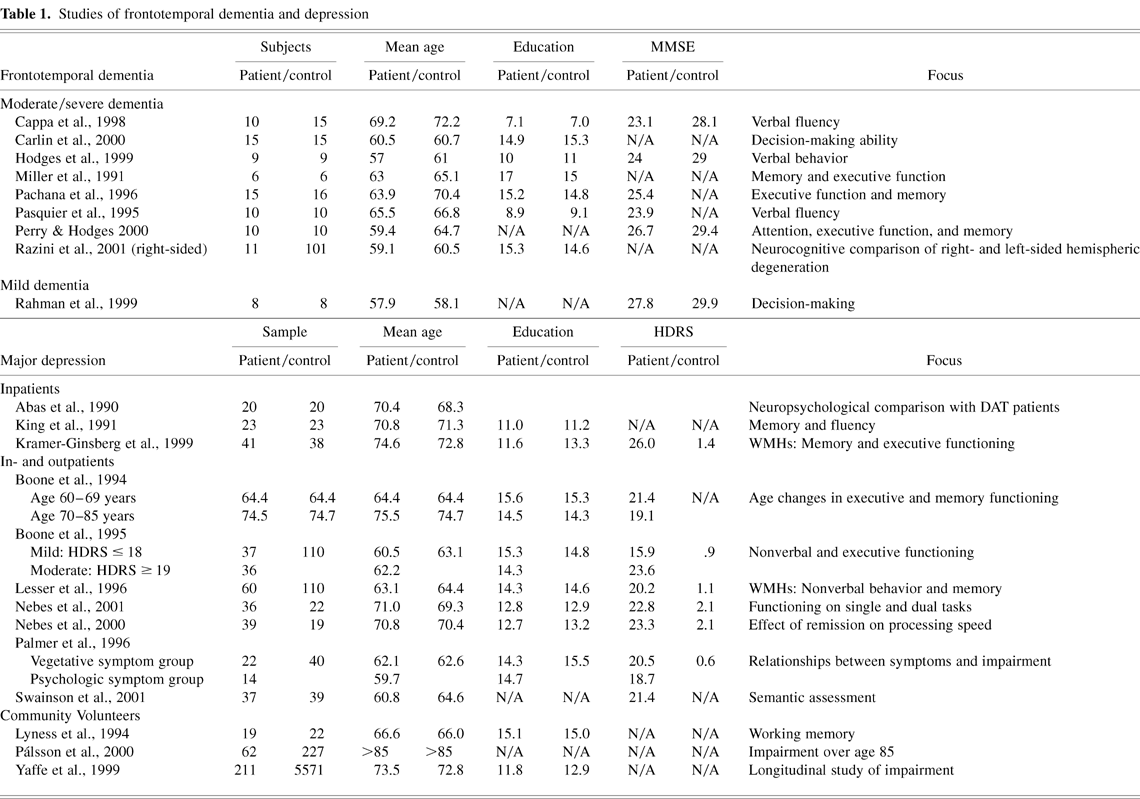
Studies of frontotemporal dementia and depression
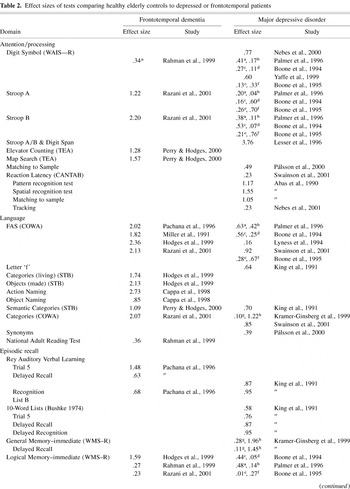
Effect sizes of tests comparing healthy elderly controls to depressed or frontotemporal patients

Table 2 continued

Table 2 continued
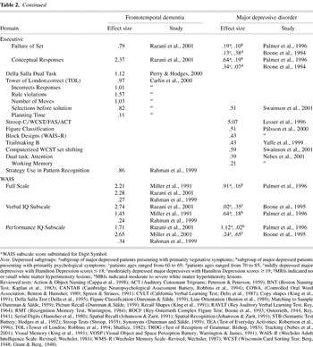
Table 2 continued

Weighted average effect sizes per cognitive domain among moderately advanced FTD patients, early FTD patients, major depressed inpatients, and major depressed community volunteers. The graph depicts the range of scores for each group.

Weighted effect sizes for three subgroups of FTD patients and four subgroups of major depressed patients. Note: Lesser et al. (1996) study was omitted because scores represented composite scores rather than individual test scores.
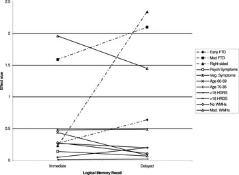
Change in FTD and MDD patient subgroup performance in the Logical Memory Test (WMS–R) between immediate recall and delayed recall. FTD groups are represented by dotted lines and MDD groups by solid lines, and both groups had patients who ranged from mildly to severely ill. For all subgroups of FTD patients, their performance was worse at the delayed recall than at the immediate recall. MDD patients tended to perform consistently across recall points.
 You have
Access
- 43
- Cited by