Introduction
A number of techniques have been described for the surgical correction of choanal atresia. This case report gives the first published description of transoral use of a transnasal flexible laryngoesophagoscope during surgical correction of choanal atresia.
Case report
A four-month-old boy was referred to the ear, nose and throat department by his general practitioner with a two-week history of left-sided, watery, green, nasal discharge.
On examination, the cold spatula test revealed absent fogging from the left nostril, and an attempt to pass a paediatric catheter transnasally failed.
A subsequent computed tomography scan demonstrated a unilateral, left, membranous choanal occlusion with a narrow posterior nasal aperture.
The child was taken to the operating theatre, where the left membranous blockage was punctured and the atresia sequentially dilated with Lister sounds. The posterior septum was then trimmed to further improve the posterior nasal airway.
During this procedure, a transnasal flexible laryngoesophagoscope was employed as a substitute for the rigid endoscope normally used in our practice. The current practice of employing a four-handed surgical technique was followed, but patient positioning was improved for the surgeon performing the resection, whilst instrument crowding was almost entirely resolved as the endoscope operator was able to visualise the operative field without encroaching on the primary surgeon's field of movement. The 5.1 mm diameter endoscope was employed solely via the oral cavity, in order to provide a retrograde view of the choanae from the nasopharynx. This provided adequate space within the child's nasal passage through which to pass the necessary instruments to perform the surgery, with the omnidirectional visualisation of the endoscope being utilised to view the most posterior aspect of the surgical field. Other features which aided the surgeon's progress included the suction and insufflation functions of the endoscope, both of which facilitated a clear operative field. An additional benefit was the extra port, which allowed the surgeon to insert additional instruments into the operative area to aid any further resection if needed.
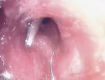
One month after surgery, examination under anaesthesia revealed a small degree of granulation tissue (Figure 1) occluding the neo-aperture. This tissue was excised by judicious use of monopolar diathermy, under the guidance of a transnasal flexible laryngoesophagoscope introduced transorally.
Transoral endoscopic view, using a transnasal flexible laryngoesophagoscope, showing the left posterior choana one month after the first operation. An instrument tip is clearly demonstrated passing posteriorly out of the left posterior choana to engage a small collection of granulation tissue.

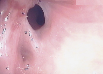
During a third and final procedure, similar endoscopic examination under general anaesthesia demonstrated a patent aperture with no sign of granulation tissue recurrence (Figure 2).
Transoral endoscopic view, using a transnasal flexible laryngoesophagoscope, showing the left posterior choana during the third and final operation. A patent posterior choana is clearly demonstrated.

Discussion
Choanal atresia is congenital stenosis or complete occlusion of one or both posterior nasal apertures, and was first described by Roederer in 1755. Choanal atresia occurs in approximately one in five to eight thousand live births, and is more commonly seen in preterm babies and multiple births.Reference Deutsch, Kaufman and Eilon 1 There is a higher incidence in females than males.Reference Deutsch, Kaufman and Eilon 1 Congenital unilateral obstruction is more common than bilateral obstruction, whilst bony or cartilaginous tissue is more likely to be the cause of obstruction than completely membranous tissue.Reference Stankiewicz 2 The incidence of other associated congenital abnormalities concomitant with choanal atresia is reported to be as high as 72 per cent.Reference Morgan and Bailey 3 Developmental malformation of the choana is thought to be due to the persistence of the oronasal membrane past the sixth week of gestation.Reference Hengerer, Brickman and Jeyakumar 4
Over the years, a variety of different approaches have been reported for the management of choanal atresia. Transnasal puncture, transpalatal resection and transnasal endoscopic resection are all established methods of treatment. Transnasal puncture is no longer the preferred method, as it may only provide temporary patency and there is a risk of cerebrospinal fluid leakage and midbrain trauma.Reference Theogaraj, Hoehn and Hagan 5 Blind puncture is associated with the need for repeated dilatation and a high re-operation rate.Reference Hengerer, Brickman and Jeyakumar 4 Early methods of surgical correction focused on transpalatal resection.Reference Owens 6 The directly visualised transpalatal approach provides excellent exposure of the operative space, but the removal of the posterior hard palate and midline suture can leave the patient with a high palatal arch and a cross-bite deformity; intra-oral fistulae and wound dehiscence have also been reported as complications.Reference Pirsig 7 , Reference Durmaz, Tosun, Yldrm, Sahan, Kvrakdal and Gerek 8
With the advent of improved endonasal endoscopic technology and techniques, the transnasal approach became feasible. Benjamin was the first to report endoscopic repair of choanal atresia, in 1985.Reference Benjamin 9 Since then, a great deal of literature has demonstrated the improved outcomes available with a transnasal technique. Current techniques utilise endoscopic visualisation, both transnasal (alongside the surgical instruments) and also transoral (using an angled endoscope to visualise the choana from the nasopharynx whilst the operating instruments are passed transnasally).Reference Deutsch, Kaufman and Eilon 1 , Reference Benjamin 9 – Reference Hassan, AboEl-Ezz and Youssef 13 Others have described the benefit of creating a posterior septal window so the endoscope can be used on one side and the surgical instruments on the other.Reference Hall, Watanabe, Kenan and Baylin 14 , Reference Rudert 15
Transnasal endoscopic resection is currently the most accepted treatment option for choanal atresia. A meta-analysis of 238 cases demonstrated an 85.3 per cent success rate, a 14.7 per cent restenosis rate and a 14.2 per cent minor complication rate.Reference Durmaz, Tosun, Yldrm, Sahan, Kvrakdal and Gerek 8 Minor complications included mucosal bleeding, the formation of granulation tissue and the creation of a septal perforation. The largest single unit cohort study, assessing 50 patients over a 6-year period, demonstrated an 84 per cent success rate with a 12 per cent restenosis rate and a 6 per cent incidence of palatal fistula.Reference Morgan and Bailey 3
More recently, complementary adjuncts to surgical resection have been proposed. These include the use of intranasal stents, laser ablation and topical mitomycin. The use of a laser is thought to provide an instant haemostatic effect, rapid re-epithelialisation and avoidance of scar formation; however, there is no clear evidence of its benefit as yet.Reference Healy, McGill, Jako, Strong and Vaughan 16 , Reference D'Eredita and Lens 17 There is still debate over the role of post-operative stenting. Shortened periods of nasal stenting are thought to reduce the likelihood of granulation tissue formation and post-operative infection; however, prolonged use seems to promote the opposite.Reference Pasquini, Sciarretta, Saggese, Cantaroni, Macri and Farneti 18 – Reference Samadi, Shah and Handler 20 Topical mitomycin has been shown to successfully reduce restenosis and granulation tissue formation, albeit in a small cohort of patients.Reference Bozkurt, Keles, Azimov, Ozturk and Arbag 21
The design of the transnasal flexible laryngoesophagoscope has progressed as the result of advances in endoscopic digital technology. The current design consists of an 80 K digital video camera incorporated into a 5.1 mm diameter endoscope (Pentax, Tokyo, Japan). The endoscope incorporates a high definition, colour, charged-couple device chip at the tip which provides 1080p Full HD resolution. The instrument provides a 180° range of movement over the distal 60 mm, and comes equipped with suction, insufflation and a 2 mm instrument port. The transnasal flexible laryngoesophagoscope has been shown to be useful in the management of various ENT conditions, including foreign body removal and head and neck cancer surveillance.Reference Price, Jones and Montgomery 22 – Reference Snelling, Price, Montgomery and Blagnys 24
-
• Surgical correction of choanal atresia generally involves a rigid endoscope
-
• In this case, transoral use of a transnasal flexible laryngoesophagoscope proved a useful improvement
-
• This endoscope's omnidirectional movement improved operative vision
-
• The suction and insufflation functions ensured clarity of the operative field
-
• The extra instrument port was also useful
This case report highlights the benefits of transoral use of the transnasal flexible laryngoesophagoscope during transnasal repair of choanal atresia. This endoscope provides greater flexibility, aiding the operator's use of the other surgical instruments. During our patient's first operation, this endoscope was utilised solely via the transoral route, enabling tissue resection with a microdebrider via the nose and affording an excellent view of the nasopharynx. Transoral use of the transnasal flexible laryngoesophagoscope benefits the surgeon by enabling a clearer operative field and by supplying high definition picture clarity. The incorporated suction and insufflation functions, and the option of passing extra instruments via the endoscope port, are of particular advantage during surgical choanal atresia correction.