Introduction
Hybrid vigour has been known for a long time in agriculture. Specifically, hybrid offspring obtained by mating two different strains show superior traits than their parents (Castle, Reference Castle1926). Although the mechanism causing hybrid vigour has been discussed, this mechanism is still unclear (Ishikawa, Reference Ishikawa2009). There are various strains of laboratory mice and MII stage oocytes from these mice have various traits (Pfeiffer et al., Reference Pfeiffer, Taher, Drexler, Suzuki, Makałowski, Schwarzer, Wang, Fuellen and Boiani2015). For intracytoplasmic sperm injection, an MII stage oocyte is injected with sperm using a specialized glass capillary. The oocytes of B6D2F1 (BDF1) mice, obtained by mating female C57BL/6 (B6) and male DBA/2 (D2) mice, show higher survival rates after injection than those of B6 mice (Kawase et al., Reference Kawase, Iwata, Toyoda, Wakayama, Yanagimachi and Suzuki2001; Kaneko and Ohno, Reference Kaneko and Ohno2011). Additionally, in the production of somatic cell nuclear transfer embryos, it is well known that BDF1 oocytes as recipients show a better developmental rate than other strains (Gao et al., Reference Gao, Czirr, Chung, Han and Latham2004). Therefore, BDF1 mice have been used for various studies (Inoue et al., Reference Inoue, Noda, Ogonuki, Miki, Inoue, Katayama, Mekada, Miyoshi and Ogura2007a, Reference Inoue, Ogonuki, Miki, Hirose, Noda, Kim, Aoki, Miyoshi and Ogura2007b; Miki et al., Reference Miki, Wakisaka, Inoue, Ogonuki, Mori, Kim, Ohta and Ogura2009). As a reason for this superior trait of BDF1 MII stage oocytes, it is assumed that BDF1 mice have the superior traits of each parent by hybrid vigour. However, the responsible factors have not been clarified. Here, we focused on the cell membrane of oocytes as a phenotype to characterize differences between strains. One approach to verify the phenotype of the cell membrane was examination of tolerance to osmotic pressure changes. Previous studies had cultured oocytes in various osmotic media and reviewed the differences in resistance to various osmolalities (Pedro et al., Reference Pedro, Sakurai, Edashige and Kasai1997a, Reference Pedro, Zhu, Makino, Sakurai, Edashige and Kasai1997b). Regardless of reciprocal crossings, in terms of BDF1 and DBF1 mice (female D2 × male B6 mice), MII stage oocytes of DBF1 mice are rarely used in experiments compared with BDF1 mice. The reason is that it is considered that there is qualitative difference between oocytes of BDF1 and DBF1 mice. If differences between BDF1 and DBF1 mice exist, inheritance of oocyte quality is different by the combination of parent’s sex, and it will be the key to make hybrid vigour mechanism clear. Therefore, we used DBF1 oocytes in this study; we prepared MII stage oocytes of BDF1, B6, D2, and DBF1 mice, and compared their resistance to osmotic changes.
Materials and methods
Animals
All mice were purchased from Japan SLC (Shizuoka, Japan) and bred in-house. B6, D2, BDF1, and DBF1 female mice at 4 weeks of age were used. The mice were maintained in a temperature- and humidity-controlled room with a 12 h light/12 h dark cycle and had free access to food and water. This study was approved by the Tohoku University Institutional Animal Care and Use Committee.
Preparation of culture medium
The basal medium was Leibovitz’s medium (L-15) (Gibco, Life Technologies Japan Ltd, Tokyo, Japan). Medium preparation was conducted according to the studies of Pedro et al. (Reference Pedro, Sakurai, Edashige and Kasai1997a, Reference Pedro, Zhu, Makino, Sakurai, Edashige and Kasai1997b) with some modifications. Briefly, for isotonic medium preparation, L-15 containing 1 g/l polyvinyl alcohol (0.1% PVA L-15 medium) was used. For hypotonic medium preparation, L-15 medium was diluted with double-distilled water containing 1 g/l polyvinyl alcohol at a v/v ratio of 1:4 for tonicity of 0.2×. Hypertonic medium was prepared by adding sucrose to isotonic medium at a concentration of 1 M. The osmolalities were measured by osmometer based on depression of the freezing point (OM-815, VOGEL). The osmolality of isotonic and hypotonic medium was 161 and 324 mOsm/l, respectively. However, this device was not able to measure hypertonic medium. Theoretical osmolality of the L-15 medium is 331 mOsm/l. The osmolality of 1 M sucrose solution is 1000 mOsm/l because sucrose is a non-electrocyte. In this study, sucrose was added in hypertonic medium at the concentration of 1 M. Thus, theoretical osmolality of hypertonic medium was 1331 mOsm/l. Media (4 ml) were placed in culture dishes (inner diameter 35 mm × inside depth 14 mm) and covered with mineral oil. All experiments were performed at 25°C on a warm plate.
Collection of oocytes
For oocyte collection, female mice from each strain were administered with 5 IU pregnant mare serum gonadotropin (PMSG) (Serotropin; ASKA Animal Health Co., Ltd, Tokyo, Japan) intraperitoneally. At 48 h after PMSG injection, the mice were administered with 5 IU human chronic gonadotropin (hCG) (Mochida Pharmaceutical Plant Co., Ltd, Tokyo, Japan) intraperitoneally to induce superovulation. Ovulated oocytes were collected from the ampulla of oviducts at 16 h after hCG injection and freed from cumulus cells by suspending in 0.1% PVA L-15 medium containing 0.1% hyaluronidase, followed by washing in fresh 0.1% PVA L-15 medium. After washing oocytes, polar body exclusion was checked under a microscope and their stage was determined. Only MII stage oocytes were used in subsequent experiments. Oocytes were pooled in fresh 0.1% PVA L-15 medium before treatment. The numbers of oocytes of BDF1 and B6, D2, DBF1 used in this experiment were 453, 355, 166 and 457, respectively.
Culture of oocytes in osmotic modified media
The collected oocytes were cultured at 25°C for 30 min in isotonic, hypotonic or hypertonic media. After 30 min of culture, they were washed with isotonic medium and transferred to fresh isotonic medium.
Evaluation of osmotic pressure resistance in oocytes
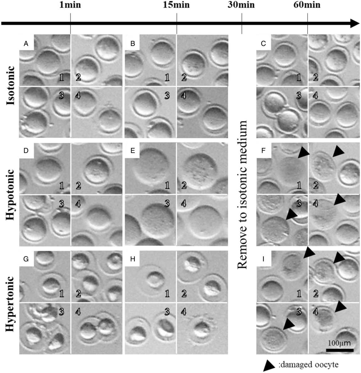
After 30 min of culture, oocytes were observed under a stereoscopic microscope (Olympus, Tokyo, Japan) to evaluate their resistance to osmotic pressure changes. Oocytes with a broken membrane were judged as ‘damaged’ as shown in Fig. 1 (black arrowheads). The number of damaged oocytes was counted to calculate the ‘normal rate’ of oocytes. The normal rate of BDF1 oocytes was set to 1, and those of B6, D2, DBF1 oocytes were compared.
Morphological changes of MII oocytes after culture under various osmotic pressures. MII stage oocytes that morphologically changed in hypotonic or hypertonic media are shown. These images were obtained during culture in osmotic modified medium or at 30 min after transfer to isotonic medium (60 min after culture). In each image, 1 shows BDF1 oocytes, 2 shows B6 oocytes, 3 shows D2 oocytes and 4 shows DBF1 oocytes, respectively. Oocytes with a broken membrane, as indicated by black arrowheads, were judged as ‘damaged’, while the other oocytes were judged as ‘normal’. One minute after the culture had started in the medium (A, D and G). Thirty minutes after culture in medium (B, E and H). Thirty minutes after returning to isotonic medium from the experimental media (C, F and I).

Average numbers of live births of BDF1 and DBF1 mice
To product BDF1 mouse, two female B6 mice and one male D2 mouse were coupled for more than 3 weeks and less than 2 months until the first pups were obtained. To product DBF1 mouse, two female D2 mice and one male B6 mouse were coupled for the same duration as the BDF1 production. The average number of live birth was calculated as the average number of pups per mother.
Statistical analysis
Data were tested statistically using the Tukey–Kramer multiple comparison test with JMP 11 software (SAS Institute Inc., Cary, NC, USA). A P-value < 0.05 was considered as statistically significant.
Results
Changes in the oocyte volume, inflation or shrinking, were observed immediately after oocytes were transferred into osmotic modified media as shown in Fig. 1. During culture in isotonic medium, the volumes of MII stage oocytes of each strain did not change (Fig. 1 a–c). The volumes of oocytes in hypotonic medium increased compared with those of oocytes in isotonic medium (Fig. 1 d, e). Additionally, the volumes of oocytes in hypertonic medium decreased compared with those of oocytes in isotonic medium (Fig. 1 g, i). However, the volumes of oocytes in hypotonic and hypertonic media returned to their original volumes, while some oocytes were damaged and their membranes could not be observed (Fig. 1 f, i, black arrowheads).
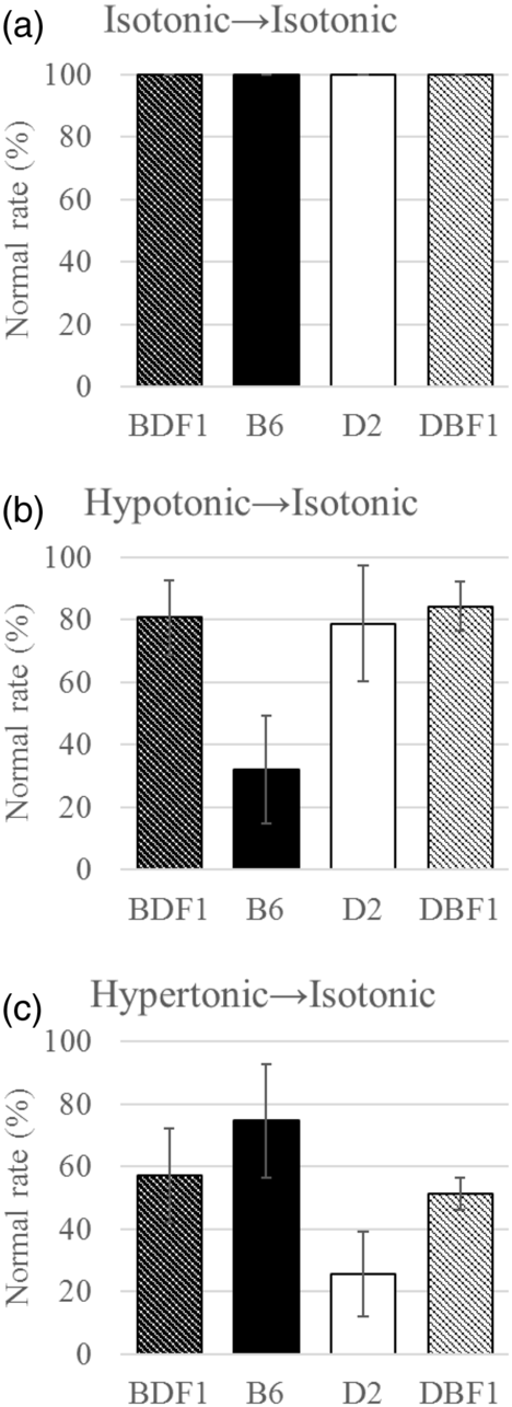
The normal oocyte rate in isotonic medium culture was 100% for each strain (Fig. 2 a), whereas those of BDF1, B6, D2 and DBF1 oocytes in hypotonic medium culture were 80.7, 32.1, 78.8 and 84.3%, respectively (Fig. 2 b). Normal rates of BDF1, D2, and DBF1 oocytes were significantly higher than that of B6 oocytes. The normal rates of BDF1, B6, D2 and DBF1 oocytes in hypertonic medium were 57.2, 74.7, 25.6 and 51.3%, respectively (Fig. 2 c). There were no significant differences between the normal rates of BDF1 oocytes and other strains. The normal rate of B6 oocytes was significantly higher than that of D2 oocytes. DBF1 oocytes showed no significant difference in the normal rate compared with BDF1 oocytes in each osmotic medium.
Table 1 shows the average number of live births of BDF1 and DBF1 mice. The average number of live births was calculated as the number of pups per number of mothers. Data were tested statistically using a T-test with JMP 11 software (SAS Institute Inc., Cary, NC, USA). A P-value < 0.05 was considered as statistically significant. The average pups number of BDF1 and DBF1 mice was 5.6 and 2.9, respectively (Table 1). The number of DBF1 pups was significantly less than that of BDF 1.
Comparison of normal oocyte ratios in each mouse strain. The ratios of normal oocytes obtained by culture in osmotically modified media, isotonic (a), hypotonic (b), and hypertonic (c), are shown. Data were tested statistically using the Tukey–Kramer multiple comparison test with JMP 11 software (SAS Institute Inc., Cary, NC, USA). Mean ± standard error (SE) with different letters are significantly different (P < 0.05).

Average number of live births of BDF1 and DBF1 mice.

P < 0.01.
Discussion
It is well known that red blood cells burst because of water inflow into the cytoplasm upon suspension in hypotonic medium (Yang and Kamino, Reference Yang and Kamino1995; Yamaguchi et al., Reference Yamaguchi, Terada and Ideguchi1997). For this reason, it is considered that the same phenomenon damages oocytes cultured in hypotonic medium. Bursting is a phenomenon in which the initial breach grows instantly (Sakumichi and Okumura, Reference Sakumichi and Okumura2017). Cell bursting in hypotonic medium and by capillary glass injection have similar characteristics at point of instant growth of initial breach. Previous studies have reported that BDF1 and D2 oocytes showed higher survival rates after capillary glass injection than B6 oocytes (Ogonuki et al., Reference Ogonuki, Mori, Shinmen, Inoue, Mochida, Ohta and Ogura2010; Kaneko and Ohno, Reference Kaneko and Ohno2011). This finding is very similar to the strain-dependent difference observed in the hypotonic environment of this study. In terms of tolerance to a hypertonic environment, a different system that may work in the hypotonic environment might be appropriate.
We considered two factors for strain-dependent differences. One is actin distributed just under the membrane of MII stage oocytes. Strain-dependent differences observed in this study might have been caused by differences in factors related to the actin layer. Actin is one of the cytoskeleton proteins. F-actin, which is a polymerized actin filament, exists just under the cell membrane in many animal cells and plays a role in the morphology and strength of cells (Alberts et al., Reference Alberts, Johnson, Lewis, Morgan, Raff, Roberts and Walter2014). In MII stage oocytes of mice, the actin layer exists directly under the oocyte membrane (Mao et al., Reference Mao, Lou, Lou, Wang and Jin2014).
Previous studies have shown that culturing oocytes with cytochalasin, an inhibiter of actin polymerization, attenuates their fluorescence strength (Zampolla et al., Reference Zampolla, Spikings, Rawson and Zhang2011) and decreases the elasticity of the oolemma (Grasland-Mongrain et al., Reference Grasland-Mongrain, Zorgani, Nakagawa, Bernard, Paim, Fitzharris, Catheline and Cloutier2018). Based on these reports, it may be considered that oolemma elasticity is associated with the degree of actin polymerization, which increases with the increasing degree of actin polymerization. Cell membranes consist of a phospholipid bilayer with both hydrophilic and hydrophobic sites. When a breach occurs in the cell membrane, hydrophobic sites of phospholipids are exposed to water. This situation is energetically unfavourable. Thus, a breach on the cell membrane is repaired by the natural ‘self-organizing capacity’ (Araiso, Reference Araiso1994). A long time is required to repair a large breach, and there is the possibility of bursting. Therefore, it is considered that a high degree of actin polymerization facilitates elasticity of the cell membrane and a high surface tension of the cell. As a result, upon damage of a cell with a high degree of actin polymerization, cell bursting occurs because a long time is required to repair the breach. Specifically, the degree of actin polymerization is associated with the possibility of cell bursting, i.e. a high degree of polymerization actin makes cells burst easily and a low degree of actin polymerization makes cells hardy. A previous study reported that BDF1 oocytes repair scaring of the oolemma rapidly (Kawase et al., Reference Kawase, Iwata, Toyoda, Wakayama, Yanagimachi and Suzuki2001). In the BDF1 oolemma, it may be considered that the speed of initial breach propagation is relatively slow because of the low degree of actin polymerization, and cell repair before rupturing.
Another possibility is the cell volume regulation system against extracellular osmotic pressure. Cells are exposed to various osmotic changes in vivo. A previous report has shown that cell volume increases because of water flow into the cell in a hypotonic environment (Pogorelova et al., Reference Pogorelova, Golichenkov, Pogorelova, Panait, Smirnov and Pogorelov2015). Although cells are forced to change their volume according to extracellular environments with varying osmolalities, cells have a system to resist volume variations and maintain a stable volume. In this cell volume regulation system, entirely different pathways operate according to the difference in the extracellular osmolality. One is the cell volume regulation system after swelling osmotically in a hypotonic environment, which is called the regulatory volume decrease (RVD), and the other is the cell volume regulation system after shrinking osmotically in a hypertonic environment, which is called the regulatory volume increase (RVI) (Okada, Reference Okada2004). The results of our study showed that B6 oocytes had high resistant to the hypotonic environment, D2 oocytes had high resistant to the hypertonic environment, and BDF1 and DBF1 oocytes had high resistant to both hypotonic and hypertonic environments. It is presumed that B6 oocytes may be more contributed by RVD-related factors than D2 oocytes. Conversely, D2 oocytes may be more contributed by RVI-related factors than B6 oocytes. Their hybrid strains, BDF1 and DBF1 oocytes may have aedcquired more RVD-related or RVI-related factors by dominant inheritance and potentiate resistance to osmotic challenge. In MII stage oocytes of mice, it has been reported that volume-sensitive outwardly rectifying anion channels are important for RVD and glycine transporter 1 is important for RVI (Baltz and Zhou, Reference Baltz and Zhou2012). Therefore, there is the possibility that these proteins are related to the strain-dependent differences in our results. However, it remains unclear and requires further study.
Our results showed that tolerance to hypertonic or hypotonic changes in osmotic pressure in oocytes derived from the hybrid strain depended on the combination of parent strains. BDF1 and DBF1 mice did not differ in the normal rate of oocytes subjected to various osmotic stimuli. Differences in maternal or paternal genes did not influence the normal oocyte rate. Therefore, these results suggested that the traits tested in this study are not regulated by imprinting genes.
As shown in Table 1, the average number of live births of DBF1 mice was significantly lower than that of BDF1 mice. This difference in the number of live births between BDF1 and DBF1 mice is considered to cause the difference in their experimental usage.
Acknowledgements
We thank M. Arico from Edanz Group (https://www.edanzediting.com/ac) for editing a draft of this manuscript.
Financial support
This research received no specific grant from any funding agency, commercial or not-for-profit sectors.
Conflicts of interest
There are no conflicts of interest.