INTRODUCTION
Fascioliasis is a major parasitic disease of livestock in temperate regions of the world. There has been a marked increase and spread of the disease in recent years, largely as a result of climate change. In the UK, for example, infection levels of more than 30% in cattle and more than 20% in sheep exist in some areas (Wolstenholme et al. 2004). Fascioliasis is also emerging as a major zoonosis and is considered to be a serious human health problem in some countries (reviewed by Mas-Coma et al. 2005).
Triclabendazole (TCBZ) remains the current drug of choice to treat liver fluke infections, because of its particularly high activity against juvenile flukes (Boray et al. 1983; Smeal and Hall, 1983; Turner et al. 1984). However, resistance to the drug was first identified in the mid-1990s (in Australia: Overend and Bowen, 1995) and appears to be widespread in Europe for which the data is most comprehensive (reviewed by Fairweather, 2005). The combination of increased disease prevalence and spread of TCBZ resistance is of considerable concern to the livestock industry, because no other fasciolicides on the market have comparable activity against the damaging immature stages of fluke.
Several proposals to overcome resistance have been attempted; they include use of alternative fasciolicides, use of synergistic combinations of drugs and the development of new drugs. Several existing fasciolicides – closantel, oxyclozanide, nitroxynil, clorsulon and albendazole – have been shown to be active against TCBZ-resistant flukes (Coles et al. 2000; Moll et al. 2000; Coles and Stafford, 2001). However, their activity is limited to the adult fluke, not the juvenile stages. Recently, a derivative of TCBZ, designated compound alpha (or compound 6) has been synthesized and tested for activity against F. hepatica (Hernández-Campos et al. 2002). It has been shown to be active against both mature and immature infections in sheep and cattle, killing flukes as young as 3 days old (Hernández-Campos et al. 2002; Ibarra et al. 2004; Vera Montenegro et al. 2004). This spectrum of activity is similar to that of TCBZ itself. The isolate used in these studies was a field isolate, which is presumed to be TCBZ-susceptible. To date, compound alpha has not been tested against TCBZ-resistant isolates of F. hepatica.
The present study represents the first of a series of experiments designed to evaluate the activity of compound alpha against TCBZ-resistant F. hepatica. It was carried out in vitro, to determine the effect of the sulphoxide metabolite of compound alpha (hereafter referred to as alpha.SO) against the Sligo TCBZ-resistant isolate of F. hepatica. By analogy with TCBZ, it is likely that alpha.SO is the active metabolite of the drug. The study focuses on the tegument, one of the principal absorptive surfaces of the fluke for the uptake of drugs. Changes to the tegument surface, the tegumental syncytium and the tegumental cells were assessed by scanning and transmission electron microscopy (SEM; TEM). As TCBZ.SO is believed to target microtubule-based processes in the fluke, alpha.SO may act in a similar way. In order to examine this possibility, immunocytochemistry using an anti-tubulin antibody was also carried out, to examine changes in the level and distribution of tubulin immunoreactivity in the tegument following treatment with alpha.SO.
MATERIALS AND METHODS
Adult male Sprague-Dawley rats were each experimentally infected with 20 metacercarial cysts of a TCBZ-resistant isolate of F. hepatica derived originally from County Sligo in Ireland. The Sligo isolate has been shown to be resistant to the action of TCBZ in vivo (Coles et al. 2000; Coles and Stafford, 2001; McCoy et al. 2005) and has been used in a number of in vitro studies (Robinson et al. 2002, 2004; Alvarez et al. 2005). Adult flukes (at least 12 weeks old) were removed from the bile ducts of the rats under sterile conditions in a laminar flow cabinet. They were washed repeatedly in warm (37 °C) sterile NCTC 135 culture medium containing antibiotics (penicillin, 50 IU/ml; streptomycin, 50 μg/ml). The recovered flukes were transferred to fresh culture medium containing compound alpha sulphoxide (alpha.SO) metabolite at a concentration of 10 μg/ml and incubated for 24 h at 37 °C. Alpha.SO was initially prepared as a stock solution in dimethyl sulphoxide (Me2SO) and added to the culture medium to give a maximum solvent concentration of 0·1% (v/v). The concentration of alpha.SO used in this study was selected so as to be comparable to maximum blood levels attained in sheep following an oral dose of 12 mg/kg compound alpha: 8·3 μg/ml at 10 h (Rivero et al. 1998). Controls were prepared by incubating whole flukes in NCTC 135 medium in the absence of alpha.SO, but containing Me2SO.
For the tubulin immunocytochemistry experiment, flukes of the Cullompton isolate, which has been shown to be susceptible to the action of TCBZ in vivo and in vitro (Robinson et al. 2002; McCoy et al. 2005) were treated in vitro for 24 h at 37 °C in 15 μg/ml TCBZ.SO; 10 μg/ml albendazole sulphoxide (ABZ.SO); and 10 μg/ml alpha.SO. Sligo flukes were also incubated for 24 h at 37 °C in ABZ.SO at 10 μg/ml; and TCBZ.SO at 15 μg/ml.
Tissue preparation for SEM
Flukes were lightly flat-fixed for 1 h at room temperature in a 3[ratio ]1 mixture of 4% (w/v) aqueous glutaraldehyde and 1% aqueous osmium tetroxide and subsequently free-fixed in fresh fixative for a further 3 h at 4 °C. They were briefly washed several times in distilled water, dehydrated in an ascending series of ethanol, dried in hexamethyldisilazane, mounted on aluminium stubs and sputter-coated with gold-palladium. The specimens were viewed in a JEOL 6400 SEM operating at 10 keV.
Tissue preparation for TEM
The flukes were lightly flat-fixed for 1 h at room temperature in 4% (w/v) glutaraldehyde in 0·1 M sodium cacodylate buffer (pH 7·4) containing 3% (w/v) sucrose. The specimens were dissected into apical cone (including ventral sucker), mid-body and tail regions. The mid-body was further divided into transverse sections of approximately 2 mm in width. The sections of fluke were then free-fixed for a further 3 h, after which they were washed in 0·1 M sodium cacodylate buffer (pH 7·4) containing 3% (w/v) sucrose and left overnight at 4 °C. Following post-fixation in 1% osmium tetroxide for 1 h, the tissue was washed in fresh buffer, dehydrated in an ascending series of ethanol and infiltrated and embedded in Agar 100 resin. Ultrathin sections, 60–70 nm in thickness, were cut on a Reichert Ultracut E ultramicrotome, mounted on bare 200-mesh copper grids, double-stained with uranyl acetate (10 min) and lead citrate (6 min) and viewed in a JEOL 100-CX TEM operating at 100 keV.
Tissue preparation for tubulin immunocytochemistry
Flukes were lightly flat-fixed in absolute methanol for 30 min, free-fixed in absolute methanol for 2·5 h at room temperature and infiltrated and embedded in JB4 resin. Sections, 4 μm in thickness, were cut on a pyramitone, mounted on multi-welled slides and washed in 0·1 M phosphate-buffered saline (PBS), pH 7·3. The JB4 sections were then incubated overnight in primary polyclonal anti-tubulin antiserum (1[ratio ]50) at room temperature. The antibody was raised in rabbit against 13-day-old chick embryo tubulin (Sigma-Aldrich Co. Ltd, Poole, Dorset, UK). The sections were subsequently washed in PBS (pH 7·3) and incubated in an FITC-conjugated swine anti-rabbit secondary antibody (1[ratio ]50) (Dako Ltd, High Wycombe, UK). Finally, the sections were washed in PBS, mounted in anti-fade solution [90% (v/v) glycerol, 10% (v/v) PBS (pH 7·3), 2·5% (w/v) diazabicyclo] and observed using a Leitz Epifluorescent microscope. A number of controls were carried out: they included the omission of the primary antiserum, omission of the secondary antiserum and the use of untreated specimens.
RESULTS
Scanning electron microscopy
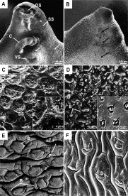
Following 24 h incubation in vitro with 10 μg/ml of alpha.SO, extensive spine loss was observed on the ventral surface of the apical cone (Fig. 1A). Severe swelling of the tegument occurred in the central region of the dorsal apical cone (Fig. 1B). On the ventral surface of the apical cone, at the base of the empty spine sockets, the basal lamina was exposed (Fig. 1C). Loss of the apical plasma membrane had occurred to the tegument surrounding the spine sockets (Fig. 1C inset). In the areas where the spines remained on the apical cone, the tegument typically adopted a ‘roughened’ appearance, particularly around the oral and ventral suckers where it had begun to slough off. The inter-spinal tegument in the central region of the dorsal cone was carpeted in blebs and the spines had become slightly obscured by the swollen tegument surrounding them (Fig. 1D). Areas with empty spine sockets were also present on the dorsal cone region; in these areas, spines that remained in situ had split along their length (Fig. 1D inset).

Fig. 1. Scanning electron micrographs (SEMs) of the tegumental surface of the liver fluke Fasciola hepatica following 24 h in vitro treatment with alpha.SO. (A) The ventral surface of the apical cone, showing furrowing and cracking of the tegument (arrows) and empty spine sockets (SS). The cirrus (C) is visible extending from the gonopore. OS, oral sucker; VS, ventral sucker. (B) The dorsal surface of the apical cone, showing swelling (arrows) of the central region. (C) Higher magnification image showing empty spine sockets (SS) on the ventral surface of the apical cone indicated by * on Fig. 1A. The inter-spinal tegument (T) has lost the apical plasma membrane. Inset showing a higher magnification image of the inter-spinal tegument where the apical plasma membrane has been lost (arrow). (D) Higher magnification image of the dorsal surface of the apical cone indicated by * on Fig. 1B, showing severe swelling of the tegument which is covered in blebs (B) of various sizes. The tegument covering the spines is also disrupted (arrows). Inset shows a higher-power image of spines of the apical cone that appear to have split along their length (arrows). (E) The central area of the anterior mid-body region on the ventral surface of the fluke showing swelling and furrowing (F) of the tegument. Blebs (B) are present on the tegument and the spines (S) appear sunken. (F) The anterior mid-body region on the dorsal surface of the fluke, showing spines (S) which appear to be sunken due to the swelling of the tegument surrounding them. Small blebs (B) are also present on the surface.
The anterior mid-body region showed evidence of disruption to the tegument on the ventral surface, in that the tegument was generally swollen with patches of blebs adorning the surface and the spines had assumed a sunken appearance due to the swollen tegument around them (Fig. 1E). On the dorsal surface in the anterior mid-body region, the tegument was again often swollen up around the spines (Fig. 1F), though not to the same extent as that seen on the ventral surface. As observed on the ventral surface, the tegument surrounding the spines often appeared swollen, with blebs on the surface.
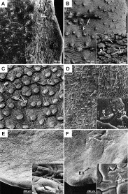
Sloughing of the tegument exposing, in some cases, the basal lamina, occurred along the posterior mid-body lateral margins of the fluke on both the ventral and dorsal surfaces (Fig. 2A). It should be noted, however, that tegumental loss occurred to a greater degree on the ventral surface. Along the posterior lateral margins on the dorsal surface, the tegument was typically swollen, with large patches of blebbing (Fig. 2B). Blebbing occurred on the ventral surface and the tegument was slightly swollen, otherwise the tegument and spines largely retained a normal morphology (Fig. 2C). On the dorsal surface the tegument was swollen and furrowed and a layer of small blebs carpeted the surface (Fig. 2D).

Fig. 2. Scanning electron micrographs (SEMs) of the tegumental surface of the liver fluke Fasciola hepatica following 24 h in vitro treatment with alpha.SO. (A) The posterior lateral margin of the ventral surface, showing sloughing of the tegument which has exposed the basal lamina (BL) in some areas. (B) The posterior lateral margin of the dorsal surface, showing swelling and furrowing (F) of the tegument. Inset shows a higher magnification image of the same region of the fluke, where the tegument is swollen and covered in blebs (B). S, spine; F, furrowing. (C) The ventral surface in the posterior mid-body region, showing blebbing (B) and a slight swelling of the tegument. (D) The dorsal surface of the posterior mid-body region, showing swelling and furrowing (F) of the tegument. Inset shows a higher magnification image of the same area. The spines (S) have sunk into the swollen tegument surrounding them and there are blebs (B) on the tegumental surface. (E) The ventral tail region, showing a slight swelling of the tegument. Inset shows a higher-power image of the tegumental surface, which bears microvillus-like projections (MV) and loss of tegument from the tips of the spines (arrow). (F) The dorsal surface of the tail region shows swelling and furrowing (F) of the tegument. The excretory pore (EX) is also visible. Inset shows very small blebs (B) on the tegumental surface.
A predominantly normal morphology was observed on the ventral surface of the tail region, though a slight swelling of the tegument was apparent in a number of flukes (Fig. 2E). The tegument covering the tips of the spines was disrupted and the inter-spinal tegument occasionally assumed a ‘roughened’ appearance due to the presence of microvillus-like projections (Fig. 2E inset). The dorsal surface of the tail region displayed patches of swelling and furrowing of the tegument (Fig. 2F). When viewed at a higher magnification, very small blebs were visible (Fig. 2F inset).
The surface architecture of the control specimens appeared normal.
Transmission electron microscopy
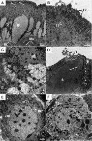
Sections for TEM were taken from the anterior mid-body region of the fluke. Within the tegumental syncytium, there was a dark band running just below the apical plasma membrane in all the specimens examined; swelling of the basal infolds was a common feature as well (Fig. 3A). At a higher magnification, the dark band was seen to be made up of an accumulation of secretory bodies, predominantly T2 secretory bodies (Fig. 3B). ‘Open’ bodies were observed below the apical plasma membrane, although they were not a typical feature. Blebs were also seen in a number of specimens projecting from the apical surface (Fig. 3B). The mitochondria within the syncytium were swollen, with distinct cristae, and had assumed a rounded shape rather than the normal cylindrical form. The typical alignment of the mitochondria (vertically stacked from the basal lamina to approximately the centre of the syncytium) was also disrupted (Fig. 3C). Swelling of the basal infolds occurred in the majority of specimens (Figs 3A and C). The tegument covering the spines was observed sloughing away from the spine tips and the spines themselves were often cracked (Fig. 3D).

Fig. 3. Transmission electron micrographs (TEMs) of the tegumental syncytium and tegumental cells of Fasciola hepatica following 24 h in vitro treatment with alpha.SO. (A) Transverse section through the tegumental syncytium. A dark band of secretory bodies (arrowed), predominantly T2 bodies, have accumulated below the apical plasma membrane. The basal infolds (BI) and mitochondria (m) are swollen. Below the basal lamina (BL) the muscle blocks (Mu) remain unchanged. (B) Accumulations of T1 (T1) and T2 (T2) secretory bodies are present below the apical plasma membrane of the tegument. The T1 bodies are not as abundant as the T2 bodies. Blebbing (B) of the plasma membrane is also seen. (C) TEM showing the basal region of the tegumental syncytium. Above the basal lamina (BL) the basal infolds (BI) are swollen and throughout the tegumental syncytium swollen mitochondria (m) are abundant. The T1 secretory bodies (T1) are distributed evenly throughout the syncytium. (D) TEM image showing disruption to the tegument (T) covering the spine (S). Damage to the spine itself is also evident, in the form of cracking (arrow). (E) The nucleus (n) of this T1-type of tegumental cell appears normal, although the mitochondria (m) are swollen, as are the cisternae of the granular endoplasmic reticulum (ger). The T1 secretory bodies (T1) are less numerous than normal as are the Golgi complexes. (F) TEM image of a T2-type of tegumental cell in which the nucleus (n) and nucleolus (nu) both retain a normal morphology. The mitochondria (m) are swollen and the T2 secretory bodies (T2) have accumulated within the cell. Inset shows a collection of T2 secretory bodies (T2) in the cytoplasm of the cell.
T1 and T2 secretory bodies were present in their respective tegumental cells (Figs 3E and F). Golgi complexes, though present in both T1 and T2 cells, occurred infrequently and the complex was typically reduced with few discernable cisternae. In the T1 tegumental cells, the mitochondria were swollen and some had assumed a circular appearance; the cisternae of the granular endoplasmic reticulum (ger) were also swollen (Fig. 3E). The number of T1 secretory bodies appeared to be slightly depleted in comparison with control cells (Fig. 3E). The mitochondria were swollen, though not to the extent as seen within the T1 tegumental cells. T2 secretory bodies appeared to have accumulated within the cell (Fig. 3F inset).
The tegumental ultrastructure of the control specimens was normal.
Tubulin immunocytochemistry
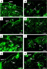
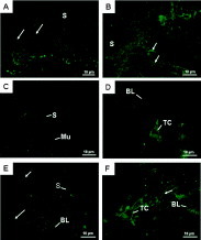
In the control TCBZ-resistant material, tubulin immunostaining was evident as a strong fluorescent band running below the apical plasma membrane of the tegumental syncytium. Fluorescence occurred throughout the syncytial layer and often, a distinct increase in the intensity of fluorescence occurred in the syncytium just above the basal lamina. Strong fluorescence was frequently seen in the syncytium surrounding the spines, specifically, the syncytium around the base of the spine (Fig. 4A). Tubulin immunoreactivity (IR) was localized within the tegumental cells situated below the muscle blocks and within the cytoplasmic connections linking tegumental cells to the syncytium (Fig. 4B). The control material for the Cullompton isolate displayed the same distribution and intensity of tubulin IR as described for the Sligo isolate.

Fig. 4. Micrographs showing tubulin immunostaining in the tegumental syncytium and tegumental cells of the TCBZ-resistant Sligo isolate of Fasciola hepatica. (A) 24 h control. The tegumental syncytium shows a strong band of fluorescence running beneath the apical plasma membrane (apm) and a more dispersed band just above the basal lamina (BL). The tegumental syncytium in the vicinity of the spines (S) displays strong fluorescence (arrow). Mu, muscle. (B) 24 h control. The tegumental cells (TC) are immunopositive for tubulin, as are the cytoplasmic connections (arrow). BL, basal lamina. (C) 24 h incubation with alpha.SO (10 μg/ml). The tegumental syncytium displays tubulin immunoreactivity (IR) beneath the apical plasma membrane (apm) and dispersed throughout the lower half of the syncytium (arrow) and in association with the spines (S). (D) 24 h incubation with alpha.SO (10 μg/ml). The cytoplasmic connections (arrow) and tegumental cells (TC) display tubulin-IR. Mu, muscle. (E) 24 h incubation in ABZ.SO (10 μg/ml). Tubulin-IR is present throughout the syncytium and occurs as a distinct band running beneath the apical plasma membrane (apm). Below the muscle (Mu) the tegumental cells (TC) are immunopositive for tubulin. (F) 24 h incubation in ABZ.SO (10 μg/ml). Tubulin immunostaining can be seen throughout the syncytium (arrows) and in close association with the spines (S). BL, basal lamina. (G) 24 h incubation in TCBZ.SO (15 μg/ml). Tubulin-IR is present as a band running beneath the apical plasma membrane (apm) and also throughout the syncytium (arrows). S, spine; Mu, muscle. (H) 24 h incubation in TCBZ.SO (15 μg/ml). The tegumental cells (TC) are immunopositive for tubulin. BL, basal lamina; apm, apical plasma membrane.
Sligo isolate treated with alpha.SO
The distribution of tubulin-IR observed in the Sligo isolate following 24 h treatment with alpha.SO resembled that present in the control specimens. The distinct band of fluorescence remained below the apical plasma membrane and fluorescence occurred throughout the tegumental syncytium. No discrete band of fluorescence was found at the base of the syncytium of the Sligo isolate treated with alpha.SO; rather, the tubulin-IR observed was more diffuse, resulting in an increase in fluorescence within the lower half of the syncytium (Fig. 4C). Frequently, an increase in tubulin-IR was observed in the syncytium in close proximity to the spines, particularly, around the base of the spine. The tegumental cells in alpha.SO-treated flukes were immunopositive for tubulin, as were the cytoplasmic connections (Fig. 4D).
Sligo isolate treated with ABZ.SO
The distribution of tubulin-IR in the Sligo isolate treated with ABZ.SO was similar to that observed in the Sligo isolate following treatment with alpha.SO. The intense band of fluorescence running below the apical plasma membrane remained. Fluorescence occurred throughout the entire syncytium, though the lower half of the syncytium often displayed an increase in tubulin-IR. Both the cytoplasmic connections and tegumental cells remained positive for fluorescence (Fig. 4E). Again, as is the case in the control specimens, tubulin-IR within the syncytium often increased in intensity in the vicinity of the spines. In a number of specimens, tubulin-IR was dispersed uniformly throughout the syncytium; in these cases the band of immunofluorescence running beneath the apical plasma membrane was not apparent (Fig. 4F).
Sligo isolate treated with TCBZ.SO
The distribution of tubulin-IR did not appear to be significantly altered following treatment with TCBZ.SO. Immunofluorescence occurred throughout the syncytium and as a distinct band running beneath the apical plasma membrane. An increase in fluorescence frequently occurred throughout the lower half of the syncytium (Fig. 4G). The tegumental cells and cytoplasmic connections remained positive for fluorescence (Fig. 4H).
Cullompton isolate treated with alpha.SO
Treatment with alpha.SO resulted in a significant reduction in tubulin-immunostaining throughout the entire tegumental syncytium (Fig. 5A). Where tubulin-IR occurred in the syncytium it did so just above the basal lamina, often in the vicinity of cytoplasmic connections and spines. The intensity of the tubulin immunostaining was significantly reduced compared to the control material. The tegumental cells and cytoplasmic connections were immunopositive, though the number of cytoplasmic connections displaying tubulin-IR was significantly reduced in comparison to the untreated control tissue (Fig. 5B).
Cullompton isolate treated with ABZ.SO
Treatment with ABZ.SO resulted in the loss of tubulin-IR within the tegumental syncytium in approximately 80% of specimens examined (Fig. 5C). In those specimens where fluorescence occurred within the syncytium, it was in the form of very weak thread-like bands running beneath the apical plasma membrane and just above the basal lamina. In all the specimens examined, the fluorescence was massively reduced in the cytoplasmic connections, occurring infrequently and as very weak thread-like bands. Tegumental cells remained immunopositive for tubulin in all specimens (Fig. 5D).

Fig. 5. Micrographs showing tubulin immunostaining in the tegumental syncytium and tegumental cells of the TCBZ-susceptible Cullompton isolate of Fasciola hepatica. (A) 24 h incubation in alpha.SO (10 μg/ml). Weak immunostaining can be seen just above the basal lamina (arrows). The remaining tegumental syncytium is negative for tubulin-IR. S, spine. (B) 24 h incubation in alpha.SO (10 μg/ml). Tubulin immunostaining is present within the cytoplasmic connections (arrows) and occurs in association with spines (S). (C) 24 h incubation in ABZ.SO (10 μg/ml). Tubulin-IR is absent from the tegumental syncytium. S, spine; Mu, muscle. (D) 24 h incubation in ABZ.SO (10 μg/ml). The tegumental cells (TC) are immunopositive for tubulin. BL, basal lamina. (E) 24 h incubation in TCBZ.SO (15 μg/ml). The syncytial layer is largely devoid of immunofluorescence. However, a weak tubulin-IR may be seen running parallel to both the apical (small arrow) and basal (large arrow) plasma membranes. S, spine; BL, basal lamina. (F) 24 h incubation in TCBZ.SO (15 μg/ml). The tegumental cells (TC) and cytoplasmic connections (arrow) are immunopositive for tubulin. BL, basal lamina.
Cullompton isolate treated with TCBZ.SO
A weak layer of tubulin-IR occurred along the tegument apex and base, otherwise fluorescence was absent from the tegumental syncytium (Fig. 5E). The tegumental cells were immunopositive for tubulin. The tubulin-IR associated with the cytoplasmic connections and spines, though still present, was very weak and observed less frequently than the untreated control material (Fig. 5F).
In the control sections following the omission of primary and/or secondary antibody, immunofluorescence was not observed.
DISCUSSION
It is evident from this study that compound alpha.SO exerts a greater effect on the tegument of TCBZ-resistant fluke than TCBZ.SO (as described by Robinson et al. 2002), with surface disruption occurring throughout the fluke, but particularly in the apical cone region and along the lateral margins of the body. Internally, the tegumental syncytium was shown to display a number of abnormalities, but there was little disruption of tubulin immunostaining. The results will be compared to previous studies on compound alpha, ABZ.SO and on TCBZ.SO.
The SEM results agree with those from a previous study on F. hepatica following incubation in compound alpha (Rivera et al. 2004), in that the ventral surface was more severely disrupted than the dorsal. This difference has been observed in studies involving TCBZ.SO (Stitt and Fairweather, 1993a; Meaney et al. 2002), but is not a hard-and-fast rule as the opposite has been described (Walker et al. 2004). The anterior focus of alpha.SO-induced disruption seen in the present study also conflicts with the results of the study by Rivera et al. (2004), which showed that surface disruption was more severe in the mid-body and posterior regions of the fluke. Again, there is no consistent pattern as to whether the anterior or posterior region of the fluke is more disrupted by TCBZ.SO (cf Stitt and Fairweather, 1993a and Walker et al. 2004). Other features of surface disruption – sloughing, loss of spines, swelling and blebbing of the tegument – resemble those observed following incubation with TCBZ.SO and ABZ.SO (Stitt and Fairweather, 1993b; Meaney et al. 2002; Robinson et al. 2002; Buchanan et al. 2003; Walker et al. 2004). However, they are not exclusive to benzimidazoles, as they have been seen in studies with other fasciolicides (e.g. Fairweather et al. 1987; Skuce and Fairweather, 1990; McKinstry et al. 2003; Meaney et al. 2003, 2005a).
Blebbing is a stress response to drug treatment that has been observed in a number of helminth species: in the fluke, it represents a survival mechanism that enables the fluke to replace damaged tegument and has been observed following treatment with a number of fasciolicides, namely, TCBZ.SO (Stitt and Fairweather, 1993a, 1994; Walker et al. 2004); ABZ.SO (Buchanan et al. 2003); clorsulon (Meaney et al. 2003, 2004, 2005a,b); and nitroxynil (McKinstry et al. 2003). Other indicators of stress include the apical accumulation of secretory bodies in the syncytium and the presence of ‘open’ bodies (Rogan and Threadgold, 1984) which signifies their accelerated release.
One of the principal changes observed within the tegumental syncytium was a swelling of the basal infolds. The mitochondria in the syncytium and tegumental cell bodies were also swollen, as were the cisternae of the granular endoplasmic reticulum (GER) in the cell bodies. The swelling could have an osmotic foundation, as a result of disruption of ion pumps on the tegumental membranes. Similar changes have been observed following treatment with TCBZ.SO (Stitt and Fairweather, 1994), but they are not exclusive to TCBZ.SO (or indeed microtubule inhibitors: Stitt and Fairweather, 1993b), as they have also been seen in studies on other fasciolicides (Fairweather et al. 1986; Skuce et al. 1987; Skuce and Fairweather, 1990; Anderson and Fairweather, 1995; Meaney et al. 2004, 2005b). The decline in numbers of T1 secretory bodies in the T1-type of tegumental cell may be due to a decrease in their synthesis. This may be correlated with the decrease in the size of the Golgi complexes and the number of discernible GER cisternae within the cells. A reduction in the number of secretory bodies within the tegumental cells may also be due to an accelerated transport of secretory bodies to the tegumental apex as part of the stress response. The T1-type of cell is less active in the adult fluke than the T2-type, so any changes in its activity become more pronounced. In contrast, T2 secretory bodies continue to accumulate in the T2-type cells. This might be explained by an increased synthesis of T2s, as part of the stress response or by a block in their transport due to microtubule disruption. The latter, however, is not supported by the current ICC findings. The changes in secretory body numbers were also seen in TCBZ.SO- and microtubule inhibitor-treated flukes (Stitt and Fairweather, 1993b, 1994; Robinson et al. 2003). Any impairment of secretory body production and transport to, and release from, the apical plasma membrane, would disrupt the integrity of the membrane and potentially lead to the osmotic effects observed.
The extensive spine loss that occurred on the ventral surface of the oral cone and, to a lesser extent, on the dorsal surface of the fluke's anterior mid-body, indicates that the spines are a point of weakness in the tegument. This result supports previous observations on the effects of various fasciolicides (Fairweather et al. 1987; Stitt and Fairweather, 1993a; Anderson and Fairweather, 1995; Meaney et al. 2002, 2003, 2005a; Buchanan et al. 2003; McKinstry et al. 2003; Walker et al. 2004). Indeed, in the study on the Sligo TCBZ-resistant isolate, swelling of the tegument surrounding the spines was the only change seen (Robinson et al. 2002). Any disruption of the tegument covering the spines would allow a drug such as an anthelmintic to penetrate deeper into the tissues of the fluke, causing more widespread damage and eventual sloughing of the tegument. In addition to this, the spines themselves were seen to be disrupted in the present study. Damage to the spines of F. hepatica has also been observed following treatment with clorsulon (Meaney et al. 2004). The spines are crystalline structures, composed of actin, but the cause of the damage induced by alpha.SO is not known.
Though the precise mechanism of action of TCBZ remains uncertain, tubulin has been put forward as a likely target since TCBZ is a derivative of the BZM anthelmintics, which are believed to selectively bind to β-tubulin molecules (Lacey, 1988). The data for F. hepatica with TCBZ and established microtubule inhibitors is consistent with this idea (data reviewed by Fairweather, 2005). Compound alpha is a derivative of TCBZ and therefore might be expected to have a similar mode of action. The results of the immunocytochemical study are equivocal. With regards to tubulin distribution, the results show that the TCBZ-resistant Sligo isolate may hold at least partial resistance to alpha.SO and ABZ.SO, as well as to TCBZ.SO. The effects of the metabolites on the distribution of tubulin-IR in the Sligo isolate is similar to that observed following treatment with the microtubule inhibitor tubulozole-C (Robinson et al. 2003) and TCBZ.SO (Robinson et al. 2002), in that tubulin-IR does not appear to be significantly disrupted. Alpha.SO, ABZ.SO and TCBZ.SO all caused either a loss or significant reduction in tubulin-IR throughout the tegumental syncytium of the TCBZ-susceptible Cullompton isolate. The intensity of fluorescence within the cytoplasmic connections was also greatly reduced. The results from the Cullompton isolate are consistent with previous studies, indicating depolymerization of microtubules following in vitro treatment with either ABZ.SO or TCBZ.SO (Robinson et al. 2002; Buchanan et al. 2003).
Although ABZ has previously been found to be effective against TCBZ-resistant F. hepatica (Coles and Stafford, 2001), ABZ.SO does not appear to disrupt the distribution of tubulin-IR to any great extent. ABZ is a BZM anthelmintic and thus its target is suspected to be β-tubulin. Alpha.SO, while exerting an effect on the distribution of tubulin-IR within TCBZ-susceptible flukes, does not induce changes to the organization of tubulin-IR within the Sligo isolate, even though the current EM studies show alpha.SO to be active against the Sligo isolate. The lack of disruption to the arrangement of tubulin within the tegumental syncytium and tegumental cells following treatment with alpha.SO and ABZ.SO suggest that the Sligo isolate holds, at the least, a partial resistance to these two anthelmintics. The results of this study indicate a mechanism for TCBZ resistance that does not involve a change in the structure of the tubulin molecule. A number of alternative mechanisms for TCBZ resistance have been put forward, including the over-expression of tubulin (Robinson et al. 2002); enhanced efflux of drug from cells (Alvarez et al. 2005); and increased conversion of TCBZ.SO into the less potent sulphone metabolite, TCBZ.SO2 (Robinson et al. 2004).
The present study has shown that alpha. SO is active against adult TCBZ-resistant F. hepatica in vitro. Also, that alpha.SO is more active than TCBZ.SO against TCBZ-resistant fluke; indeed, TCBZ.SO lacks activity against TCBZ-resistant flukes. Perhaps surprisingly, alpha.SO causes greater disruption to the Sligo TCBZ-resistant isolate than to the (presumed) TCBZ-susceptible field isolate used in the studies of Rivera et al. (2004, 2005). It remains to be determined whether compound alpha shares a similar mechanism of action with TCBZ. The fact that it affects tubulin staining in the Cullompton isolate suggests that it may also target fluke tubulin, but the change in structure of the molecule may mean that it binds to fluke tubulin at a different binding site from TCBZ. The Sligo data, however, contradict this idea as treatment with alpha.SO does not significantly disrupt tubulin-IR, although the EM results revealed significant changes to the tegument and associated tegumental cells. ABZ.SO, too, is effective against the Sligo isolate but, as is the case with alpha.SO, the distribution of tubulin-IR is not significantly altered.
Compound alpha has been shown to be active against the juvenile stages of flukes belonging to the Mexican field isolate (Hernandez-Campos et al. 2002; Ibarra et al. 2004; Vera et al. 2004). It is important to determine whether it exerts similar activity against juvenile TCBZ-resistant F. hepatica. An in vitro study has been carried out and the results will be published separately, though early results suggest that, as is the case with mature flukes, treatment with alpha.SO does not appear to cause major changes to the distribution and level of tubulin-R within the tegument syncytium of 4-week-old TCBZ-resistant fluke. Any commercial development of compound alpha against TCBZ-resistant flukes depends on demonstration of its activity in natural sheep and cattle hosts; this study is in progress.
This work was supported by a DARDNI Postgraduate studentship to M. McConville.