Introduction
Infratemporal fossa schwannomas are extremely rare, benign, encapsulated tumours that originate from the nerve sheath of the maxillary or mandibular branch of the trigeminal nerve and are limited to the infratemporal fossa.Reference Krishnamurthy, Holmes and Powers1–Reference Arena and Hilal3 Early symptoms of infratemporal fossa schwannomas are often subtle, and the majority of the tumours become quite large before being diagnosed.Reference Krishnamurthy, Holmes and Powers1–Reference Moffat, De, Hardy and Moumoulidis5 The treatment of choice for infratemporal fossa schwannoma is surgical removal, particularly when the tumour begins to be symptomatic. Surgical access to the infratemporal fossa is challenging, as this area is deeply seated.
Infratemporal fossa schwannomas have traditionally required an extensive approach, such as the middle fossa extradural approach, infratemporal fossa extradural approach, transmaxillary approach, transmandibular approach or transcervical approach. However, these approaches may cause complications, including cosmetic problems, facial nerve dysfunction, hearing loss and dental malocclusion.
Because of the significant morbidity associated with these approaches to the infratemporal fossa, less invasive alternatives have been sought. We report two cases of infratemporal fossa schwannoma which were completely resected using mini-invasive approaches. The first case was surgically removed using a preauricular subtemporal approach and the second one using a purely endoscopic transnasal approach. There were no intra-operative or post-operative complications or morbidities in either case.
Case reports
Case one
The first case concerns a 49-year-old female patient referred to the otology clinic for the management of right-sided, persistent otitis media with effusion. She complained of right-sided aural fullness and hearing loss.
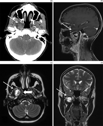
Otoscopic examination revealed right otitis media with effusion. The head and neck examination, which included nasopharyngeal endoscopy, was unremarkable except for mild diminution of sensation in the distribution of the right mandibular nerve. A cranial computed tomography scan demonstrated a well-circumscribed, homogeneously enhancing mass in the right infratemporal fossa with widening of the foramen ovale. Magnetic resonance imaging (MRI) revealed a 3 cm discrete, well-circumscribed mass, which was hypointense on T1-weighted imaging and hyperintense on T2-weighted imaging, with slight enhancement after gadolinium injection. The mass did not invade the temporal fossa and there was no extension into the nasopharynx (Figure 1).
Radiological investigation findings for case 1. (a) Axial computed tomography scan, showing a mass in the infratemporal fossa (arrow); notice the compression of the eustachian tube and the relationship of the mass to the pterygoid processes. (b) Sagittal and (d) coronal magnetic resonance imaging (MRI) scans, showing the relation of the mass with the temporal lobe (arrows). (c) Axial MRI scan, showing the pterygoid lateral and medial muscles (large arrow), which are pushed medially. (The small arrows indicate the lesion.)

Considering the size of the tumour and its symptomatic features, a surgical resection was indicated. Three months later, the tumour was surgically removed via a preauricular subtemporal approach.
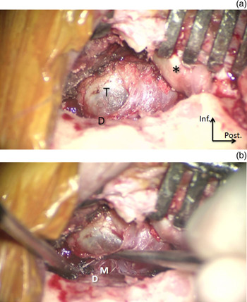
A curvilinear incision was made anterior to the tragus, from just below the level of the zygoma, and carried superiorly behind the hairline to the midline. A splitting incision of the temporalis muscle fascia was made, with preservation of the frontalis branch of the facial nerve aided by a facial nerve monitoring system. The zygoma was then stripped of its temporalis muscle, leaving the masseter muscle attached. The temporalis muscle was elevated and reflected inferiorly. A vertical osteotomy was made in the zygomatic bone just anterior to the temporomandibular joint. Another vertical osteotomy was made anteriorly behind the orbital rim to complete the zygomatic osteotomy. The zygomatic bone was then reflected with temporalis muscle inferiorly on the masseter muscle. Using a high-speed drill, the lateral part of the inferior aspect of the greater wing of the sphenoid and the floor of the temporal fossa were removed, exposing the foramen ovale. The middle fossa dura was retracted superiorly, thus exposing the infratemporal fossa and the containing tumour within (Figure 2).
Surgical view of the anterior subtemporal approach. (a) The zygomatic process and reflected temporalis muscle are retracted inferiorly (asterisk). The horizontal portion of the greater wing of the sphenoid bone is drilled out until the foramen ovale is reached, exposing the middle cranial fossa dura (D). The latter is retracted superiorly to expose the infratemporal fossa and the tumour (T). (b) Microscopic dissection of the tumour from the mandibular nerve (M), which is preserved. Inf = inferior; post = posterior

Using microscopic dissection, the tumour was completely removed en bloc, with preservation of the mandibular division of the trigeminal nerve, which was identified on the medial wall of the tumour capsule. After good haemostasis, the bony defect was sealed with a piece of fat harvested from the abdominal wall. The zygomatic osteotomy was then reattached using miniplates (Leibinger Universal Miniplates; Stryker, Freiburg, Germany). The temporalis muscle was reattached to the temporalis fossa and the skin was closed.
The surgical procedure ran smoothly, without any intra-operative or post-operative complication. The duration of the whole intervention was 2 hours. The patient was discharged home 2 days after the intervention.
Histological examination confirmed the diagnosis of schwannoma. After 12 months of follow up, the patient had complete resolution of her aural fullness and regained normal hearing and normal facial sensation. Otoscopic examination findings were normal. Magnetic resonance imaging revealed complete tumour removal without any brain herniation.
Case two
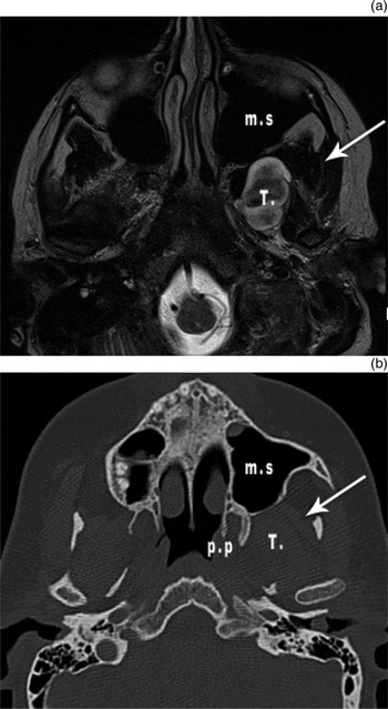
A 22-year-old female known to have neurofibromatosis type I was referred to our ENT department for the management of a left-sided infratemporal fossa schwannoma that was 20 mm in size. The mass was discovered incidentally on a screening brain MRI conducted as part of an epilepsy investigation. She underwent surgery to remove multiple lumbar and cervical spinal nerve schwannomas. Otological examination was normal. Head and neck examination showed only mild diminution of sensation in the distribution of the left mandibular nerve. Repeat MRI six months later showed an increase in the size of the infratemporal fossa schwannoma to 25 mm, which indicated surgical removal (Figure 3).
Radiological investigation findings for case 2: (a) axial magnetic resonance imaging scan and (b) axial computed tomography scan. The tumour (T) is localised in the infratemporal fossa, lateral to the pterygoid process (pp), displacing the lateral pterygoid muscle laterally (arrow). Note the relationship of the tumour to the maxillary sinus (ms), for which a transnasal endoscopic approach is favoured.

Removal of the tumour was performed via a neuronavigation-guided endoscopic transnasal approach. After endoscopic maxillary antrostomy, ethmoidectomy and sphenoidotomy, the sphenopalatine artery was cauterised at its exit from the sphenopalatine foramen. An endoscopic medial maxillectomy was performed, resulting in wide exposure of the maxillary sinus posterior wall. An additional fenestration of the maxillary sinus anterior wall was used in order to allow the introduction of a supplementary instrument by the assistant surgeon for continuous aspiration, thus enabling a three-handed technique.
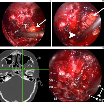
The bone of the maxillary sinus posterior wall and the pterygoid process were removed using a high-speed diamond drill, an elevator and a rongeur, to expose the boundaries of the tumour. The internal maxillary artery, which was pushed anteriorly and inferiorly by the tumour, was divided after coagulation. The tumour was separated from the surrounding structures with an elevator, and it was dissected from the mandibular nerve, which was partially preserved (Figure 4).
Photographs obtained during the endoscopic endonasal dissection of the tumour (T) in the left infratemporal fossa in case 2 before (a) and after (b) resection (arrows indicate infratemporal fossa fat and arrowhead shows the middle fossa dura). (c) Neuronavigation system display showing an axial computed tomography scan (left side) with the respective endoscopic view on the right side. The tracking instrument was used intra-operatively to localise the foramen ovale (fo), as shown by the crossing lines on the displayed image. Note: for a better orientation, we turned the camera laterally; hence, in the photographs, the right maxillary sinus is localised superiorly and the nasal septum inferiorly. Vn = vidian nerve canal; e = ethmoid; on = optic nerve canal; ca = carotid artery canal; h = hypophyseal gland; ss = sphenoid sinus; T = tumour; V3 = foramen ovale; lat = lateral; sup = superior

After tumour resection, bleeding from the pterygopalatine fossa venous plexus was controlled by bipolar cauterisation, which was time consuming. The cavity was packed with resorbable haemostatic material.
The surgery was guided by a neuronavigation system, which allowed better orientation in the operative field, and was helpful for the identification of borders and critical anatomical structures in real time (Figure 4c).
The duration of the intervention was 4 hours. The volume of intra-operative bleeding was approximately 500 ml. Post-operatively, the patient complained only of nasal crusting, which was treated by repetitive endoscopic debridement under local anaesthesia.
The post-operative histopathological findings indicated schwannoma. No residual tumour was identified by MRI 10 days after the surgery. After 18 months of follow up, the patient was doing well, but still complained of mild hypoaesthesia in the distribution of the left mandibular nerve. Magnetic resonance imaging revealed no tumour residue or recurrence.
Discussion
Trigeminal schwannomas may arise in any segment of the trigeminal nerve, but the majority develop intracranially from the Gasserian ganglion.Reference Krishnamurthy, Holmes and Powers1 Trigeminal schwannomas limited to the infratemporal fossa are extremely rare; to our knowledge, only 22 cases have been reported in the literature.Reference Krishnamurthy, Holmes and Powers1–Reference Arena and Hilal3 Most reported cases of infratemporal fossa schwannomas present during the second to fifth decades of life, with equal distribution for both sexes.Reference Krishnamurthy, Holmes and Powers1–Reference Arena and Hilal3 Trigeminal schwannomas have been reported in association with neurofibromatosis. These include bilateral trigeminal schwannomas or plexiform schwannomas of the trigeminal nerve.Reference Yamada, Ohta and Miyamoto6
Because of their low growth rate and their concealed location, diagnosis of infratemporal fossa schwannoma is often delayed. Not uncommonly, the diagnosis is incidental during radiological investigation for an unrelated problem. The most common reported symptoms are facial numbness, headache, toothache, nasal obstruction or hearing loss.Reference Whitlock, McCrea and Emerson2–Reference Samii, Migliori, Tatagiba and Babu4
A variety of benign and malignant neoplasms involve the infratemporal fossa.Reference Tiwari, Quak, Egeler, Smeele, Waal and Valk7 Magnetic resonance imaging can provide some information regarding the nature of the mass and aid differential diagnosis. Schwannomas are typically well-circumscribed, and appear isointense or hypointense on T1-weighted images, and hyperintense on T2-weighted images, with slight enhancement after gadolinium injection.Reference Yoshida and Kawase8, Reference Beauvillain, Calais, Launay, Bordure, Zanaret and Legent9 Computed tomography scans can supply useful details about the relationship of the tumour with cranial bones, and the presence of bone erosions or thinning.Reference Ogren, Wisecarver, Lydiatt and Linder10
The two cases presented conformed to the clinical description of most other cases reported in the literature. One of the two patients had neurofibromatosis type I, which reflects the association of the infratemporal fossa schwannoma with neurofibromatosis type I. The identification of an infratemporal fossa schwannoma should alert the physician to consider the diagnosis of neurofibromatosis.
Complete excision is the treatment of choice for these benign tumours. Several approaches have been proposed, reflecting the complexity of this region.
The infratemporal fossa is a deeply seated quadrangular retromaxillary space of the skull base that is situated beneath the floor of the middle cranial fossa. The medial boundary of the fossa is formed by the lateral pterygoid plate of the sphenoid. The foramen ovale is just posterior to the junction of the lateral pterygoid plate with the body of the sphenoid, and the foramen spinosum is positioned posterolaterally, a few millimetres away. The cartilaginous eustachian tube is immediately posteromedial to the lateral pterygoid plate. The average distance between the lateral pterygoid plate and the upper border of the zygomatic arch is 38.2 mm and represents the distance to the foramen ovale.Reference Tiwari11 Laterally, the fossa communicates with the temporal fossa between the zygomatic arch and the lower temple. The infratemporal fossa contains the pterygoid muscles, and important neurovascular structures that include the mandibular nerve, the chorda tympani, the middle meningeal artery, the internal maxillary artery and vein, and the pterygoid venous plexus.
The classical approaches to the infratemporal fossa can be classified as anterior, lateral and transcranial routes. The anterior approaches are categorised as transmaxillary, transoral, transfacial and transpalatal. Anterior transmaxillary approaches using the Caldwell–Luc procedure with removal of the maxillary sinus posterior wall provides limited exposure to the region, and cannot be used to treat tumours with extension to the orbit or the middle cranial fossa.Reference Rengahary, McMahon, Bigongiari, Ketcherside and Gunter12 Use of a transoral approach via the superior gingivolabial sulcus has been reported previously.Reference Shinoara, Uchida, Hiromatsu, Hida and Kikuta13 This approach did not give adequate exposure of the surgical field and was almost abandoned. Sabit et al. proposed a microsurgical model to approach the infratemporal fossa via a lateral transantral maxillotomy;Reference Sabit, Schaefer and Coldwell14 however, this approach is limited to tumours restricted to the anteromedial part of the infratemporal fossa, and could result in vascular complications from the pterygoid venous plexus and the maxillary artery.
In 1978, Fisch reported the posterolateral infratemporal fossa approach.Reference Fisch15 He and his colleagues advocated a type C approach for this purpose.Reference Fisch, Fagan and Valvanis16 However, hearing is permanently sacrificed in the ear on the affected side during this approach. His procedure has therefore been modified by others. Sekhar et al. described a subtemporal preauricular infratemporal fossa approach, which provides extensive exposure of the infratemporal fossa; furthermore, the conductive hearing apparatus is preserved.Reference Sekhar, Schramm and Jones17 Al-Mefty and Anand described the zygomatic approach, a preauricular approach achieved by displacing the temporalis muscle upward and keeping its vascularisation intact.Reference Al-Mefty and Anand18 However, there is a reasonable risk of frontal branch injury with this approach.
The transcranial route is a frontotemporal or subtemporal approach, which is achieved through a limited temporal or frontotemporal craniotomy and a zygomatic osteotomy.Reference Krishnamurthy, Holmes and Powers1 The entire surgical procedure is extradural, and is useful for tumours with extension into the middle cranial fossa. The procedure provides excellent exposure of the infratemporal fossa, but is attained at the price of craniotomies and high invasiveness.Reference Krishnamurthy, Holmes and Powers1, Reference Yoshida and Kawase8
Because of the significant morbidity associated with the classical approaches to the infratemporal fossa, less invasive alternatives have been sought. Kassam and co-workers proposed a fully endoscopic, expanded endonasal approach for selected cases of tumours in the infratemporal fossa, but the lesion must be medial and inferior to the principal neurovascular structures.Reference Kassam, Gardner, Snyderman, Mintz and Carrau19 The potential advantages of the endoscopic endonasal approach to the infratemporal fossa include shorter hospital stay, reduced risk of facial nerve injury and favourable wound healing. Limitations of this approach include a steep learning curve and narrow corridor for the working space. Moreover, there is no proximal vascular control via the endoscopic technique, and an additional external incision may be necessary for this purpose.
With this variety of surgical approaches, the surgeon has to choose the most appropriate technique. The technique should provide maximum exposure, with preservation of the neighbouring structures and minimal morbidity, so as to preserve the patient's quality of life.
In the first reported case, the approach used was a modification of the preauricular subtemporal approach. Complete resection with preservation of all neurovascular components of the infratemporal fossa was achieved without any morbidity within 2 hours. The preauricular subtemporal approach is suitable for the resection of tumours arising in the infratemporal fossa, and for intracranial tumours arising in the boundaries of the anterior temporal bone or greater wing of the sphenoid and extending into the infratemporal fossa. The expansion of the approach can be tailored according to disease extensions. The approach does not, however, allow the safe resection of any portion of the tympanic bone, and thus is not suitable for ear or temporomandibular joint extension.
The preauricular approach respects the auditory apparatus, and provides a direct route and the shortest approach to the infratemporal fossa. It is entirely extradural and enables wide exposure of the contents of the infratemporal fossa compared with the other approaches described above. Such wide exposure permits bimanual manipulation, easy management of large vessels and cranial nerves, and resection of eventual intracranial extension. The simplicity and the mini-invasiveness of this approach permit the reduction of operation time and hospitalisation stay of patients with lesions in this region.
In the second patient, the tumour was located on the anteromedial part of the infratemporal fossa. Complete removal was achieved using an endoscopic, combined transnasal and transmaxillary two-surgeon technique, guided by neuronavigation with computerised tomography. The only problem we faced during this procedure was continuous bleeding from the pterygoid plexus in the infratemporal fossa, which was time consuming and significantly prolonged the operative time.
• Complete excision of infratemporal fossa tumours is challenging and associated with high morbidity
• Several surgical approaches to the infratemporal fossa have been proposed; approach selection is influenced by neoplasm dimensions, and surgeon's preferences and experience
• Preauricular subtemporal and endoscopic transnasal approaches allow non-invasive and wide exposure of the infratemporal fossa
• Surgical approach should be selected according to the tumour's anatomical location with respect to the maxillary sinus posterior wall
• A preauricular subtemporal approach is indicated for infratemporal fossa tumours localised posterolaterally to the maxillary sinus posterior wall and/or with mild intracranial extension
• An endoscopic transnasal approach is indicated for tumours localised in the anteromedial part of the infratemporal fossa with respect to the maxillary sinus posterior wall
The clear reduction of invasiveness, without any external scar and without osteotomies, along with guaranteed good exposure of the surgical field, represents, in our opinion, real progress for surgery in this region. In addition, the use of neuronavigation increases the safety and accuracy of this procedure.
Serious complications may include vascular injuries to branches of the internal maxillary artery and internal carotid artery. However, with careful dissection and the use of neuronavigation, these complications are theoretical. Sacrifice of the vidian nerve may occur with this approach, which may result in hypolacrimation and keratitis.
Conclusion
The choice of surgical approach has up until now been influenced more by the dimensions of the neoplasm, and the surgeon's preferences and experience. Nowadays, the preauricular subtemporal approach and the transnasal endoscopic approach are the two main ones that allow non-invasive and wide exposure of the infratemporal fossa, as compared to the classical approaches. The surgical approach should be selected according to the anatomical location of the tumour, with respect to the posterior wall of the maxillary sinus.
For tumours localised posterolaterally with respect to the maxillary sinus posterior wall and/or with mild intracranial extension, we propose the preauricular subtemporal approach. A tumour in this location is considered relatively superficial and thus easy to resect using the preauricular subtemporal approach. However, a transnasal approach from the maxillary sinus to such a location necessitates deep dissection in the infratemporal fossa, which could be time consuming and may even be hazardous to neurovascular elements, especially in a bloody surgical field. Medial and anterior tumours near the posterior wall of the maxillary sinus can be best removed using a transnasal endoscopic approach, thus avoiding unnecessary deep dissection in the infratemporal fossa from lateral to medial directions.