Introduction
Cystic fibrosis (CF) is one of the UK's commonest inherited life-threatening diseases, affecting over 8500 individuals.1 The disease is an autosomal recessive genetic disorder resulting in exocrine gland dysfunction with various clinical manifestations.
Although targeted newborn screening is usually effective in identifying those with the condition, children are also often diagnosed following the development of respiratory or digestive tract symptoms. Sinonasal symptoms are almost universal, resulting in chronic sinusitis or polyposis.Reference Gysin, Alothman and Papsin2 However, these symptoms themselves are rarely the disorder that results in a diagnosis of CF.
We present here the case of a five-month-old child diagnosed with CF following an initial presentation with bilateral maxillary mucoceles.
Case report
A five-month-old baby girl was referred to our ENT department with symptoms of nasal congestion. The symptoms appeared following her first vaccination and improved, but then deteriorated again 10 days following her second routine vaccination. Despite being constantly ‘snuffly’ the child was reasonably well, feeding and gaining weight normally, with no morphological abnormalities. She had recently recovered from an ear infection, and examination of the ears was normal.
Flexible nasendoscopy showed her nasal airway to be patent bilaterally. However, both middle turbinates were grossly enlarged, occluding the middle portion of the nasal cavity and touching the septum. A computed tomography (CT) scan was arranged to assess the extent of the problem.
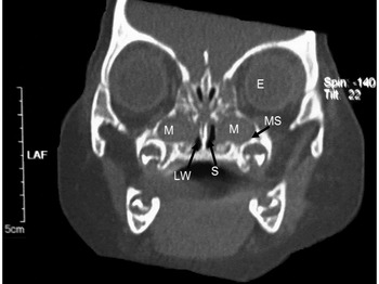
The scan (Figure 1) demonstrated bilateral maxillary mucoceles. Considering the rarity of this diagnosis, there was a clinical suspicion of CF, and the child was referred for a sweat conductivity test. This confirmed the diagnosis of CF. Of interest, a paediatric review confirmed that the child was doing well otherwise, and there were no other symptoms or family history. The child weighed 8.88 kg, which was between the 75th and 91st percentiles for her age.
Coronal computed tomography scan of the five-month-old patient demonstrating bilateral maxillary mucoceles. There is expansion of the maxillary sinus bilaterally, with medial displacement of the lateral nasal wall, consistent with a slowly expansile lesion, i.e. a mucocele. E = eye; M = mucocele; MS = maxillary sinus; LW = lateral nasal wall; S = nasal septum.

The child was treated for her maxillary mucoceles by endoscopic bilateral uncapping of the maxillary mucocele, uncinectomy and middle meatal antrostomy.
In the anaesthetic room, the nasal mucosa was pre-operatively prepared with 1:30 000 topical adrenaline on neurosurgical patties.
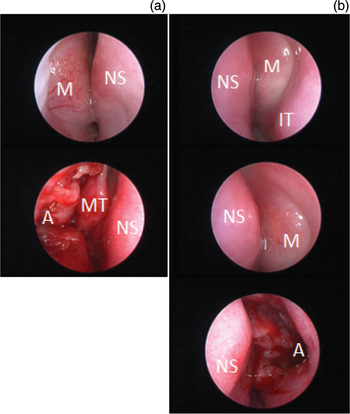
During surgery, a 0° view, 2.7 mm diameter paediatric endoscope with a camera system was used to visualise the surgical field. Bilaterally, the mucoceles were seen to bulge through both anterior and posterior fontanelles, obscuring the view of the uncinate process. An endoscopic uncinectomy was impractical given limited space. An anterior inferior incision was made through the anterior fontanelle to uncap the mucocele, using a ‘lancet’ blade myringotomy knife (Figure 2). Mucus was drained from the mucocele, confirming the diagnosis and origin. Suction was used to clear the field, and a paediatric sinus back-biting rongeur was used to perform the uncinectomy and antrostomy.
Intra-operative endoscopic images showing the maxillary mucoceles, and the appearance following uncapping and middle meatal antrostomy, on the (a) right side and (b) left side. M = mucocele; NS = nasal septum; A = antrostomy; MT = middle turbinate; IT = inferior turbinate.

Post-operatively, no nasal packing was used. Betnesol nasal drops (Recipharm, Ashton-under-Lyne, Lancashire, UK) were administered for 10 days. The child was well after surgery, and was discharged the following day.
Further treatment for CF was initiated following surgery, and included physiotherapy, dietician review, paediatric review, Creon (Abbott Products GMBH, Hannover, Germany), vitamin E, multivitamins and flucloxacillin. There was no recurrence of the presenting symptoms upon review at six weeks.
Discussion
Sinus mucoceles are common complications of chronic sinusitis in adults but are rare in the paediatric population, and no prevalence data currently exist for children with cystic fibrosis.Reference Di Cicco, Costantini, Padoan and Colombo3 Although an association is known between the two conditions, mucoceles are rarely the presenting feature of the underlying disease.Reference Guttenplan and Wetmore4
The frontal sinus is most commonly affected by mucoceles and is involved in approximately two-thirds of cases, the ethmoids are the next most common site, and the maxillary and sphenoidal sinuses are less likely to be involved.5
It is thought that mucoceles probably occur as a result of sinus ostium obstruction, resulting in mucus accumulation and expansion of the sinus. Others believe they develop secondary to a gradually enlarging mucus-retention cyst filling the sinus.5 As the mucoceles expand, they distort local anatomy and apply pressure to surrounding structures.
The symptoms and signs experienced reflect the size and sinus affected. For example, frontal sinus involvement presents as a mass, whereas maxillary sinus mucoceles elevate the orbital floor causing proptosis. In cases of infection within the mucocele, as in our case, symptoms are similar to acute sinusitis and pus is often present. If the diagnosis is suspected then imaging is best done by combining CT and magnetic resonance imaging to assess for any bony changes or intra-orbital or intracranial extension.
• Patients with cystic fibrosis (CF) commonly experience sinonasal symptoms, but usually in later childhood and after diagnosis
• Bilateral mucoceles are rare in children
• We report the first case of bilateral mucoceles in an infant, as the first presentation of CF
• Modified surgical procedures were required due to the small nasal cavity and underdeveloped sinuses
• Children with bilateral mucoceles should be suspected of CF and referred promptly
Current treatment involves functional endoscopic sinus surgery (FESS) for drainage and marsupialisation of the mucocele. Despite early concerns about acquired maxillary sinus hypoplasia,Reference Kosko, Hall and Tunkel6 the procedure appears to be safe and effective. The recurrence rates are low in adults and children with and without CF, both in the short and long term.Reference Di Cicco, Costantini, Padoan and Colombo3, Reference Sciarretta, Pasquini, Farneti and Ceroni7–Reference Bockmühl, Kratzsch, Benda and Draf9
In paediatric patients, FESS is ideal as it involves minimal scarring, low morbidity, direct visualisation and a short in-patient stay.Reference Olze, Matthias and Degenhardt8 Unfortunately, there are few randomised, controlled trials evaluating the surgical treatment of sinus disease in CF.Reference Robertson, Friedman and Rubin10
Although the maxillary sinus is present at birth, it continues to develop into adolescent life. In very young patients, the sinus is undeveloped and there is relatively little space to operate; therefore, using the conventional approach of uncinectomy, middle meatal antrostomy and uncapping of the mucocele poses additional risks to surrounding structures. Without direct visualisation, these may be damaged, resulting in abnormal growth or recurrence of the mucocele. We recommend intra-operative endoscopic evaluation of the maxillary mucocele to determine the order of surgery. Given the size of the mucoceles in our patient, their initial uncapping allowed easier access for uncinectomy and antrostomy, and avoided the risk of orbital damage and cerebrospinal fluid leakage.
Conclusion and recommendations
Although maxillary sinus mucoceles are extremely rare in the paediatric population, they are associated with CF.Reference Kosko, Hall and Tunkel6 The majority of cases are likely to be secondary presentations following chronic nasal symptoms. When presenting in a child without a previous diagnosis, clinicians should be aware of the possibility of CF.
To the best of our knowledge, we present here the first report of an infant (five months) presenting with nasal congestion due to sinus mucoceles, as the solitary indicator of CF. We recommend that clinicians be aware that mucoceles can present in this very young age group, and may indicate an underlying diagnosis of CF. A high index of clinical suspicion will help avoid delays in diagnosis and long-term management.