Introduction
Recent advances in molecular diagnosis have promising applications to the investigation of carcinogenesis pathophysiology. The survival rate for laryngeal cancer has remained unchanged for the past 30 years.Reference Almadori, Bussu, Cadoni, Galli, Paludetti and Maurizi1 Moreover, the standard tumour–node–metastasis (TNM) staging system is known to be inadequate in predicting the prognosis of this cancer.Reference Almadori, Bussu and Paludetti2 A better understanding of the molecular events involved in malignant transformation may help to classify tumours based on predicted clinical behaviour, to evaluate treatment responses and to direct treatment in order to improve clinical outcomes.Reference Loyo and Pai3
The disintegrin and metalloproteinase gene family codes for a group of recently discovered transmembrane and secreted proteins which include disintegrin and metalloproteinase domains.Reference Wolfsberg, Primakoff, Myles and White4 Studies have shown that some of these proteins participate in multiple physiological processes (e.g. cell adhesion, cell migration, and the release of cell surface molecules such as cytokines and growth factors), while others play a role in such complex events as smooth muscle development, fertilisation and the immune response.Reference Yamamoto, Higuchi, Yoshiyama, Shimizu, Kataoka and Hijiya5
These proteins may play a role in the promotion of tumour invasion and metastasis, via cleavage of extracellular matrix proteins or by direct modulation of tumour cell adhesion.Reference Edwards, Handsley and Pennington6 Furthermore, the disintegrin and metalloproteinase domain containing protein 33 (also known as ADAM-33) has been shown to promote angiogenesis, which is an important feature of carcinogenesis.Reference Puxeddu, Pang, Harvey, Haitchi, Nicholas and Yoshisue7
This study aimed to evaluate the expression of “a disintegrin and metalloproteinase-33” in laryngeal squamous cell carcinoma, and to determine its correlation with patients' clinicopathological characteristics.
Materials and methods
The study was approved by the Başkent University institutional review board.
We evaluated retrospectively the medical records of 98 patients suffering from laryngeal squamous cell carcinoma diagnosed and surgically treated in the Başkent University otolaryngology department. Those patients whose records contained complete follow-up information, and whose tissue blocks were stored in the archives of the Başkent University pathology department, were chosen as the study group.
We excluded from the study any patients with multiple primary cancers, a history of pre-operative radiotherapy and/or chemotherapy, or inadequate follow up.
The final study group comprised 40 surgically treated stage T2 to T4 laryngeal carcinoma patients with a follow-up period of at least two years. These patients' case records were reviewed and used to assess the patients' clinical characteristics. Clinical staging was done according to the American Joint Cancer Committee/Union Internationale Contre le Cancer 2002 recommendations.8
For statistical analysis, TNM classification, clinical stage and tumour differentiation were grouped as follows: T1 and T2 tumours versus T3 and T4 tumours; clinical stage I and II tumours versus stage III and IV tumours; moderately and poorly differentiated tumours versus well differentiated tumours, and metastatic node negative tumours versus node positive ones.Reference Figueiredo, Mamede, Spagnoli, Silva, Zago and Neder9
Immunohistochemical staining
Expression of “a disintegrin and metalloproteinase-33” was evaluated using laryngeal squamous cell carcinoma tissue specimens mounted in paraffin blocks, following the tissue microarray technique as previously described.Reference Erbek, Erinanc, Erbek, Topal and Kiyici10 This technique allows large numbers of small, punched-out tissue cores from different cases to be analysed in a single immunohistochemical staining experiment.
Briefly, formalin-fixed, paraffin-embedded tissue blocks were retrieved from the archives of the Başkent University pathology department. Areas containing epithelial and stromal components were identified on corresponding haematoxylin and eosin stained slides; these areas were cored and the cores transferred to a new, ‘recipient’ block. Two cores were taken for each case, each approximately 0.6 mm in diameter. After construction, three-micrometer-thick sections were obtained from the recipient paraffin blocks and placed onto poly-l-lysine covered slides. These sections were incubated at 56°C for 24 hours and rested for 1 hour in xylene and 10 minutes in graded alcohols for de-paraffinisation. Antigen retrieval was performed by boiling in citrate buffer solution (pH 6) for 20–25 minutes in a pressure cooker. After being incubated for 30 minutes at room temperature and then placed into 0.3 per cent hydrogen peroxide solution for 5 minutes, slides were washed again in phosphate-buffered saline for 2–3 minutes.
Rabbit polyclonal antibodies to “a disintegrin and metalloproteinase-33” (C-20; dilution 1:100; Santa Cruz Biotechnology, Santa Cruz, California, USA) were added at appropriate dilutions and incubated for 24 hours at room temperature. After incubation with primary antibody, slides were washed with phosphate-buffered saline for 5 minutes. Biotinylated goat anti-polyvalent (Lab Vision, Fremont, California, USA) was applied and again washed in phosphate-buffered saline. Streptavidin peroxidase (Lab Vision) was then applied and slides were incubated for 15 minutes at room temperature. Finally, after the application of 3-amino-9-ethylcarbazole (AEC) (Dako, Glostrup, Denmark) for 5 minutes, slides were washed with tap water. The procedure was then completed and the slides mounted.
The slides were examined and the level of immunoreactivity assessed. Assessment of “a disintegrin and metalloproteinase-33” staining was performed by a single pathologist who was blinded to patient outcome. “A disintegrin and metalloproteinase-33” expression within areas of laryngeal squamous cell carcinoma was compared with that in the surrounding normal, healthy tissue, and the staining intensity graded as: 0 = negative; 1+ = weak; 2+ = moderate; and 3+ = strong. The intensity of immunostaining in the epithelial cells and stromal and vessel mesenchymal cells was compared with that in normal laryngeal tissue.
Statistical assessment
Statistical analysis was performed using the Statistical Package for the Social Sciences version 11.0 software program (SPSS Inc, Chicago, Illinois, USA). Differences were considered to be statistically significant when the p value was less than 0.01. The chi-square test and Pearson analysis were used to evaluate the associations between the “a disintegrin and metalloproteinase-33” immunostaining scores and the following parameters: T stage, N stage, clinical stage, histological grade, perineural invasion, extracapsular spread, status of surgical margins, local recurrence, distant metastasis, tobacco consumption and alcohol consumption.
Results and analysis
Patients' clinical characteristics
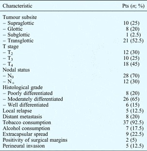
The study group included 38 men and two women diagnosed with laryngeal carcinoma of squamous cell origin (median age, 61 years; age range, 38–78 years). Patient characteristics are summarised in Table I.
Laryngeal SCC patients' clinical characteristics

SCC = squamous cell carcinoma; pts = patients; T = tumour stage; N = node stage
Immunohistochemical staining
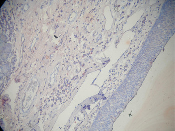
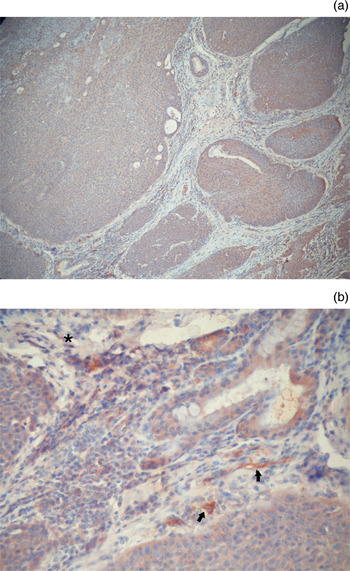
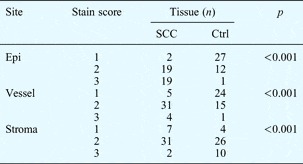
Figure 1 shows “a disintegrin and metalloproteinase-33” immunohistochemical staining in normal laryngeal tissue. Figure 2 shows increased “a disintegrin and metalloproteinase-33” immunostaining in the epithelial cells and stromal and vessel mesenchymal cells of laryngeal cancer tissue. Immunostaining scores for epithelium, stroma and vessels were significantly higher in tumour tissue compared with controls (p < 0.001) (Table II).
Photomicrograph of normal laryngeal mucosa prepared with “a disintegrin and metalloproteinase-33” antibody immunohistochemical staining; a general pattern of weak staining is seen. (×20)

Photomicrograph of laryngeal squamous cell carcinoma tissue prepared with “a disintegrin and metalloproteinase-33” antibody immunohistochemical staining, showing increased (brown) immunostaining of epithelial cells, stroma and vessels at (a) ×20 magnification and (b) ×40 magnification. The arrows indicate positive immunostaining of vessels, and the asterisk indicates positive immunostaining of stromal mesenchymal cells.

ADAM-33 immunohistochemical staining in laryngeal SCC and controls

ADAM-33 = “a disintegrin and metalloproteinase-33”; SCC = squamous cell carcinoma; stain score = immunohistochemical ADAM-33 staining score; ctrl = control; epi = epithelium
No correlation was observed between the tumour tissue “a disintegrin and metalloproteinase-33” immunostaining scores and the patient's clinical and histopathological characteristics (p > 0.05).
Discussion
Members of the disintegrin and metalloproteinase family mediate the extracellular matrix remodelling and cellular adhesion alteration which characterise the development of certain neoplastic and non-neoplastic pathologies. Recent studies have shown that “a disintegrin and metalloproteinase-33” over-expression is associated with airway remodelling (seen in asthma) and angiogenesis (a key step in tumour progression and metastatic dissemination).Reference Puxeddu, Pang, Harvey, Haitchi, Nicholas and Yoshisue7, Reference Erbek, Erinanc, Erbek, Topal and Kiyici10 To date, the association of “a disintegrin and metalloproteinase-33” with cancer has been studied in gastric and breast carcinomas.Reference Kim, Song, Hahm, Yoon, Park and Lee11, Reference Seniski, Camargo, Ierardi, Ramos, Grochoski and Ribeiro12 Kim et al. have reported that disintegrin and metalloproteinase protein 33 plays an important role in the process of vascular endothelial growth factor D induced interleukin 18 secretion and promotes tumour growth and metastasis, suggesting that it may constitute an effective therapeutic target in gastric cancer.Reference Kim, Song, Hahm, Yoon, Park and Lee11 In the human lung, “a disintegrin and metalloproteinase-33” has been shown to be expressed, predominantly and strongly, in basal epithelial cells, which supports its possible involvement in the remodelling process.Reference Dijkstra, Postma, Noordhoek, Lodewijk, Kauffman and Hacken13
Thus, our study aimed to evaluate the expression profile of “a disintegrin and metalloproteinase-33” within laryngeal squamous cell carcinoma, and to determine its association with the disease extent and clinical characteristics at the time of diagnosis. To our best knowledge, this study is the first to report increased expression of “a disintegrin and metalloproteinase-33” in laryngeal cancer. All epithelial, stromal and vascular staining scores were found to be significantly increased in tumour tissue compared with controls (p < 0.001).
The expression of “a disintegrin and metalloproteinase-33” was first observed in cells of mesenchymal origin, and was considered to predominantly occur in fibroblasts.Reference Van Eerdewegh, Little, Dupuis, Del Mastro, Falls and Simon14, Reference Powell, Wicks, Holloway, Holgate and Davies15 However, more recent publications have reported that expression occurs predominantly in the basal epithelial cells, and that the vascular endothelium is also strongly positive.Reference Dijkstra, Postma, Noordhoek, Lodewijk, Kauffman and Hacken13 In line with recent studies, and in addition to the expected staining of stromal and vessel mesenchymal cells, we also found strong staining in the tumour tissue epithelial cells.
• “A disintegrin and metalloproteinase-33” is a member of the disintegrin and metalloproteinase family of transmembrane and secreted proteins
• Over-expression of this protein is associated with several pathological processes
• These include airway remodelling, angiogenesis, and tumour promotion and metastasis
• This study found a possible association between “a disintegrin and metalloproteinase-33” and laryngeal cancer
“A disintegrin and metalloproteinase-33” has strong catalytic activity as a matrix metalloproteinase.Reference Edwards, Handsley and Pennington6 This catalytic activity had been shown to be related to angiogenesis.Reference Roy, Zhang and Moses16 In the human lung, Dijkstra et al. observed strong expression of “a disintegrin and metalloproteinase-33” in vascular endothelial cells, whereas other disintegrin and metalloproteinase proteins were more weakly expressed.Reference Dijkstra, Postma, Noordhoek, Lodewijk, Kauffman and Hacken13 Our study found significantly greater expression of “a disintegrin and metalloproteinase-33” in tumour tissue vessels, compared with controls (p < 0.001). Based on this, it can be speculated that the over-expression of “a disintegrin and metalloproteinase-33” in the vascular endothelial cells of laryngeal cancer may be associated with the angiogenetic effects of this protein. Angiogenic vessel formation is crucial to support tumour growth, and plays a central role in tumour extension.Reference Loyo and Pai3
However, our results revealed no association between the “a disintegrin and metalloproteinase-33” expression and the extent of patients' laryngeal carcinoma. In fact, our study was limited by a small number of patients and by their TNM stage distribution. This was because our clinic initially treats most patients with early stage cancer using radiotherapy. So, after the exclusion of these patients, our study group comprised mostly late stage cancers, together with some T2 tumours. In order to speculate logically about the correlation between “a disintegrin and metalloproteinase-33” expression and cancer progression, knowledge about expression patterns in early stage cancers is needed. A prospective study including patients suffering from laryngeal dysplasia and carcinoma in situ may give additional information about the carcinogenesis process.
Our study represents a preliminary report suggesting a possible association between “a disintegrin and metalloproteinase-33” and laryngeal cancer. Prospective studies focussing on genetic coding and messenger RNA expression may supply additional knowledge about the exact pathogenesis of laryngeal cancer.
Conclusion
This study suggests that increased expression of “a disintegrin and metalloproteinase-33” may play a role in the pathogenesis of laryngeal carcinoma. However, the disease extent and patient clinical characteristics at the time of diagnosis did not correlate significantly with the “a disintegrin and metalloproteinase-33” expression profile.
Acknowledgement
This study was approved by the Başkent University institutional review board (project number KA10/78) and supported by the Başkent University Research Foundation.