1. Introduction
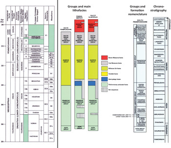
The Middle and Upper Mississippian of southern Pembrokeshire has long been a subject of study for different reasons: lithology, stratigraphy, sedimentary facies, tectonics and the existence of remarkable breccias linked to the Visean limestones (Dixon, Reference Dixon1921; George, Reference George1958, Reference George1970, Reference George and Owen1974, Reference George1979; Thomas, Reference Thomas1971; Kelling, Reference Kelling and Owen1974; Owen, Reference Owen and Owen1974; Jones, Reference Jones and Owen1974; Leeder, Reference Leeder, Duff and Smith1992; Dunning, Reference Dunning, Duff and Smith1992; Kelling & Collinson, Reference Kelling, Collinson, Duff and Smith1992; Walsh et al. Reference Walsh, Battiau-Queney, Howells, Ollier and Rowberry2008). Waters et al. (Reference Waters, Waters, Barclay and Davies2009) have recently given a synthesis of the lithostratigraphy of the Carboniferous successions of southern Britain. They used the international chart and not the historical nomenclature based on lithofacies. The same choice is made in this paper (Fig. 1). Recently, a particular type of isovolumetric weathered rock (‘ghost-rock’ in Quinif, Reference Quinif2010) has been described in the Carboniferous limestones of southern Pembrokeshire (Rowberry et al. Reference Rowberry, Battiau-Queney, Walsh, Błażejowski, Bout-Roumazeilles, Trentesaux, Křížová and Griffiths2014). These formations are well observed in Bullslaughter Bay.
Chronostratigraphic framework for the Carboniferous successions and southern Pembrokeshire groups and formations (source: Waters et al. Reference Waters, Waters, Barclay and Davies2009).

The purpose of our research was to explain the weathering processes. After five years of field campaigns and a set of various laboratory analyses, we are able to present new data on the Late Mississippian environment that propose a complex sequence of events. Sedimentological, petrographic and isotopic (carbon and oxygen) analyses of the Visean limestones of Bullslaughter Bay give evidence of multiple effects of sea-level oscillations on the depositional environment and early weathering of carbonate rocks.
2. Geological setting
The Mississippian sedimentary environment depended on local and regional contexts but also on global climate and eustatic sea-level changes. In southern Britain, and especially in South Wales, regional correlations between different formations are not easy because sedimentary successions frequently evolved in isolation, especially in late Visean and early Serpukhovian times (Waters et al. Reference Waters, Waters, Barclay and Davies2009). The Visean carbonate platforms of southern England and Wales evolved from gently sloping ramps in Asbian time to flat-topped shelves in Brigantian time (D. I. Gray, unpub. Ph.D. thesis, Univ. Newcastle upon Tyne, 1981; Walkden, Reference Walkden, Miller, Adams and Wright1987). The depositional environment in southern Pembrokeshire was that of a carbonate platform in a coastal or nearshore position. It was flanked to the south by a steep continental slope, and northwards it abutted the continental area known as St George’s Land, which extended westward from the London–Brabant Massif.
The sedimentation was cyclothemic and reflected sea-level oscillations driven by glaciation on Gondwana (Wanless & Shepard, Reference Wanless and Shepard1936; Ramsbottom, Reference Ramsbottom1973, Reference Ramsbottom1977; Davies, Reference Davies1984; Horbury, Reference Horbury, Arthurton, Guteridge and Nolan1989; Isbell et al. Reference Isbell, Miller, Wolfe, Lenaker, Chan and Archer2003; Davies, Reference Davies, Fielding, Frank and Isabell2008; Rygel et al. Reference Rygel, Fielding, Frank and Birgenheier2008; Fielding et al. Reference Fielding, Frank, Isbell, Fielding, Frank and Isbell2008). The emergence and submergence of the carbonate platforms began abruptly at around 330 Ma with an approximate periodicity of 100 ka linked to Milanković eccentricity effects (Wright & Vanstone, Reference Wright and Vanstone2001). In southern Britain, the thickness of individual cyclothems increased from a few metres during Asbian time to as much as 30 m during Brigantian time (Walkden, Reference Walkden, Miller, Adams and Wright1987), while eustatic sea-level oscillations increased from 10 to 50 m in Asbian–early Brigantian times to a maximum of 95 m by the close of the Visean period (Smith & Read, Reference Smith and Read2000; Wright & Vanstone, Reference Wright and Vanstone2001). Each cyclothem started with subtidal sediments and was bounded at the top by exposure surfaces associated with palaeokarstic features, rhizocretions, laminar calcretes and clay palaeosols (Walkden, Reference Walkden1972, Reference Walkden1974; Somerville, Reference Somerville1979; Wright, Reference Wright1982 a; Walkden & Davies, Reference Walkden and Davies1983; Davies, Reference Davies1991; Wright et al. Reference Wright, Vanstone and Marshall1997; Vanstone, Reference Vanstone1998). Glacial lowstands were characterized by a regular transition from humid to semi-arid climatic conditions (Vanstone, Reference Vanstone, Strogen, Somerville and Jones1996, Reference Vanstone1998). Well-preserved gymnosperm wood has growth rings indicative of tropical seasonality (Falcon-Lang, Reference Falcon-Lang1999).
As with other Asbian strata in southern Britain, the Oxwich Head Formation of southern Pembrokeshire hosts a range of surface exposure phenomena such as palaeosols near St Govan’s Head (George, Reference George2008) and palaeokarstic pits near Stack Rocks and Stackpole Head (Rowberry et al. Reference Rowberry, Battiau-Queney, Walsh, Błażejowski, Bout-Roumazeilles, Trentesaux, Křížová and Griffiths2014). Nevertheless, evidence of surface exposure phenomena had never been described in the Oxwich Head and Oystermouth formations of Bullslaughter Bay before the present study.
The South Wales Serpukhovian sequence (Lower Namurian in the old nomenclature) reflects the rapid southward development of a paralic-facies complex, fringing the uplifted St George’s Land massif (Kelling, Reference Kelling and Owen1974; Kelling & Collinson, Reference Kelling, Collinson, Duff and Smith1992). Sea-level oscillations driven by the development of a major ice-sheet in the southern hemisphere continued to characterize the period (Ramsbottom, Reference Ramsbottom1977; Waters & Davies, Reference Waters, Davies, Brenchley and Rawson2006). During Westphalian time, paralic or deltaic environments persisted in South Wales, which was ‘marked by a low relief in which even minor adjustments of the relative sea level resulted in substantial lateral migration of the strand-line and associated facies deltas’ (Kelling, Reference Kelling and Owen1974). In Pembrokeshire, the Westphalian crops out in a thin belt extending from Carmarthen Bay to St Brides Bay. The Westphalian is totally absent southwards in the Castlemartin Peninsula and was probably never deposited. The Westphalian marks the onset of Variscan deformation with considerable changes in the regional topography of southern Pembrokeshire (Battiau-Queney, Reference Battiau-Queney1984). As a major consequence, St George’s Land was peneplanated, and an uplifted land emerged in the Bristol Channel (Kelling, Reference Kelling and Owen1974).
3. Study area
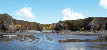
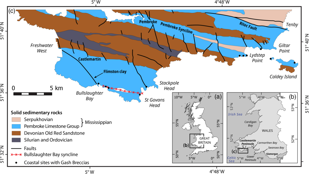
Bullslaughter Bay is located on the south coast of the Castlemartin Peninsula within an ‘Area of Outstanding Natural Beauty’ in the Pembrokeshire Coast National Park (Fig. 2). The 250 m wide bay is surrounded by subvertical 35–40 m high cliffs by the edge of a flat plateau lying at 35–50 m above sea level. The plateau truncates the Visean (Pembroke Limestone Group) and Serpukhovian folded strata (Fig. 3).
Northward view of Bullslaughter Bay showing the Variscan folded limestone strata. In the middle, the yellowish rock marks the axis of the Bullslaughter Bay syncline. OHF – Oxwich Head Formation; OYMF – Oystermouth Formation; PM – Pendleian mudstones.

Simplified geological map of southern Pembrokeshire. (Sources: Thomas, Reference Thomas1971; British Geological Survey, 1977; Walsh et al. Reference Walsh, Battiau-Queney, Howells, Ollier and Rowberry2008; design: D. Marin.)

Two limestone formations are exposed in the cliffs of Bullslaughter Bay (Fig. 2): (i) the Oxwich Head Formation with thick-bedded recrystallized and bioturbated skeletal packstones/grainstones deposited during Asbian time; and (ii) the Oystermouth Formation with thin- to medium-bedded bioclastic grainstones, argillaceous limestones and mudstones deposited during Brigantian time (George et al. Reference George, Johnson, Mitchell, Prentice, Ramsbottom, Sevastopulo and Wilson1976).
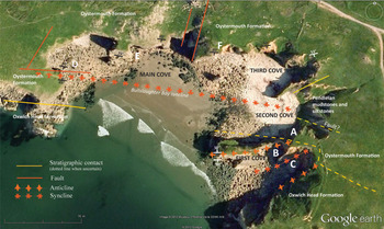
Besides the general lithological characteristics of these Visean limestones, Bullslaughter Bay is known to host huge masses of breccias (Dixon, Reference Dixon1921; Thomas, Reference Thomas1971; Walsh et al. Reference Walsh, Battiau-Queney, Howells, Ollier and Rowberry2008; Woodcock et al. Reference Woodcock, Miller and Woodhouse2014). Less known and not yet studied is the presence in many places of yellow, red or black loose formations which contrast with the white/grey solid limestone. They form a heterogeneous patchwork with the parent rock and are sometimes laminated, especially in the first and second coves of the bay (Fig. 4). The properties and origin of these loose formations are the main topic of the present paper.
Geological sketch of Bullslaughter Bay showing the main significant structures. The Oxwich Head Formation crops out on the west side of the bay south of the Bullslaughter Bay Syncline, and also on the southeast side. ‘Gash-Breccias’ are not indicated. A, B, C, D, E and F mark the sampling sites. (Sources: Dixon, Reference Dixon1921; Thomas, Reference Thomas1971; background from Google Earth © 2012 Bluesky, Infoterra Ltd & COWI A/S.)

4. Material and methods
One hundred and fifty samples were analysed from both formations in the Laboratory of Oceanology and Geosciences of the University of Lille (France), using a range of techniques including calcimetry, particle size distribution, scanning electron microscopy (SEM) analysis, thin-section petrography, X-ray diffraction (XRD) and Raman spectroscopy of selected particles.
The carbonate-content analyses were performed using a Bernard Calcimeter. X-ray diffraction was performed with a Bruker Endeavor DA system. Particle size distribution (PSD) was obtained by a Malvern Mastersizer 2000 laser diffraction instrument. The thinly laminated sediments were analysed using the sieve method. The samples were separated in sieves of three different sizes (>200 µm, between 200 and 125 µm, and between 125 and 40 µm) to identify the grains and select some of them to obtain SEM micrographs and conduct energy-dispersive X-ray spectroscopy (EDX) analyses using an Environmental FEI Quanta 200 SEM coupled with a Bruker Quantax EDXS spectrometer. The exoscopy of 50 quartz grains was performed at Charles University (Czech Republic). Grains between 250 and 500 µm were boiled for 10 minutes in concentrated hydrochloric acid, washed in distilled water and dried. They were subsequently mounted on carbon tape, gold plated and photographed using a scanning electron microscope (JEOL 6380 LV).
Thin-sections were made on more or less solid rocks and also on non-cohesive laminated sediments, which were impregnated. In the field the samples of non-cohesive material were pushed into rectangular 10 × 7 cm boxes to keep the undisturbed structure of the sediment. Thin-sections were realized at the thin-section lab in Toul (France) and at the University of Lille (France). Micropetrography was performed with a BX60 microscope and a Flex camera. The mineralogy, particularly of iron oxides, was identified using a LabSpec HR800UV Raman microspectrometer (Jobin Yvon labs).
Thirty-two samples were selected for whole-rock carbon and oxygen isotopic analyses, in order to evaluate the effects of the diagenetic alteration as revealed through the petrographic study. They were prepared at the Free University of Brussels (Belgium). Analysed samples consisted of unaltered solid limestones and partly decalcified limestones. In one case (sample BB39) the altered carbonate and its parent limestone were analysed at their contact, at a centimetre scale.
Carbonate powders were reacted with 100 % phosphoric acid at 70 °C using a Gasbench II connected to a ThermoFinnigan Five Plus mass spectrometer at the University of Erlangen-Nuremberg (Germany, Prof. M. Joachimski). All values are reported in per mil relative to the Vienna Pee-Dee Belemnite (V-PDB) by assigning δ13C and δ18O values of +1.95 ‰ and −2.20 ‰ to international standard NBS19 and −46.6 ‰ and −26.7 ‰ to international standard LSVEC, respectively. Reproducibility and accuracy were monitored by replicate analysis of laboratory standards calibrated to NBS19 and LSVEC.
5. Results
5.a. Lithostratigraphy
The Visean formations consist of a diverse range of rock types including thin- to medium-bedded argillaceous mudstones–wackestones and thicker-bedded packstones and grainstones with brachiopods, echinoderms, foraminifers and algae. It is also common to encounter silicified limestones, chert nodules and cherty beds. Dixon (Reference Dixon1921) noted frequent rapid lateral variations in the succession.
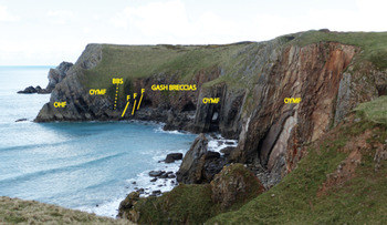
On the west side of the bay (Fig. 5), the southern limb of the main syncline exposes medium-bedded limestones of the Oxwich Head and Oystermouth formations. The northern limb is faulted, and a huge mass of breccias (the so-called ‘Gash-Breccias’ of Dixon, Reference Dixon1921) replaces the original beds. Forty metres to the north, the cliff exposes nearly vertical thin-bedded limestones (5–10 cm thick) of the Oystermouth Formation dipping southwards.
Westward view of the western cliffs of Bullslaughter Bay. ‘Gash-Breccias’ separate the syncline axis and the near vertical limestones of the Oystermouth Formation. Small caves, dug by waves, expose good sections of the Oystermouth Formation with different rock types and rapid lateral or vertical facies. Several samples of the left cave (close to the fault zone) have been analysed (photo M. Rowberry, 2012). OHF – Oxwich Head Formation; OYMF – Oystermouth Formation; BBS – Bullslaughter Bay syncline.

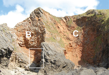
The eastern part of the bay exposes medium-bedded limestones (Fig. 6). According to Dixon (Reference Dixon1921), they belong to the D2 Dibunophyllum Subzone, equivalent to the Oystermouth Formation, in site A, and to the D1 Dibunophyllum Subzone, equivalent to the Oxwich Bay Formation, in sites B and C. Nevertheless, Dixon noted that the contact between both formations is uncertain in the area, because of lack of zonal fossils of value, and also owing to the presence of breccias. In the second cove of Figure 4 (site A) a conodont assemblage was found in laminated sediments. They give a typical early Brigantian age (Błażejowski & Walsh, Reference Błażejowski and Walsh2013) and strengthen the attribution of these sediments to the Oystermouth Formation.
The ‘East Cove’ of Figure 4 with sites B and C marked (view to the NE). The limestone belongs to the Oxwich Head Formation, according to Dixon (Reference Dixon1921). A syncline axis sharply cuts site C. Placement of samples in the white frame. See detail on Figure 17.

The Serpukhovian stage is poorly observed in the Castlemartin Peninsula and difficult to recognize with precision. In accordance with Dixon (Reference Dixon1921), the British Geological Survey (1977) attributed to the Namurian an outcrop of black shales with cherts and brown fine-grained sandstones, close to the Newton farmyard, 1 km east of Bullslaughter Bay. Plant fragments were found but could not be determined (Dixon, Reference Dixon1921). A coastal deltaic or lagoonal sedimentary environment was suggested. The outcrop follows the W–E Bullslaughter Bay syncline (Fig. 3). In the eastern part of Bullslaughter Bay, in the main syncline axis, light yellowish siliceous mudstones with black cherts have been attributed to the same formation by Thomas (Reference Thomas1971) who described them as ‘down-faulted Millstone Grit shale’, although the northern contact with the Oystermouth Formation does not seem clearly fault-controlled. The southern limit cannot be seen properly in the field, but a fault zone is likely to exist at the contact with the limestones of the Oystermouth Formation exposed at site A. These mudstones are curiously indicated as Triassic Gash-Breccias on the geological map of the British Geological Survey (1996), although it is much more coherent and plausible to attribute them to the Pendleian (lower Serpukhovian). They are strongly weathered and folded.
5.b. Structure
The Variscan orogeny in South Wales began with preliminary movements through the Visean age (Owen, Reference Owen and Owen1974). The late Visean unrest was widespread. In eastern Bullslaughter Bay it is evidenced by slight angular unconformities in the Brigantian sequence (see Fig. 16 below). Nevertheless, the main movements took place in late Stephanian – early Permian times (Leveridge & Hartley, Reference Leveridge, Hartley, Brenchley and Rawson2006). In southern Pembrokeshire, as in Gower and Glamorgan, Variscan structures are typically E–W trending. In the Castlemartin Peninsula, the Visean limestone beds have been strongly folded and rarely dip less than 45°. The fold axes are displaced by several SSW–NNE-trending cross-faults. The main regionally significant tectonic feature is the W–E Bullslaughter Bay syncline, which coincides with a fault zone in the western part of the bay. In the eastern part of the bay, the structure is very complex with a series of tight WSW–ENE-trending anticlines and synclines south of the main syncline axis.
5.c. Sedimentary facies
The field observations of weathered rocks show two main facies, mosaic and laminated.
5.c.1. Mosaic facies of weathered limestone
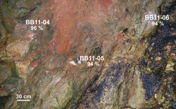
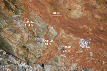
Weathered rocks are present in all places in the cliffs of the bay. The alteration rate changes rapidly from place to place giving a mosaic facies with a patchwork of strongly, weakly and unweathered rocks. It is particularly well seen on the northern side of the Bullslaughter Bay syncline, at site D. The patches of weathered rock are a few decimetres thick and may incorporate residues of the parent limestones (Fig. 7). Owing to the differential alteration, the facies can appear brecciated-conglomeratic with various sized and coloured blocks of host limestones embedded in a loose porous matrix (Fig. 8). The residual limestone blocks can form irregular discontinuous but bedding-concordant lenticular sheets.
Mosaic of solid and more or less weathered limestone at site D. For each sample the percentage of calcium carbonate is given (see also Table 1).

More or less weathered limestone at site D with slight laminated structure of the reddish loose material (see also Table 1).

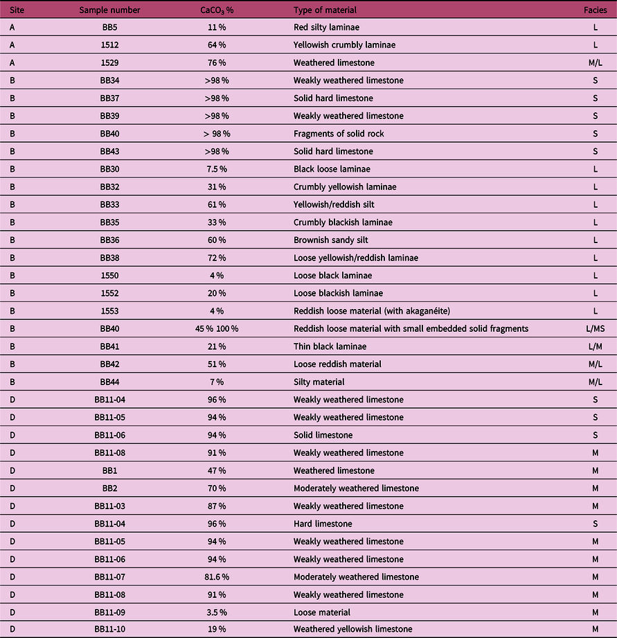
In several places, it is possible to see the gradual transition from the solid to weathered rock, proving that nothing was significantly displaced during the alteration process. Weathered rocks can be seen in sheltered places having no connection with the present topographic surface. In two places, the weathered rocks have been found in small natural caves embedded in the solid rocks: the first one in a few metres wide cave of site D on the western side of the bay (seen on Fig. 5, below ‘G’ of the Gash-Breccias) and the other one in a metre-scale cavity at the base of site B (Fig. 6 and ‘a’ in Fig. 17 below). More generally, although the alteration rate may change rapidly, it is not related to a vertical gradient linked to the present topography but rather to the bedding planes. Carbonate-content analyses of several samples of solid and more or less weathered rocks reveal a close relationship between the rate of weathering and the loss of calcium and rock density (tested in the field with a Schmidt hammer) (Figs 7, 8; Table 1). This type of weathered limestone is similar to the ‘ghost-rock’ defined by Quinif (Reference Quinif2010). It occupies the same space as the solid rock but it is more or less crumbly and easily crushed into silt and fine sand. The related loss of calcium of this ‘ghost-rock’ has been reported in the studied area by Rowberry et al. (Reference Rowberry, Battiau-Queney, Walsh, Błażejowski, Bout-Roumazeilles, Trentesaux, Křížová and Griffiths2014). Solid limestone rocks are more than 94 % CaCO3; weakly weathered limestones, which are still relatively hard, display CaCO3 values of between 91 % and 94 %; moderately to deeply weathered rocks, which easily crumble, contain between 81 % and 3.5 % CaCO3 (Table 1).
Carbonate content of several samples of sites A, B and D (see text for explanation)

L – laminated; M – mosaic; S – solid rock.
5.c.2. Loose laminated facies
The loose laminated facies concern, in particular, the southern limb of the Bullslaughter Bay syncline, in the first and second coves of the bay, where the Oxwich Head and Oystermouth formations crop out (Fig. 4). Forty-five samples from sites A and B were analysed. Here, the laminated sediments contrast strongly with the host limestone with their yellow, red or grey colour, loose consistency, silty texture and poor CaCO3 content (4–72 % in Table 1). They may incorporate solid or more or less weathered residues of the parent limestones.
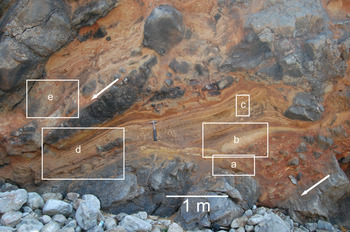
At site A, a spectacular formation – although not reported in detail by Dixon (Reference Dixon1921) and ignored by Thomas (Reference Thomas1971) – has aroused interest over the past 40 years (Battiau-Queney, Reference Battiau-Queney1980; Błażejowski & Walsh, Reference Błażejowski and Walsh2013; Woodcock et al. Reference Woodcock, Miller and Woodhouse2014) (Fig. 9). In fact, the origin of this formation was never made clear owing to the lack of analyses.

Although the studied area is just a few square metres, it provides interesting data to reconstruct the story of sedimentation. The thin- to medium-bedded parent limestone steeply dips northwards (45° to 50°) (white arrows in Fig. 9).
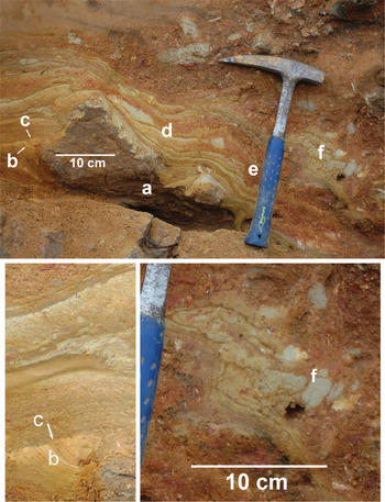
In Figure 10, a block of limestone (Fig. 10 ‘a’) is locally laminated and weakly weathered. On the left side (Fig. 10 ‘b’), very thin millimetric curved laminae, made of silt and fine sand, abut on the block (see detail in Fig. 10). It could be an aeolian deposit. It has been truncated (Fig. 10 ‘c’) by a 3 cm thick bed of coarser sand, which is roughly laminated and covered by a 1 cm thick fine sand bed. This bed is covered by a series of a few centimetres wide undulated beds showing imbricated superposition, which skirts the limestone block (Fig. 10 ‘d’). Masses of heterometric coarse sand, angular gravels and small blocks of transported weathered limestone lie on these beds, sometimes in centimetre-sized furrows (Fig. 10 ‘e’). To the right of the hammer in Figure 10 (Fig. 10 ‘f’), there is the presence of an enterolithic-type structure with bulging or ‘boudinage’ (Fig. 10, detail). Fossils of the local parent rock are abundant in this heterometric material. The structural disposition of these sediments suggests a slope deposit in a metric-sized open cavity formed during a period of emergence, with evaporitic-type sliding and slumping. Halite and gypsum have been discovered in these contorted laminae (see Section 5.d below, site A).
Site A: laminated sediments (box ‘a’ of Fig. 9) with detailed pictures below.

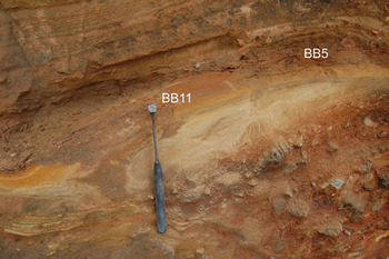
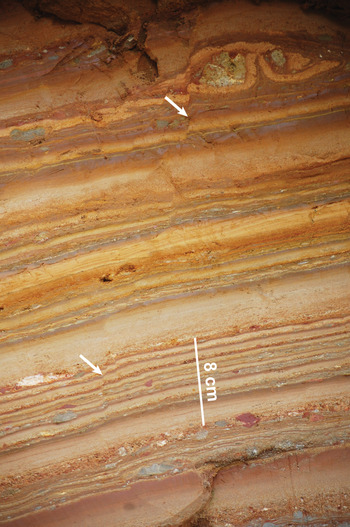
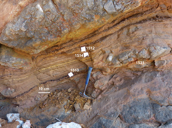
They are overlain by a lenticular bed containing 54 % silt and 36 % fine sand (less than 125 µm) (sample BB11 in Fig. 11). On the left, curved laminae are present, but they disappear to the right. The mineralogy of this sample is described below. It passes upwards to a planar thinly laminated sediment (Fig. 12). The laminae are mainly composed of silt (50–55 %) and fine sand (~35 %). The finer ones contain ~80–85 % silt and 15–20 % clay. A few laminae are coarser (coarse sand and small gravel). Laminae are a few millimetres to 4 cm thick. The lamination is planar with upwards fining of the grain size. Synsedimentary microfaults (Fig. 12, white arrows) and flame structures are present. The same type of thin laminated sediment is observed in Figure 13, from which several samples were analysed (Section 5.d below).
Site A: contact of silty-sandy bed with laminated sediment (box ‘b’ of Fig. 9). Hammer for scale is 32 cm long.

Site A: graded planar laminae (box ‘c’ of Fig. 9).

Site A: graded laminae (box ‘d’ of Fig. 9), with position of samples.

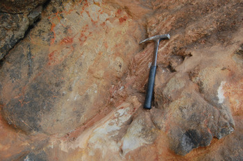
In some places, the contact of the loose laminae with the solid rock seems relatively sharp. Locally, they wrap the parent limestone rock (Fig. 14).
Site A: laminae wrapping the weakly weathered parent rock (box ‘e’ of Fig. 9). Hammer for scale is 32 cm long.

In other cases, the lamination continues into the weathered rock (Fig. 15). Dixon (Reference Dixon1921) already noted the presence of ‘silty laminated limestones’ in the Oystermouth Formation (D2 Dibunophyllum Subzone), which crops out here.

A picture showing the reconstructed setting in Brigantian time, before the Variscan folding (Fig. 16), helps to understand the disposition of the sediments.
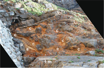
Weathered rock and laminated sediment of site A. The picture has been straightened to show the site as it was before the Variscan folding: the limestone bed at the base of the picture has been set as horizontal. See text for comments concerning areas ‘a’, ‘b’ and ‘c’ and sample 1528.

The area with weathered rock and laminae is ~2 m thick (1.6 to 2.5 m). It is covered in a slight angular unconformity (Fig. 16, dotted line) by a hard-grey limestone bed, which underlies reddish moderately weathered thinner limestone beds (sample 1528, see Section 5.d below, site A). The sediments, which have been identified as possible slope deposits with enterolithic-type structures (Fig. 16 ‘a’) are nearly perpendicular to the underlying horizontal bed and wrap a 1 m high rocky slope (Fig. 16 ‘b’). The laminae (Fig. 16 ‘c’) skirt the residual fragments of solid or moderately weathered parent rock. But they also pass gradually to laminated in situ rock (Fig. 15). Their structural arrangement favours the hypothesis of deposition in non-turbulent water on a rough rocky platform.
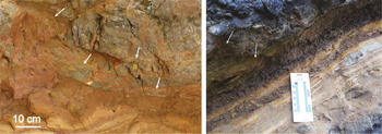
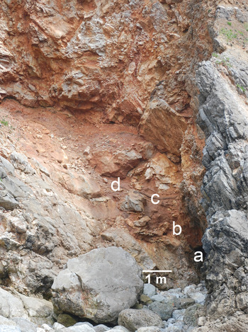
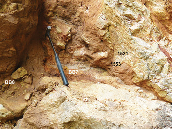
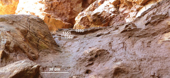
At site B (Fig. 17) several patches of laminated sediments have been discovered during the field survey. They have never been described before, probably because they were not visible when Dixon (Reference Dixon1921) investigated the area. In fact, the active current slope processes can conceal or reveal some features at any time. Here the cliffs expose the Oxwich Head Formation (D1 Dibunophyllum Subzone of Dixon). The laminar units show similarities with those of site A, although planar graded thin laminae are less frequent (Figs 18–20). The structural continuity from solid to laminated rock is seen in several places (Figs 18, 19).

Detail of site B (‘b’ in Fig. 17). Hammer for scale is 32 cm long.

Detail of site B (‘c’ in Fig. 17). Note the lamination in the nearby solid rock.

Detail of site B (‘d’ in Fig. 17). Hammer for scale is 28 cm long.

5.d. Petrography and mineralogy
To better understand the process of limestone alteration and the laminar depositional environment, petrographic (thin-sections), mineralogical (XRD), chemical (calcimetry, Raman spectroscopy, EDX) and SEM analyses were carried out on several samples to complete the field observations.
Approximately 50 samples were taken for petrographic analysis from the limestones at the different sites of the bay. They consist of two main microfacies as follows:
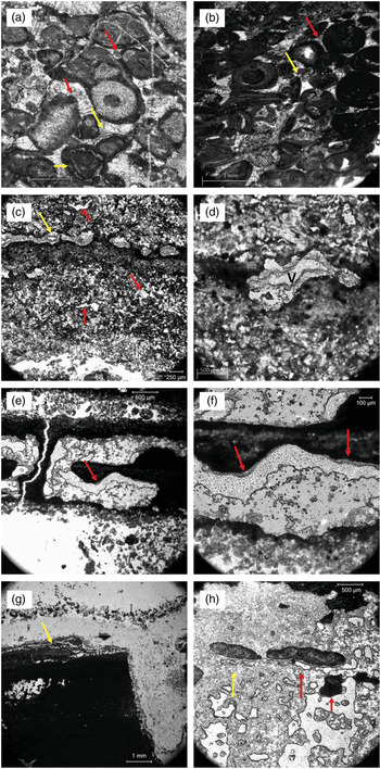
Microfacies 1 or packstones/grainstones: this microfacies consists of poorly laminated echinodermal (crinoids, sea urchin spines) and bioclastic (algae, benthic foraminifers, bryozoans, pelecypods, brachiopods, ostracods) facies with micritized well-sorted oolites (Fig. 21a, b). Bioclasts and oolites are frequently cross-bedded with thin (millimetre to centimetre) low-angle laminae. A thin irregular lamellar calcitic cement followed by blocky and granular cements is observed between the micritized oolites. Pisolites (up to 2 mm) and oncoids (up to 1 mm) are observed in the grainstones and bordered by irregular calcitic meniscus cements (Fig. 21a, b). Most of the grains are coated by a dark micritic cortex leading to the formation of composite ‘ooidic–oncoidic’ grains. The distribution of the cements is heterogeneous (Fig. 21b). A peloidal fenestral fabric is commonly observed in the packstone facies. Fenestrae are mostly irregular or tubular, sometimes developing a laminoid fabric with gravitational cements on their tops (Fig. 21c–h). Cylindrical fenestrae with the long axes subparallel to the bedding is regularly observed (Fig. 21d). The largest fenestrae form centimetre-sized cavities with micrite relics bordered by at least two lamellar calcitic cements. Anastomosing cracks are commonly observed and associated with strong alteration of the matrix and precipitation of a thin bladed calcite cement (Fig. 21g). The matrix of the packstone facies consists commonly of a fine-grained microsparite replacing totally or partly the primary facies. Silicification is sometimes observed.
Thin-section photomicrographs of grain coatings and spar-filled voids (A. Préat, 2018). (a) Sample BB43/a, site B. Bioclastic grainstone with micritized (mollusc) and pitted (crinoid) grains coated by dark micrite with irregular thicknesses around the lumpy grains (large grain in centre). Thin irregular rim of equant (microblocky) calcite cement inside or around micritic grains (yellow arrows). It is followed by wide lamellar (red arrows) and coarse or granular calcite cements (white zones in the centre of the former voids). The bioclasts are totally or partly micritized. Compaction is very weak. (b) Sample BB11, site D. Bioclastic (crinoids, molluscs) and oolitic grainstone with a discrete oblique stratification, showing strong micritization of the oolites (lower part of the photo) and other grains (micritized grains). Same irregular microblocky (red arrow) and large bladed calcite cements as observed in (a), and calcitic meniscus cements (yellow arrow), followed by white granular calcitic cements (centre of the former voids). Most of the grains are coated by a dark micritic cortex leading to the formation of ‘ooidic–oncoidic’ grains. The distribution of the cements is heterogeneous. Compaction is very weak. (c–h) Sample 1512, site A: (c) Microsparitized peloidal packstone showing a small-sized irregular and tubular fenestral fabric (red arrow). Most of the microspar consists of acicular and lamellar calcite crystals. Laminoid fenestrae (upper part, yellow arrow) are filled with an equant calcite cement. Scale bar in lower left corner is 250 µm; (d) clotted peloidal microsparitic packstone with an irregular fenestra representing a calcified root inside a darker micrite (not microsparitized). Individual small-sized blocky and isometric calcitic crystals bordered by a thin internal microsparitized layer constitute the ‘cortex’ of the fenestra. The void (V) (centre of the fenestra-root) is empty; (e, f) large irregular laminoid fenestral cavities with gravitational cements (red arrows) composed of very fine blocky calcitic crystals. The cavities were formed in a dark mudstone and are interconnected through a thin subvertical microchannel (e); (g) very large irregular cavity, with a general tubular subvertical cylinder (whitish area bordering the right side of the photograph) affecting the dark micrite matrix. The micritic relic is bordered by two medium-grained lamellar calcitic cements separated by a dark micritic discontinuity (with solution features) forming a thin layer, more irregular on the vertical wall. This cement is also bordered by a very thin greyish micrite lamina, which fills a probable shrinkage crack. Other anastomosing very thin empty shrinkage cracks are on the top of the micritic relic block (yellow arrow); (h) greyish to darkish microsparitized mudstone relics (yellow arrow, below an elongated relic) in a homogeneous microsparite with abundant large irregular and tubular empty vugs or fenestrae. The vugs are bordered and interconnected by thin laminar coatings, sometimes with pendant cements consisting of very fine-grained microblocky calcite below an empty vug (red arrow in the centre) and below a mudstone relic (right red arrow).

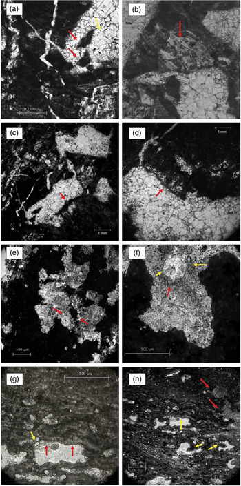
Microfacies 2 or mudstones/wackestones: this microfacies contains a few bioclasts (mainly echinoderms) in a microsparitized micritic matrix containing large-sized irregular sparmicritized (Kahle, Reference Kahle1977) fenestral-like cavities connected to thin tubules or veins (Fig. 22a–f). The cavities in the micrite are lined by two replacive calcite cements, the first one being lamellar or bladed, the second one being blocky equant and sometimes centripetal (Fig. 22a–d). Microcodium clasts composed of prismatic grains are probably present (Fig. 22b). The matrix also contains small-sized pyrite and sometimes displays an alveolar structure sensu Reference WrightWright (1982b ). Small-sized dolomite rhombs are also observed. Sometimes they are abundant in site D. The matrix suffered in-place recrystallization and was partly replaced by large-sized yellowish calcitic crystals.
Thin-section photomicrographs of fenestral fabrics in dark mudstones (A. Préat, 2018). (a–d) Sample BB39, site B. Slightly microsparitized (bioclastic) packstone with large-sized irregular fenestral-like cavities connected to thin tubules or veins. The cavities in the micrite are lined by two replacive calcite cements, the first one being lamellar or bladed (red arrow in (a)), the second one being blocky or equant (yellow arrow in (a)), and sometimes centripetal ((b) and lower cavity in (c)). Remnants or relics of former matrix are present in the cavity and generally bordered by the first lamellar calcitic cement phase (a). Partly leached and micritized grains are also observed ((a) lower right corner). The veins are sometimes empty, or cemented with walls consisting of small-sized equant calcitic crystals (d). Long acicular prismatic calcite crystals could also replace differently the matrix or are present in the matrix as possible Microcodium ((b) red arrow). The grain is slightly micritized and contains irregular peloids. Irregular and limited networks of micrite walls are present in the cavities ((c) and (d) red arrows) and could represent deposition of fine-grained carbonate around decaying organic matter (rootlets)? (e, f) Sample BB11-05, site D. Dark mudstone with irregular/tubular fenestral fabric showing intense sparmicritization. Peloidal relics of the mudstone are preserved ((e) red arrows) as a calcite void or tube (rootlet or some organism? in (f), yellow arrow), which pushed small-sized pellets aside (red arrow). A very thin micritic layer surrounds the tube. (g, h) Sample BB2, site D. Microsparitized dark mudstone with abundant more or less stratified irregular and fingered ((h) yellow arrows) fenestrae. The tubular fenestrae are sometimes complex, containing other small-sized tubes ((g) yellow arrow). Thin microbladed cement is preferentially observed in the roof of the fenestrae ((g) red arrows).

Microfacies 1 and 2 are present together at each study site (A, B and D). They are altered and lose their original characteristics, especially in sites A and B, which transforms the parent rocks into crumbly sediments in the loose laminated facies. In this case, it is often impossible to determine whether the original microfacies is type 1 or type 2, especially when the dissolution has been very extensive. In general, the processes of alteration are marked mainly by dissolution and/or cementation, micro- or macro-sparitization and ferruginization. They are analysed below on the basis of representative samples.
At site A, sample BB5 from a red lamina (Fig. 11) has a PSD of 82 % silt (between 2 µm and 63 µm) and 15 % clay (less than 2 µm). The mineralogical composition of the total rock (XRD) is dominated by quartz and goethite with 11 % CaCO3 (Table 1). XRD analysis of the clay fraction gives 90 % illite/muscovite. Sample BB11 taken from a yellowish lamina (Fig. 11) has a PSD of 54 % silt and 36 % fine sand (less than 125 µm) and a mineralogy dominated by calcite and quartz (XRD). Sample 1512 (Figs 13, 21) contains 64 % CaCO3 (Table 1). A thin-section from this sample has been analysed in detail (Fig. 21). It consists of a well-developed fine- to medium-grained greyish dirty microsparitic matrix replacing partly or totally the bioclasts (molluscs, brachiopods, algae, bryozoans, foraminifers, echinoderms). Fenestrae, gravitational cements, calcified roots and shrinkage cracks have been observed. Peloids are common, sometimes very abundant, and associated with silty quartz grains coated by goethite (Raman spectroscopy). Columnar palissadic gypsum (sometimes pseudomorphed by calcite) are locally observed in silty and clayey mudstones (Figs 24, 25) They are associated with abundant small euhedral bipyramidal quartz crystals (Figs 24b, 26). In the most-altered facies, replacement and ferruginization of the grains is common. Echinoderm fragments show dissolution vugs and are microsparitized or partly replaced by orange and brown clay aggregates. Sub-rounded to well-rounded reddish to blackish Fe–(Mn?) pisolites (up to several millimetres), with irregular asymmetric cement, are also observed (samples 1512, 1513; Fig. 13). Sample 1528 is microsparitized and contains abundant Fe-pisolites with a few preserved pelecypods in an irregular brownish laminar crust. Abundant gypsum patches forming hemi-pyramids and rosettes (Logan, Reference Logan1987), up to 200 µm, grew in the sediment. Fractures, open and filled, are widespread, mostly between the pisolites.
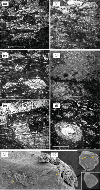
Photomicrographs of (a–h) weathered limestones and (g, h) quartz grains. (a–e) Thin-section of sample BB8, site B (A. Préat, 2018). Strongly recrystallized (aggrading neomorphism) (whitish zones in (a, b)) poorly laminated packstone with coated grains and micritized and bioclasts (mainly pelecypods in (a, b)) and algae (central part of (c)). The micritic matrix is microsparitized (irregular greyish dirty zones, lower part of picture and upper left corner of (a), main part of (b)) with irregular peloids, and irregular and surrounded relics of the former dark micrite upper half of (d). Coarser crystals, associated within the microspar, indicate that sparmicritization occurred (upper part of (b)). Recrystallized larger (millimetre-sized) calcite crystals, with strong varying direction of twin lamellae, highlight intensive stress tectonic processes (c), also highlighted in (e) with calcite-filled (two generations) fracture. (f) Thin-section of sample BB37, site B (A. Préat, 2018). Bioclastic packstone dominated by large crinoid pieces. Although the pieces are more resistant than other bioclasts, they suffered dissolution (white central part, bottom) and varying direction of twin lamellae. (g, h) BB7, site B (© Lenka Křižovaʾ, 2016). Gypsum rosettes (diameter 100–200 µm) and isolated gypsum laths (length 20–50 µm), which have precipitated on rounded quartz grains.

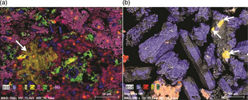
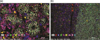
SEM photomicrographs and EDX analysis of samples of site A. (a) Thin-section of sample 1513 with fibrous gypsum (white arrow), small euhedral quartz and iron oxides in a matrix of clay minerals. (b) Selected particles of sample BB11. Euhedral quartz. Note a cluster of very small euhedral quartz with micro-patches of (residual?) gypsum (white arrows).

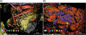
SEM photomicrographs and EDX analysis. (a) Sample BB7: gypsum laths. (b) Sample BB8: gypsum crystals and silica.

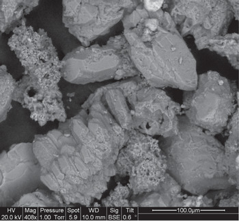
SEM photomicrograph of euhedral quartz (here in sample BB11).

In SEM, samples 1512 and 1513 contain abundant bipyramidal euhedral quartz, gypsum crystals and micas flakes (Fig. 24). The euhedral quartz grains are always characterized by a pitted surface and a vacuolar structure (Fig. 26). Their length rarely exceeds 50 to 80 µm. Most of them are smaller. They are found in all laminated formations of site A. In sample BB11, an aggregate of small bipyramidal quartz is mixed with micro-patches of residual gypsum (Fig. 24b).
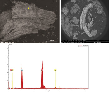
Sample 1532, taken from a weathered laminated limestone, close to the loose laminae 1512, 1513 and 1514 (Fig. 13), contains abundant millimetric pristine crystals of fibrous material. Some of them are curved and look like ram’s horn gypsum crystals. In SEM the chemical analysis shows that they are made of pure halite. In fact, they are halite pseudomorphs after gypsum (Fig. 27).
Halite pseudomorphs after gypsum, in sample 1531 (SEM photomicrographs and EDX analysis).

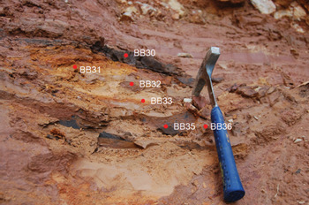
At site B, several samples were analysed. Sample BB30 (Fig. 20 and Fig. 17 ‘d’) corresponds to a black loose laminated material with small solid rock fragments. BB37 is a solid hard limestone. BB39 is a moderately weathered limestone, still cohesive but easily crumbled. Both are seen on Figure 17 (‘d’) and Figure 22. BB43 and BB44 were taken from a small cavity in a roughly laminated material (Fig. 17 ‘a’). BB43 is a weathered limestone with different grades of alteration (Fig. 21). BB44 is a loose sandy rock. BB7 and BB8 are well laminated. They are seen on Figures 18 and 17 (‘b’).
The most-altered facies are related to the fine-grained mudstones/wackestones and muddy packstones. They consist of a fine microsparitic mudstone with micritized grains and abundant irregular veins and small-sized cavities filled with coarse-grained calcite (sample BB39, Fig. 22a–d). The micritic coatings are sometimes thick and asymmetric (Fig. 22a). Their main characteristics are extensive microsparitization and the abundance of various calcitic cavities with medium- to coarse-grained sparry calcite interconnected or in association with a fracture pattern. Large calcitic deformed crystals embedded in the microsparitized matrix are common (Fig. 22c). Peloids are common, sometimes abundant and associated with silty quartz grains coated by iron oxides. In SEM, bipyramidal euhedral quartz with a vacuolar structure is present in all laminated facies and especially abundant in BB44, BB7 and BB8. Gypsum crystals are abundant in samples BB7 and BB8 (Fig. 25). In BB39, calcite crystals have replaced sulfates before being dissolved and silicified. The matrix also shows strongly deformed microsparitic veins, with multiplication of the primary thickness by folding or sliding and microslumps (sample 1521 in Fig. 18).
Another characteristic of site B is the presence of laminae very rich in iron oxide, identified as goethite by XRD. Sample BB30 contains 7.5 % CaCO3 (Table 1), and the main minerals are goethite and quartz. The secondary minerals are calcite, aluminosilicates and gypsum. Idiomorphic quartz grains are abundant (Fig. 28), but the most spectacular aspect of sample BB30 is the presence of numerous goethite nodules observed in thin-section (Fig. 29a). There is also a sharp contact between two laminae, one rich in goethite and the other rich in clay minerals (Fig. 29b). Brownish root mats are also present (1550 in Fig. 19). These weathered mudstones and wackestones formed crusts, which exhibit common sparmicritization and micritization. All stages of gradation between micritization of grains and sparmicritization from the parent limestones to the crusts are possible.
SEM photomicrograph and EDX analysis of a thin-section of sample BB30. Abundant quartz grains. Many of them are idiomorphic.

SEM photomicrographs and EDX analysis of a thin-section of sample BB30. (a) Goethite nodules. (b) Sharp contact between two different mineralogical laminae.

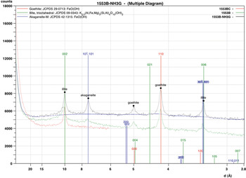
Sample 1553, close to samples BB7 and BB8, was taken from a weathered reddish laminated rock (Fig. 18). It contains 4 % CaCO3 (Table 1). XRD analysis of the total rock shows that the main mineral is goethite associated with illite/muscovite. The clay fraction (Fig. 30) is dominated by illite/muscovite, associated with relatively abundant goethite and akaganéite, an iron oxyhydroxide (β-FeOOH) reported for the first time in the studied area. Akaganéite is known to form in acid sulfate soils by oxidation of sulfide-rich sediments (Bibi et al. Reference Bibi, Singh and Sivester2011).
XRD diagram of sample 1553 containing illite, akaganéite and goethite (source: Viviane Bout-Roumazeilles, CNRS-LOG-University of Lille).

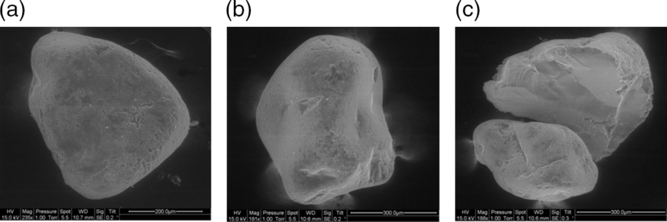
Sample BB7 (Fig. 23g, h) shows smooth rounded quartz grains partially covered by thin gypsum rosettes and twinned crystals observed in SEM. Sample BB44, taken from the nearby small cavity, also contains the same type of rounded quartz grains (Fig. 31). Their surfaces have a very low relief with small shock impact marks. A few grains are fractured, and the fracturing occurred after the round-shaping.
SEM photomicrographs of sample BB44: (a) rounded quartz grains with smooth surface, (b) v-shaped shock marks and (c) conchoidal fracture.

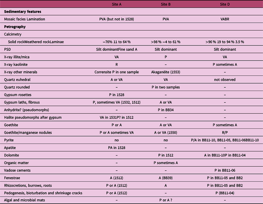
At site D, laminated formations are not frequent and the mineralogy of the weathered rocks significantly differs from sites A and B in the absence of gypsum and euhedral quartz, and the presence of pyrite (sometimes abundant) and dolomite in some samples (Table 2). Field evidence and petrographic observations of the weathered rocks indicate that the limestone parent rock suffered an intense alteration. There is no evidence of displacement in the weathered rocks. The weathered limestone host rocks consist mainly of sparmicritized matrices, as a result of calcretization of the host rocks, which suffered strong recrystallization leading to complex fabrics of ‘sparmicritization’ of larger calcite crystals. Sparmicritization is well developed in most of the weathered mudstones and wackestones (example of BB11-05, Fig. 7) as yellowish patchy silicification (example BB11-10, Fig. 8).
Compared petrographic, calcimetric, mineralogical and sedimentological characteristics in sites A, B and D

Abbreviations: R – rare; P – present; A – abundant; VA – very abundant.
A brief summary of the table shows that sites A and B (laminated facies) are quite similar, with numerous gypsum (pseudomorphic or not), euhedral quartz and goethite (and Fe-pisolite) nodules. Site B contains akaganéite. Site D (mosaic facies) contains dolomite and no sulfates nor euhedral quartz.
This produced different mosaics, which may be interpreted as the result of multistage replacement of calcite at the expense of micrite. This analysis was not carried out systematically, but the rocks initially altered by calcretization processes behave very differently from those of hard limestone rocks. It is likely that the initial clay content was an important parameter, because it guided variations in microporosity.
5.e. Stable carbon and oxygen isotopes
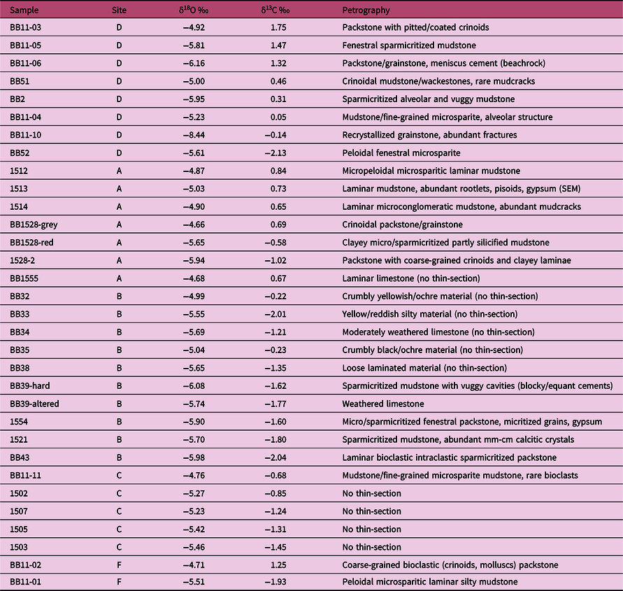
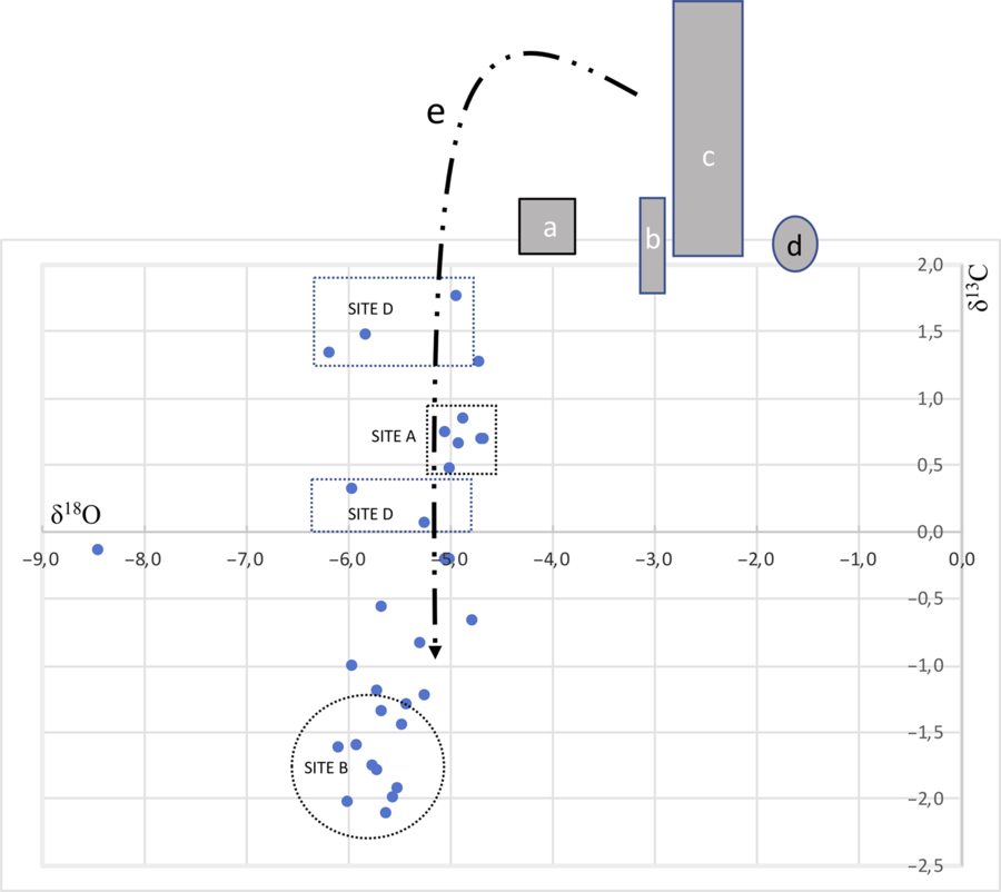
The bulk oxygen isotopic average of the middle Carboniferous samples (Visean/Serpukhovian) is −5.49 ‰ (n = 32). The values are well grouped, with no systematic trend, and range from −6.16 ‰ to −4.66 ‰ (except for one sample, BB11-10). The average carbon isotopic value (apart from BB11-10) is −0.91 ‰, ranging from −2.13 ‰ to 1.75 ‰ (Table 3) (Fig. 32). Such values are in accordance with the oxygen and carbon isotopic records of seawater for different middle Carboniferous series in low latitudes (Mii et al. Reference Mii, Grossman and Yancey1999; Wendt et al. Reference Wendt, Kaufmann and Belka2001; Grossman et al. Reference Grossman, Yancey, Jones, Bruckschen, Chuvashov, Mazzullo and Mii2008; Saltzman & Thomas, Reference Saltzman, Thomas, Gradstein, Ogg, Schmitz and Ogg2012). CaCO3 contents of the analysed samples range from 19 % to >98 % (Table 1).
Isotopic composition (δ18O and δ13C) values of the Upper Mississippian Pembrokeshire limestones, with sample numbers, site of sampling (see Fig. 4) and main petrographic characteristics. CaCO3 contents are given in Table 1

Five samples from site C and two from site F (location on Fig. 4) have been added to this table. Sites C and F have not been studied in detail yet, but it is interesting to note that the samples are in accordance with the diagenetic interpretation of sites A, B and D.
Oxygen and carbon isotopic records. Shaded rectangles mark the suggested stable isotopic composition of Upper Visean/Serpukhovian seawater from: a – Wendt et al. (Reference Wendt, Kaufmann and Belka2001); b – Grossman et al. (Reference Grossman, Yancey, Jones, Bruckschen, Chuvashov, Mazzullo and Mii2008); c – Mii et al. (Reference Mii, Grossman and Yancey1999); d – Saltzman & Thomas (Reference Saltzman, Thomas, Gradstein, Ogg, Schmitz and Ogg2012) (only isotopic carbon composition). e – ‘J inverted curve’ of Lohmann (Reference Lohmann1982). See Table 3 for our values from 32 samples selected in Bullslaughter Bay.

6. Interpretation
6.a. Palaeoenvironment
6.a.1. Petrographic data
Microfacies 1 is characteristic of an open marine (echinoderms, brachiopods and algae), shallow subtidal environment, with high degrees of agitation and slightly elevated salinity (Carew & Mylroie, Reference Carew, Mylroie, Vacher and Quinn1997). In this shallow environment, the sediment was frequently reworked by currents as shown by the cross-stratification of the oolites and bioclasts, which formed sandy bars or shoals (Fig. 21a, b). Peloidal irregular and laminoid fenestral fabrics in the packstone facies (Fig. 21c–h) display evidence of intertidal–supratidal diagenesis related to temporary subaerial exposure, as also highlighted by meniscus cements in the fabrics (Tanner, Reference Tanner, Alonso-Zarza and Tanner2010). Cementation of intergranular pore spaces between oolites and bioclasts led to the formation of beachrocks in the grainstone (Fig. 21a, b). The tiny irregular lamellar and meniscus cements in both facies (packstone and grainstone) point to a vadose influence, followed by equant blocky calcite in the phreatic environment (Land, Reference Land and Moore1989; Moore, Reference Moore1989), as highlighted by larger calcite crystals around the pore walls or inside the dissolution cavities. These blocky calcites follow the irregular bladed rim or pendant cements of the first calcite precipitation (James & Choquette, Reference James, Choquette, Mclleath and Morrow1990). Epitaxial growth on echinoderm plates is also observed in this phreatic environment. Micritic grain coats, tubiform rootlet moulds associated with anastomosing cracks and pisolites suggest that the bioclastic and oolitic substrate was calcretized and led to a crudely laminar fabric with distinctive darker and denser laminae. Evidence of subaerial exposure is also highlighted by the gravitational cements associated in the vadose diagenetic environment with the tubular voids, which could represent fine root tubules (Fig. 21d–h).
Microfacies 2 consists of fine-grained carbonates (mudstones/wackestones) with a few bioclasts of reduced diversity, which formed in a lagoonal setting in very quiet conditions. They probably represent protected coastal lagoons behind offshore barriers (with bioclastic shoals, see microfacies 1 in Section 5.d) (Hardie, Reference Hardie1977). The common fenestral fabrics (Fig. 22) point to intertidal and supratidal environments (Flügel, Reference Flügel2004). The facies records the influence of pedogenesis (rootlets, alveolar structure; Fig. 22g, h) according to Wright & Tucker (Reference Wright, Tucker, Wright and Tucker1991). This is supported by the large-sized irregular sparmicritized fenestral fabrics (Fig. 22a–f) connected to thin tubules or veins leading to the formation of calcrete crusts. Thin fibrous to microbladed calcite cements on the roof of the fenestrae (Fig. 22g) also suggest a vadose influence. Blocky equant calcite (often centripetal) crystals are also commonly observed in the fenestrae and cavities (Fig. 23a, b), as probable Microcodium clasts.
Based on the above facts, microfacies 1 and 2 recorded deposition in shallow water (respectively high and low energy). Then, they were subjected to significant subaerial exposure (beachrocks and calcretes) and later passed into the meteoric phreatic environment, as suggested by the blocky equant cement (Fig. 23c, h) filling dissolution cavities and residual intergranular porosities. Subsequent sparmicritization of the cemented cavities points to multiple diagenetic vadose–phreatic phases (Fig. 23e, f).
6.a.2. Laminated facies of sites A and B
At site A, the graded lamination characterizes the sediment, which is dominantly silty, with temporary inputs of coarser sediment. This type of feature can be observed in various aqueous environments; for example, in the swash-backwash zone of beaches, but also in supratidal flats (lagoons and sabkha) with laminar flow and abundant suspended sediment, with possible short flash flooding. The good preservation of lamination and the absence of bioturbation, the fenestral fabric and vadose features, and the mineralogy (abundant euhedral microquartz and gypsum, halite pseudomorphs after gypsum) suggest high-salinity conditions, in intertidal or supratidal salt flats (sabkha) during hot and arid climates with comparatively low humidity and rapid evaporation (Logan, Reference Logan1987; Schreiber & Walker, Reference Schreiber and Walker1992). All the fossils found in the laminae are the same as those of the solid rock. They are well dated from the early Brigantian, thanks to a conodont assemblage (Błażejowski & Walsh, Reference Błażejowski and Walsh2013). Late Visean conodonts have been recognized in shallow-water platform limestones in Ireland (Somerville & Somerville, Reference Somerville and Somerville1998). Thus, it is not excluded that the fossils present in the laminae could date the sedimentation itself.
The arrangement of the loose laminae, either parallel and in continuity with laminated solid rock, or either oblique and in sharp contact with the solid parent rock, cannot be explained in a single sedimentation event but by a complex sequence of successive events: (i) meteoric erosion during a low sea-level and emergence period leading to a rough rocky platform; (ii) laminar sedimentation in an intertidal, supratidal or lagoonal environment, which skirts and wraps the small-sized rocky stumps of the platform; (iii) early diagenesis and weathering of laminated rock in hypersaline and oxidizing conditions in a sabkha-type environment. More than one period of emergence and temporary surface exposure is possible in this sequence as highlighted by Fe-pisolites.
This sequence was followed by a transgressive phase of marine sedimentation marked by a slight unconformity of a limestone bed over the laminated weathered formations. Site A is the only place in Bullslaughter Bay where it is possible to recognize such a complex sequence of events.
At site B, despite their small size, the patches of laminated sediments provide valuable palaeoenvironmental information. As in site A, the ubiquitous bipyramidal quartz grains and the abundance of gypsum strengthen the hypothesis of one or several phases of sedimentation in a hypersaline environment (supratidal salt pan or sabkha), which is also reinforced by the presence of akaganéite (see Section 6.a.3 below). On the other hand, the rounded and larger quartz grains observed in two samples of site B (Figs 23g, h, 31) have a different origin. They are detrital. Their shape and surface features suggest marine abrasion on a beach (Krinsley & Doornkamp, Reference Krinsley and Doornkamp1973; Le Ribault, Reference Le Ribault1975). Such an origin is reinforced by the microscopic characteristics of samples BB43 (Fig. 21a) and BB8 (Fig. 23a–e). A temporary beachrock environment is compatible with these observations.
The good crystalline shape of gypsum in sample BB7 (collected in loose material close to the more or less weathered ‘BB8’ limestone) proves that the sediment was not disturbed after the formation of gypsum despite the presence of a nearby slightly collapsed limestone and fault zone.
De-dolomitization has also been observed in site B (thin-section 1521) in a ferruginous peloidal wackestone with large-sized macrosparitized calcite crystals (up to 1 mm) and calcitized dolomite (up to 500 µm). This latter consists of large rhombs (up to 1 mm), isolated or contiguous, comprising a fine-grained mosaic of euhedral calcite. Calcite pseudomorphs after prismatic evaporite crystals (gypsum?) are also observed as millimetric irregular vugs, with pendant calcite cements. Meteoric water can dissolve former evaporite crystals including dolomite. The dissolution and replacement of evaporite can also occur by undersaturated waters from underlying and adjoining aquifers, without recharge from the surface (Schreiber & El-Tabakh, Reference Schreiber and El-Tabakh2010). The calcitization of gypsum requires a supply of Ca2+ and CO3 2− and the removal of SO4 2−. However, these processes can occur either at different times (i.e. with earlier formation of voids by dissolution) or simultaneously (Armenteros, Reference Armenteros, Alonso-Zarza and Tanner2010).
6.a.3. Hypersaline environment in sites A and B
The bipyramidal quartz grains are characterized by an imperfect habit with numerous small vacuoles due to disturbance during the crystallization process. It suggests a rapid precipitation after former silica solution. Different sources of silica were possible (hydrolysis of clay minerals, solute load in fluvial or deltaic influx, solution of cherts, silica released from skeletal grains). Thus, the problem is not the source of silica but the conditions of crystallization. Euhedral quartz can form at a high temperature owing to deep burial or a hydrothermal environment. Here, such a context can be excluded, because of the perfect preservation of halite pseudomorphs and gypsum crystals in some laminae, and the limited compaction and absence of burial diagenesis in all the samples that were analysed. On the other hand, this type of euhedral quartz habit has been described in hypersaline sedimentary environments, in supratidal or inland areas such as sabkhas or salt pans (Grimm, Reference Grimm1962; Friedman & Shukla, Reference Friedman and Shukla1980; Paszkowski & Szydłak, Reference Paszkowski and Szydłak1986; Gundu Rao, Reference Gundu Rao1986; Viczian, Reference Viczian1992; Ulmer-Scholle et al. Reference Ulmer-Scholle, Scholle and Brady1993; Albright & Lueth, Reference Albright and Lueth2003; Flügel, Reference Flügel2004). According to the latter authors, the neo-crystallization of quartz is possible in the presence of salt solutions. At sites A and B of Bullslaughter Bay, the process of quartz crystallization by the replacement of sulfate is strongly suggested by SEM micrographs (Fig. 24b) showing juxtaposition of bipyramidal quartz and gypsum crystals. The absence of bipyramidal microquartz and gypsum in the more recent Brigantian limestone strata of site D (north of the Bullslaughter Bay syncline) is an argument to constraint the time of their formation and to relate them to a period of sea-level low-stand and emergence prior to late Brigantian time.
Our hypothesis of a hypersaline environment is also supported by the presence of akaganéite associated with goethite and gypsum in sample 1553 at site B. Akaganéite has been described in a highly saline inland wetland (the Bottle Bend Lagoon) in the southwest of New South Wales (Australia) (Bibi et al. Reference Bibi, Singh and Sivester2011). The precipitation of akaganéite followed a sharp fall in pH (from 8 to 3) owing to a severe drought event in 2002. Halite and gypsum crystals are always very abundant in the Australian samples containing akaganéite. It is interesting to note that the thin-section of sample 1553 revealed a very fine-grained whitish homogeneous microsparite with Fe-peloids (goethite) and enterolithic-like microveins filled with thin palissadic gypsum. Moreover, sample 1553, which contains only 4 % CaCO3 (Table 1), is close to BB7 and BB8, which are rich in gypsum crystals. Finally, one of the best arguments supporting the hypersaline environment hypothesis is the presence of pristine halite pseudomorphs after gypsum. Although infrequent in the geological record, the replacement of gypsum by halite has long been known, especially in Permian rocks. The process was analysed in relationship to the environmental context and the solubility relationships of gypsum, halite, anhydrite and brine (Schreiber & Walker, Reference Schreiber and Walker1992; Hovorka, Reference Hovorka1992). Despite some divergent explanations, these authors agreed that the replacement takes place in shallow-water hypersaline basins. For Schreiber & Walker (Reference Schreiber and Walker1992), the process implies hot and arid climates and needs contrasting temperatures between the brine surface water (which must be ‘overheated’ up to 55–57 °C) and the underlying sediment. It involves brine convection and provides the saturation relationships necessary to both dissolve gypsum and precipitate halite. It occurs most easily in shallow-water bodies with solar heating of the brine favoured by blooms of red-pigmented bacteria. The replacement takes place just below the sediment surface and concerns a very thin layer (generally less than 10 cm).
The hypersaline environment suggested by our petrographic and mineralogical analyses may be compared with the present salt pans called ‘tannes’ in Africa. They are situated in the tropical climate zone with contrasting dry and rainy seasons on the landward side of mangroves. Their formation needs a dry season lasting over three months, but the climate must be wet enough to allow the mangrove development. The acid sulfate soils of these ‘tannes’ have been studied in Senegal (Sadio, Reference Sadio1991). Marine pyrite is the main source of sulfur. Oxidation of pyrite gives jarosite, lepidocrocite, goethite and haematite according to the topographic and hydrological environment. The pH of soils may be very low (down to 3 in some cases) and the salinity five to ten times that of the seawater. A few decimetres relief is sufficient to change the pH, drainage conditions and geochemical processes. In the lower parts gypsum is abundant. On slightly higher areas with better drainage and low pH (<4) goethite forms. At the top with efficient drainage and very high salt content, ferric of ferro-manganese concretions can form. In Senegal, these acid sulfate soils are able to develop in a few dozen years. This African case helps to understand what happened in the Late Mississippian environment of Bullslaughter Bay. At sites A and B, the juxtaposition of gypsum-rich and iron/manganese-rich laminae could be explained by a type of coastal environment similar to the present Senegal salt pans. If the pH was possibly as low as in Senegal, the solution of the local limestones could have been very rapid. At sites A and B, the parent rocks were iron-rich muddy limestones. The goethite nodules are often associated with manganese. They could be compared to iron-manganese nodules observed in the semi-arid climate of northeastern Spain. Sanz et al. (Reference Sanz, Garcia-González, Vizcayno and Rodriguez1996) considered that they were formed in situ in a poorly drained soil with a high calcium carbonate content. According to these authors, Fe and Mn redistribution in soil is caused by water table oscillations.
6.b. Stable isotopes (oxygen and carbon)
The isotopic compositions of carbon and oxygen (when the decalcification was not too strong to allow analyses), which are the same in both cases (see below), confirm that the genesis of the alteration was related to a meteoric influence. The general processes could be attributed to a ‘grainification’ of the parent materials, producing secondary diagenetic matrices in the context of the general textural inversion, which is characteristic in calcrete or in pedogenic carbonates (Wright & Tucker, Reference Wright, Tucker, Wright and Tucker1991).
A crucial question in any study of carbonate chemostratigraphy is whether a primary marine signature is preserved. As we have seen in the sedimentology section, the limestones studied were deposited in a marine environment (see, for example, the bioclastic and oolite contents), but the typical cements of this environment (aragonite, fibrous high-Mg calcite (HMC) or elongated bladed HMC) have not been preserved and have been replaced by lamellar and equigranular or coarse crystals of calcite (low-Mg calcite), both in the intergranular space and in the fenestral-type dissolution cavities. The pendant cementation and vadose cavities observed in these Visean limestones, as well as the beachrocks, also show that the diagenetic alteration was early, as also attested to, for example, by the absence of compaction patterns in the alveolar structures. The primary chemical signature (marine domain) has therefore been altered in the study area. Other sedimentary basins, where this signature has been preserved, can be used as a reference for comparison purposes. From the studies of Mii et al. (Reference Mii, Grossman and Yancey1999), Wendt et al. (Reference Wendt, Kaufmann and Belka2001), Grossman et al. (Reference Grossman, Yancey, Jones, Bruckschen, Chuvashov, Mazzullo and Mii2008) and Saltzman & Thomas (Reference Saltzman, Thomas, Gradstein, Ogg, Schmitz and Ogg2012), it appears that the δ13C and δ18O isotopic compositions of the middle Carboniferous (Visean) seawater are around or higher than +2 ‰ for the δ13C values, and range from −4 ‰ to −2 ‰ for the δ18O values (see rectangles ‘a, b, c, d’ in Fig. 32). First of all, our results show that there is no correlation between δ13C and δ18O in our samples, as δ18O does not show any significant variation, excluding an alteration in the marine–meteoric mixing zone (Allan & Matthews, Reference Allan and Matthews1982). Our δ18O values (Fig. 9) are lower than those of typical marine ones and highly suggestive of diagenetic alteration in meteoric water. Samples display a narrow range of δ18O and a relatively wide range of δ13C compositions. This suggests that the middle Carboniferous samples, collected within a restricted area, were influenced in at least a single fresh water system during deposition, as highlighted by small-scale vadose vugs cemented by diagenetic equant calcite cements (Allan & Matthews, Reference Allan and Matthews1982). Depleted 18O values by several per mil can also arise from burial diagenesis. But it can be excluded here as equant cementation processes occurred early, as exemplified by the preserved former dissolved fenestrae and vugs with their small-scale gravitational cements. Compaction is also very limited with rare grain contacts. Stylolites are also not well developed. As noted previously, the Castlemartin area was never covered by thick post-Visean sediments, so deep burial diagenetic effects can be excluded in the Pembroke Limestone Group.
Our δ13C values are systematically slightly lower relative to the middle Carboniferous seawater values. These anomalously low δ13C values are consistent with the hypothesized diagenesis mentioned above. The depletion in 13C is explained by shallow vadose sedimentary environments (Land, Reference Land and Moore1989; Knauth & Kennedy, Reference Knauth and Kennedy2009; Sial et al. Reference Sial, Gaucher, Ferreira, Pereira, Cezario, Chiglino, Lima and Ramkumar2015): soil-gas CO2, which derived from oxidation of organic matter, produced depleted δ13C. The scatter plot of the variation of our δ13C and δ18O values (Fig. 32), with highly variable carbon isotopic compositions and nearly invariant oxygen ones, typically has the form of the ‘inverted J’ curve established by Lohmann (Reference Lohmann1982, Reference Lohmann, James and Choquette1988) for an idealized meteoric diagenetic system. The final isotopic composition after diagenesis depends upon the isotopic composition of the initial carbonate (here open marine), the composition of the diagenetic fluids (related to the degree of organic matter oxidation and organic productivity) and the degree of exchange.
The wide variability in our δ13C compositions and narrow variability in δ18O compositions has been documented in many limestones formations; for instance, the Holocene and upper Pleistocene of Barbados, the Lower Cretaceous Glen Rose Formation, the Pennsylvanian Strawn Formation (Texas) and the Upper Mississippian Newman Limestone (Kentucky) (see review in Allan & Matthews, Reference Allan and Matthews1982). It is interpreted as a distinctive feature of the subaerial diagenetic process. These formations have been altered by early freshwater diagenesis. The absence of a positive covariance between our δ13C and δ18O values suggests that after being deposited in shallow marine and lagoonal environments, the Castlemartin carbonates did not evolve in a marine–meteoric water mixing zone. Our interpretation of the carbon and oxygen isotope compositions of the studied area is consistent with the palaeoenvironmental reconstruction of the area. The analysis of the isotopic compositions shows that the limestones of site B have the most negative δ13C values (up to −2 ‰, with an enrichment of 3–4 ‰ compared to marine limestones; data from the literature, and Table 3 and Fig. 32). Their mineralogical stabilization has been completed in the deeper vadose or deep phreatic environments.
7. Discussion
The field observations and sample analyses give preliminary results about the sedimentary conditions and early diagenetic processes in Bullslaughter Bay. The special type of isovolumetric weathering of limestones, recently described in Bullslaughter Bay (Rowberry et al. Reference Rowberry, Battiau-Queney, Walsh, Błażejowski, Bout-Roumazeilles, Trentesaux, Křížová and Griffiths2014), and interpreted as a ‘ghost-rock’ formation sensu Reference QuinifQuinif (2010), has specifically attracted our attention. Our research shows that the process of alteration was complex besides the loss of calcium and lower density of the rock. On the microscopic scale, it was characterized by an intense micritization of grains and also pendant cementation in the vadose zone, where sparmicritization occurred as a result of dissolution and precipitation processes. Several diagenetic processes (such as ferruginization and de-dolomitization) have affected the sediments owing to probable variations in the meteoric fluxes, in relationship with sea-level oscillations and possible climate variability (length of the dry season).
Nowhere were karstic landforms related to an underground drainage system observed. At site A, at least one phase of emergence occurred. The superficial corrosion of the parent limestones was likely favoured by a very low pH in a brackish environment. It led to the development of a rocky platform with metric residual limestone blocks, which can be called ‘epikarstic’. On the other hand, dolines, solution features, palaeocaves and conduits have not been observed in Bullslaughter Bay, contrary to other areas in southern Pembrokeshire and more generally in Wales (Wright, Reference Wright1982 a; Davies, Reference Davies1991; Vanstone, Reference Vanstone1998; Rowberry et al. Reference Rowberry, Battiau-Queney, Walsh, Błażejowski, Bout-Roumazeilles, Trentesaux, Křížová and Griffiths2014).
The development of brackish and hypersaline conditions in Bullslaughter Bay implies a different flooding story of the Asbian/Brigantian platform to that of Gower and other South Wales areas (Wright et al. Reference Wright, Vanstone and Marshall1997). It suggests a slower flooding on flat-topped carbonate platforms, as has been recorded in the Bahamas (Rasmussen & Neumann, Reference Rasmussen, Neumann, James and Choquette1988, Reference Rasmussen and Neumann1990). Stagnant fresh water ponds could form during the sea-level rise. With strong evaporation in an arid or semi-arid climate, a phase of hypersaline bank waters could develop in supratidal and inland salt pans. This is in accordance with the meteoric signal given by the carbon and oxygen isotope compositions. Halite can also be concentrated from fresh continental waters, as in many salt lakes, or derived from the dissolution and recycling of older evaporites (Tucker, Reference Tucker2001; M. Al-Yousef, unpub. Ph.D. thesis, Univ. Southampton, 2003).
The noteworthy good preservation of all the features resulting from the diagenetic processes (such as, for example, the fragile halite pseudomorphs after gypsum) raises the question of the age of their formation. Most previous studies considered that the weathering processes necessarily occurred after the Variscan orogeny and probably during Triassic or Permian times (Thomas, Reference Thomas1971), if not later (Woodcock et al. Reference Woodcock, Miller and Woodhouse2014). In this study, all the processes could have developed very early, during or just after sedimentation. The Variscan folding of the limestone strata has not significantly deformed the loose laminae, because they were sandwiched between hard competent beds. Moreover, our analyses show the absence of compaction, on a metric as well as a micrometric scale, a result in accordance with the supposed non-deposition of thick upper and post-Carboniferous sediments in southern Pembrokeshire. The late Visean dating of the diagenetic processes results not only from our small-scale field observations and microscopic analysis, but also from the isotopic analyses, as was explained before. An interesting point, which has to be investigated in detail, is that a mixing of blocks of weathered and solid limestone is usually found in the nearby ‘Gash-Breccias’. Thus, the timing of alteration is constrained, since the breccias are closely linked to the late Carboniferous – early Permian Variscan folding (Walsh et al. Reference Walsh, Battiau-Queney, Howells, Ollier and Rowberry2008).
A provisional interpretation of the sedimentation story proposes a complex Asbian/Brigantian sequence:
-
1) Marine sedimentation in shallow water, early diagenesis in the vadose or phreatic zones (site D), with possible episodic evaporite depositional conditions, as recorded by the thin palissadic veins of gypsum pseudomorphs (and also gypsum relics) in several samples from sites A and B.
-
(2) Phreatic meteoric environment with emergence and superficial corrosion, leading to the construction of an epikarstic rocky platform with numerous residual metric blocks of solid limestone (site A).
-
(3) Supratidal and/or sabkha sedimentation and meteoric diagenesis in an arid hypersaline environment (sites A and B).
-
(4) Sea-level rise with shallow-water marine or supratidal brackish sedimentation (sample 1528).
Further analyses will be necessary to refine this sequence, which might be significantly different in site D and in sites A and B. It remains also to evaluate the role of bacteria and algal mats, which were detected in some samples but not yet investigated.
8. Conclusion
Despite the limited area studied, this research gives first-hand regional-scope results on the Late Mississippian sedimentary environment of southern Pembrokeshire. Multiple scientific approaches and a set of various laboratory analyses show that the limestone sedimentation occurred in a coastal infra-, inter- or supratidal environment. The sea-level oscillations played an important part in the changing sedimentary conditions, whereas the climate seems to have been constantly hot and seasonally contrasted. Diagenetic processes, active during or just after the deposition, in the vadose or phreatic zone, induced the development of isovolumetric weathering (‘ghost-rock’ formation). One unexpected result of our research brings out the importance of at least one (but probably several) period(s) of surface exposure in a hypersaline environment, as testified to by gypsum crystallization, halite pseudomorphs of gypsum, euhedral bipyramidal quartz and ferro-manganese nodules present in laminated sediments. Calcretization at or near the sediment surface, in the vadose zone, was one of the most widespread diagenetic processes in Bullslaughter Bay. Isotopic composition (δ18O and δ13C) values of weathered and unweathered sediments from the bay show that all the diagenetic processes took place in a meteoric environment, during or very soon after sedimentation. Field observations, petrographic and mineralogical analyses agree with a Late Mississippian dating of the sedimentary features observed in Bullslaughter Bay. Further research is needed in other places in southern Pembrokeshire to refine the regional sedimentary sequence and explain the ‘Gash-Breccias’ formation.
Acknowledgements
Although the authors personally assume the ideas developed in this paper, they know what they owe to all those who helped them during the research process. YBQ is particularly indebted to Peter Walsh and Matt Rowberry for the fruitful discussions they had in the field, and their decisive help to get and carry the samples. Without both of them nothing would have been possible. It is our pleasure and duty to thank all the technical team at the CNRS Laboratory of Oceanology and Geosciences (LOG) of the University of Lille (France): Sandra Ventalon (microscopy and Raman spectroscopy), Marion Delattre (calcimetry), Romain Abraham (PSD), Cindy Maliverney (thin-sections) and also Denis Marin (CNRS–ULCO University at Dunkerque) who designed the map in Figure 3. We gratefully acknowledge Lenka Křižovaẚ of Charles University (Czech Republic) for exoscopy of quartz. Our research has profited from informal discussions with many colleagues of the LOG research team. Lastly, we are greatly indebted to Franck Delpomdor (Illinois State Geological Survey) and a second anonymous reviewer, whose comments allowed us to significantly improve our original manuscript. This research received no specific grant from any funding agency, commercial or not-for-profit sectors. All the field pictures were taken by Yvonne Battiau-Queney, unless otherwise indicated. All the SEM micrographs were taken by Philippe Recourt.