The clinical syndrome of frontotemporal dementia (FTD) is characterised by progressive changes in behaviour, personality and/or language, with relative preservation of memory (Refs Reference McKhann1, Reference Neary2, 3). Clinical subtypes include the frontal (behavioural) variant (fvFTD) and two forms of primary progressive aphasia (PPA): primary non-fluent aphasia (PNFA) and semantic dementia (SD) (Refs Reference McKhann1, Reference Neary2). FTD is often associated with an extrapyramidal movement disorder (parkinsonism or corticobasal syndrome) or with motor neuron disease (MND) (Refs Reference Padovani4, Reference Lomen-Hoerth, Anderson and Miller5). FTD accounts for 5–15% of all dementia and is the second commonest cause in the presenile age group (Refs Reference Feldman6, Reference Bird7). A family history of similar disease is present in 25–50% of patients, indicating a significant genetic influence (Refs Reference Stevens8, Reference Rosso9, Reference Chow10).
It should be noted that there are currently two conventions used for the nomenclature of this clinicopathological syndrome. Here, we use the one that is more popular in North America, in which FTD is used as the general term for the clinical syndrome (including all behavioural and language variants) and the term frontotemporal lobar degeneration (FTLD) is reserved for the associated pathology. However, in the UK, FTLD is often used as the general term for both the clinical and the pathological entities, whereas FTD refers only to the clinical subtype in which behavioural abnormalities predominate. We recognise that this situation is confusing; however, both terminologies are currently in wide use and there is no consensus as to which is more appropriate.
The molecular genetic basis of FTD is heterogeneous. Autosomal dominant FTD may be caused by mutations in several genes, including those encoding the microtubule-associated protein tau (MAPT) (Refs Reference Hutton11, Reference Poorkaj12, Reference Spillantini13), progranulin (GRN) (Refs Reference Cruts14, Reference Baker15), valosin-containing protein (VCP) (Ref. Reference Watts16) and charged multivescicular body protein 2B (CHMP2B) (Ref. Reference Skibinski17), and several families with FTD and MND have shown genetic linkage to a locus on chromosome 9p (Refs Reference Valdmanis18, Reference Morita19, Reference Vance20).
The neuropathology associated with clinical FTD is also heterogeneous (Ref. Reference Cairns21). Relatively selective degeneration of the frontal and temporal lobes (frontotemporal lobar degeneration, FTLD) is a consistent feature, which correlates with the main clinical manifestations. As with many other neurodegenerative conditions, the pathology of most cases of FTD includes the presence of abnormal intracellular protein accumulations. Traditionally, these inclusion bodies were demonstrated with special histochemical staining techniques, such as silver impregnation methods. Eponymous and syndromic names were used for clinical syndromes associated with a specific morphology and anatomical distribution of cellular inclusions. However, in many cases, these clinicopathological correlations turned out to be imperfect. Modern laboratory techniques, such as immunohistochemistry, have allowed the biochemical composition of the pathological changes to be more readily determined. In recent years, it has become popular to classify FTLD pathology based on the presumed molecular defect, in the belief that this most closely reflects the underlying pathogenic process (Ref. Reference Cairns21).
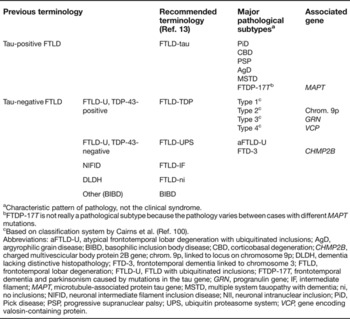
In this review, we will use the molecular-based system of nosology and nomenclature recommended in a recent consensus paper (Ref. Reference Mackenzie22). The term FTLD will be used as the general terminology for pathological conditions that are commonly associated with clinical FTD, and major subdivisions will be designated by the protein abnormality that is presumed to be pathogenic or most characteristic (Table 1). Cases are further subclassified, using traditional terminology, to define specific patterns of pathology [i.e. FTLD-tau (PiD) for Pick disease]. Existing descriptive terms are retained for rare causes of FTD that have characteristic pathological features of unknown biochemistry (such as basophilic inclusion body disease, BIBD). Finally, cases with no inclusions visible with special histochemical stains or immunohistochemistry (formerly known as dementia lacking distinctive histopathology, DLDH), will be designated FTLD-ni (no inclusions).
Recommended nomenclature for frontotemporal lobar degenerations

aCharacteristic pattern of pathology, not the clinical syndrome.
bFTDP-17T is not really a pathological subtype because the pathology varies between cases with different MAPT mutations.
cBased on classification system by Cairns et al. (Ref. Reference Cairns100).
Abbreviations: aFTLD-U, atypical frontotemporal lobar degeneration with ubiquitinated inclusions; AgD, argyrophilic grain disease; BIBD, basophilic inclusion body disease; CBD, corticobasal degeneration; CHMP2B, charged multivescicular body protein 2B gene; chrom. 9p, linked to locus on chromosome 9p; DLDH, dementia lacking distinctive histopathology; FTD-3, frontotemporal dementia linked to chromosome 3; FTLD, frontotemporal lobar degeneration; FTLD-U, FTLD with ubiquitinated inclusions; FTDP-17T, frontotemporal dementia and parkinsonism caused by mutations in the tau gene; GRN, progranulin gene; IF, intermediate filament; MAPT, microtubule-associated protein tau gene; MSTD, multiple system tauopathy with dementia; ni, no inclusions; NIFID, neuronal intermediate filament inclusion disease; NII, neuronal intranuclear inclusion; PiD, Pick disease; PSP, progressive supranuclear palsy; UPS, ubiquitin proteasome system; VCP, gene encoding valosin-containing protein.
FTLD-tau
Tau protein
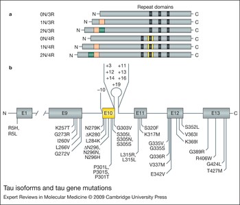
Tau is a microtubule-associated phosphoprotein (MAP) that is abundantly expressed in both the central and peripheral nervous system. By promoting microtubule (MT) assembly and stability, tau plays a fundamental role in maintaining neuronal integrity and axoplasmic transport (Refs Reference Cleveland, Hwo and Kirschner23, Reference Weingarten24). The tau gene (MAPT) is located on chromosome 17q21 and has two major haplotypes, H1 and H2, which are defined by a set of single nucleotide polymorphisms and a 238 base pair deletion in intron 9 (Ref. Reference Baker25). The H2 haplotype is also associated with an inversion of a ~900 kb region that includes MAPT (Ref. Reference Stefansson26). MAPT contains 16 exons, 11 of which encode the major tau protein isoforms (Ref. Reference Andreadis, Brown and Kosik27). Through alternative mRNA splicing of exons 2, 3 and 10, a set of six isoforms, ranging from 352 to 441 amino acids, are generated in the adult human brain. Exons 9 to 12 encode four microtubule-binding motifs, which are imperfect repeats of 31 or 32 amino acids, in the C-terminal half of the tau molecule. It is these binding domains that mediate the interaction between tau and MTs. Alternative splicing of exon 10 generates tau isoforms with either three (exon 10 missing) or four (exon 10 present) repeat domains, known as 3R and 4R tau, respectively (Fig. 1a) (Refs Reference Goedert and Spillantini28, Reference Goedert29). In Alzheimer disease (AD), abnormally hyperphosphorylated tau is the major component of the neurofibrillary lesions (known as neurofibrillary tangles, neuropil threads and dystrophic neurites), whereas β-amyloid peptide is the major component of senile plaques. Abnormal intracellular accumulation of tau is also characteristic of a number of other neurodegenerative disorders, collectively known as ‘tauopathies’ (Refs Reference Tolnay and Probst30, Reference Lee, Goedert and Trojanowski31). Comparison of the tau aggregates in these disorders reveals differences in both phosphorylation and the content of different tau isoforms. Thus, tauopathies might be subdivided into disorders with inclusions made predominantly of 3R or 4R tau or an admixture of both (Ref. Reference Delacourte32). A number of these tauopathies may be associated with clinical FTD (FTLD-tau), including Pick disease (PiD), progressive supranuclear palsy (PSP), corticobasal degeneration (CBD), argyrophilic grain disease (AGD) and hereditary frontotemporal dementia and parkinsonism linked to chromosome 17, as a result of MAPT mutations (FTDP-17T) (Ref. Reference Cairns21).
Tau isoforms and tau gene mutations. (a) Six isoforms of the tau protein found in the adult human brain result from alternative splicing of exons 2 (red), 3 (green) and 10 (yellow). Exons 9–12 encode imperfectly repeated microtubule-binding repeats (shown in black). (b) Mutations in the tau gene in frontotemporal dementia and parkinsonism linked to chromosome 17 (FTDP-17T). Thirty-six coding region mutations in exons (E) 1 and 9–13 and eight intronic mutations flanking exon 10 are shown.

FTLD-tau (PiD)
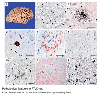
PiD is the clinicopathological prototype of FTD (Refs Reference Onari and Spatz33, Reference Alzheimer34, Reference Pick35). The most distinctive gross pathological feature is marked atrophy of the frontal and anterior temporal lobes, also known as ‘knife-edge’ lobar atrophy (Fig. 2a). The main histological features consist of severe neuronal loss, ballooned neurons (Pick cells) and pathognomonic spherical argyrophilic neuronal inclusions, called Pick bodies (PBs) (Fig. 2b). PBs are found mainly in neocortical neurons of layers II–IV, the granule cells of the dentate gyrus, pyramidal cells of the hippocampus, subiculum and entorhinal cortex, and subcortical nerve cell populations. PBs are composed predominantly of 3R tau isoforms (Ref. Reference Delacourte32), whereas 4R tau deposits might be present in glia in a subset of cases (Refs Reference Zhukareva36, Reference Mott37).
FTLD-tau (PSP)
Also known as Steele-Richardson-Olszewski syndrome (Ref. Reference Steele, Richardson and Olszewski38), PSP is the second most-common parkinsonian disorder, after classical Parkinson disease. Clinical features typically include early postural instability, axial dystonia, supranuclear vertical gaze palsy, pseudobulbar palsy and parkinsonism that is resistant to levodopa therapy. Although cognitive dysfunction is often mild, some cases of PSP present with dementia and only mild motor features (Ref. Reference Sha39). The pathology of PSP is characterised by gross atrophy of the midbrain (especially the pretectal region), superior cerebellar peduncles, subthalamic nucleus and substantia nigra. Fibrillar tau pathology occurs in both neurons and glia. Glial inclusions include tufted astrocytes (Fig. 2c), which are fairly specific for PSP, as well as thorn-shaped astrocytes, coiled bodies in oligodendrocytes and neuropil threads, all of which are also found in other tauopathies. Neurofibrillary tangles typically have a round or globose appearance (Fig. 2d). Many subcortical regions are affected, including the striatum, pallidum, subthalamic nucleus, substantia nigra, oculomotor complex, periaqueductal grey, superior colliculi, basis pontis and dentate nucleus. Cases of PSP with dementia are also found to have significant tau pathology in the cerebral cortex (Ref. Reference Bigio, Brown and White40). Biochemically, the inclusions in PSP consist predominantly of 4R tau isoforms (Ref. Reference Dickson, Rademakers and Hutton41). Inheritance of the H1 tau gene haplotype predisposes to the sporadic 4R tauopathies, PSP and CBD (Refs Reference Pittman42, Reference Di Maria43).
FTLD-tau (CBD)
CBD is an increasingly recognised neurodegenerative disease with both motor and cognitive dysfunction (Ref. Reference Rebeiz, Kolodny and Richardson44). The initial motor symptoms are akinesia, rigidity and apraxia. There is often parkinsonism, which is resistant to dopaminergic drugs. Recent studies have found that FTD is also a common clinical presentation of CBD (Refs Reference Sha39, Reference Grimes, Lang and Bergeron45). Gross examination typically shows depigmentation of the substantia nigra and frontoparietal atrophy that is most severe in the pre- and post-central areas and often asymmetrical. Swollen, achromatic neurons were initially thought to be the most characteristic histological change (Ref. Reference Rebeiz, Kolodny and Richardson44); however, it is now recognised that the affected cortex and subcortical regions have abundant glial and neuronal intracytoplasmic tau pathology. In addition to various neuronal inclusions, abundant thread pathology, coiled bodies and thorn-shaped astrocytes, a particularly characteristic feature of CBD is the presence of circular rings of short tau-positive cell processes, referred to as ‘astrocytic plaques’ (Fig. 2e) (Ref. Reference Feany and Dickson46). The biochemical profile of the pathological tau in CBD is similar to that in PSP, and consists predominantly of 4R tau isoforms (Refs Reference Tolnay and Probst30, Reference Lee, Goedert and Trojanowski31).
FTLD-tau (AGD)
AGD is a sporadic late-onset dementia that accounts for approximately 5% of all cases of dementia (Refs Reference Tolnay and Probst47, Reference Braak and Braak48). The clinical features are still poorly understood, and it can be difficult to distinguish from AD (Refs Reference Steuerwald49, Reference Togo50). However, a subset of AGD patients present with clinical features of FTD (Refs Reference Ishihara51, Reference Tsuchiya52). The hallmark lesions are abundant argyrophilic grains (AGs) in neuronal processes (Fig. 2f) and coiled bodies in oligodendrocytes. AGs are abundant in various limbic structures, including the CA1 sector of the hippocampus, layers II and III of the entorhinal and transentorhinal cortices, the amygdala and the hypothalamic lateral tuberal nuclei (Ref. Reference Tolnay and Probst47). Recent biochemical studies have revealed that AGD is also a 4R tauopathy (Refs Reference Zhukareva53, Reference Togo54, Reference Tolnay55). At present, there is controversy over whether the H1 tau haplotype is also a risk factor for AGD (Ref. Reference Tolnay and Probst47).
Other sporadic tauopathies
Some additional rare forms or variants of tauopathies may be associated with clinical FTD and/or demonstrate a frontotemporal lobar distribution of tau pathology. These include the frontal variant of AD (fvAD) (Fig. 2g) (Refs Reference Johnson56, Reference Taylor57), neurofibrillary tangle dementia (reviewed in Ref. Reference Jellinger and Attems58), sporadic multiple system tauopathy with dementia (MSTD) (Ref. Reference Bigio59), and white matter tauopathy with globular inclusions (Ref. Reference Kovacs60). Although insoluble tau in fvAD and neurofibrillary tangle dementia contains both 3R and 4R tau isoforms, sporadic MSTD and white matter tauopathy with globular glial inclusions are 4R tauopathies. Hippocampal sclerosis (HS) is a descriptive term for near-complete, selective loss of pyramidal neurons in the CA1 sector of the hippocampus and subiculum. HS can be the primary pathology in some cases of late-onset dementia (Ref. Reference Dickson61), but it more frequently occurs in association with tauopathies (Fig. 2h) (Ref. Reference Beach62) or FTLD-TDP (see below) (reviewed in Ref. Reference Probst, Taylor and Tolnay63).
Pathological features in FTLD-tau. (a) Severe atrophy of the frontal and temporal lobes and (b) Pick bodies in the temporal cortex of a patient with Pick disease. (c) Tufted astrocyte and (d) a globose tangle in a case with progressive supranuclear palsy. (e) Astrocytic plaque as a hallmark lesion of corticobasal degeneration. (f) Abundant argyrophilic grains in the hippocampus of a patient with argyrophilic grain disease. (g) Neurofibrillary tangles and neuropil threads in the frontal cortex of a case with frontal variant Alzheimer disease. Arrow indicates a neuritic plaque. (h) Tau pathology in sector CA2 of the hippocampus in hippocampal sclerosis dementia. (i) Neuronal and glial tau pathology in the frontal cortex of a patient with an intron 10 +3 splice-site mutation in the tau gene. b, d, e, f, h, tau immunohistochemistry; c, g, i, Gallyas-Braak silver stain. Scale bars: 50 µm.

FTDP-17T
In 1994, a familial form of FTD and parkinsonism was linked to chromosome 17q21-22 (Ref. Reference Wilhelmsen64). Over the following years, other families with autosomal dominant FTD were identified with linkage to the same region and, based on similarities in the clinical, neuropathological, and genetic findings, these were grouped under the umbrella of FTD and parkinsonism linked to chromosome 17 (FTDP-17) (Ref. Reference Foster65). About half of the original FTDP-17 families were found to have MAPT mutations (FTDP-17T) (Refs Reference Hutton11, Reference Poorkaj12, Reference Spillantini13), whereas the genetic cause in the others remained unknown until recently (see section on FTLD-TDP). At present, 44 MAPT mutations have been described in 131 families (Fig. 1b) (AD&FTD Mutation database: http://www.molgen.ua.ac.be/FTDMutations). Clinically, FTDP-17T is typically characterised by personality changes, motor symptoms and cognitive decline. The earliest symptoms are usually disinhibition, loss of initiative, obsessive-compulsive behaviour, and/or psychosis, followed by cognitive decline leading to profound dementia (Ref. Reference van Swieten and Spillantini66). However, some MAPT mutations result in a clinical phenotype that more closely resembles PiD, CBD, PSP, AGD or AD. In all cases of FTDP-17T, the neuropathology includes various types of fibrillar tau pathology. Some mutations are associated with inclusions in both neurons and glia (Fig. 2i), whereas others lead to a predominance of neuronal tau pathology. Depending on the mutation site, tau aggregates can be composed predominantly of 3R or 4R tau isoforms, or a mixture of both. MAPT mutations are located in the coding region or in the intron flanking the alternatively spliced exon 10 (Fig. 1b) and fall into two main functional categories: those that affect the alternative splicing of tau pre-mRNA, and those whose primary effect is at the protein level. The mechanisms by which the various MAPT mutations impair tau functions and generate filamentous tau inclusions are beyond the scope of this review, but they have recently been reviewed elsewhere (Refs Reference Goedert and Spillantini28, Reference van Swieten and Spillantini66, Reference Goedert67).
FTLD-tau due to other gene abnormalities
In addition to FTDP-17T, there remain many familial FTLD-tau cases for which no MAPT mutation has been discovered. Recently, an interesting association has been described between familial FTLD-tau and presenilin 1 (PS-1). Mutations in the genes encoding PS-1 and PS-2 (PSEN1 and PSEN2) account for the majority of early-onset familial AD (Ref. Reference Roberson and Mucke68). However, PSEN1 mutations have also been identified in patients with an FTD presentation (Ref. Reference van der Zee, Sleegers and Van Broeckhoven69). In one patient with familial FTD caused by the G183V PSEN1 mutation, the neuropathology was found to be consistent with PiD in the absence of β-amyloid deposits (Ref. Reference Dermaut70), whereas another FTD family with the M146L mutation had both PBs and AD pathology (Ref. Reference Halliday71). However, some FTD patients with PSEN1 mutations have turned out to have typical AD pathology (Refs Reference Zekanowski72, Reference Rippon73). Finally, a family with FTD that was originally attributed to the PS-1 insR352 change was recently shown to have a GRN mutation and characteristic FTLD-TDP pathology (Refs Reference Boeve74, Reference Pickering-Brown75). Currently, the mechanisms by which PSEN1 mutations contribute to neurodegeneration in a subset of FTD subjects are still elusive (Refs Reference Kumar-Singh and Van Broeckhoven76, Reference Hutton77).
FTLD-TDP
Until recently, most cases of tau-negative FTLD were thought to have no cellular inclusions and were classified as DLDH (Ref. Reference Knopman78). However, in the early 1990s, a novel pattern of pathology was first identified in patients with MND and dementia (MND-dementia), characterised by neuronal cytoplasmic inclusions (NCIs) and dystrophic neurites (DNs), in the superficial layers of frontotemporal neocortex and dentate granule cells of the hippocampus, that are immunoreactive for ubiquitin (ub-ir) but negative for tau and α-synuclein (Refs Reference Wightman79, Reference Okamoto80). Subsequently, similar ub-ir pathology was identified in a subgroup of patients with clinically pure FTD without motor symptoms (FTD-MND type or MND inclusion dementia) (Ref. Reference Jackson, Lennox and Lowe81). The pathology in cases of MND-dementia and FTD-MND type was found to be similar, regardless of the clinical phenotype (Ref. Reference Mackenzie and Feldman82), and became known as FTLD with ubiquitinated inclusions (FTLD-U) (Ref. Reference Lowe, Rossor and Dickson83). With growing knowledge of this entity amongst neuropathologists and improvements in immunohistochemical techniques, FTLD-U became appreciated as the most common neuropathological subtype of FTLD (Refs Reference Lipton, White and Bigio84, Reference Mackenzie85). The recognition of distinct patterns of FTLD-U pathology, raised the possibility that this might actually be a heterogeneous group of disorders, each with a different ubiquitinated protein (Refs Reference Mackenzie86, Reference Sampathu87). However, this issue was largely resolved in 2006, when the transactive response (TAR) DNA-binding protein with a mass of 43 kDa (TDP-43) was identified as the pathological protein in the vast majority of clinical and pathological FTLD-U subtypes as well as sporadic amyotrophic lateral sclerosis (ALS) (Refs Reference Neumann88, Reference Arai89). The pathological group, now referred to as FTLD-TDP (Ref. Reference Mackenzie22), includes sporadic cases and three autosomal dominant familial forms of FTLD (Table 1).
Normal function of TDP-43
TDP-43 is a protein comprising 414 amino acids, which is encoded by the TARDBP gene on chromosome 1 (Fig. 3a). It was first cloned as a human protein capable of binding to the transactive response DNA of human immunodeficiency virus Type 1 (Ref. Reference Ou90), and later identified as part of a complex involved in splicing of the cystic fibrosis transmembrane conductance regulator gene (Ref. Reference Buratti91) and the apolipoprotein-A-II gene (Ref. Reference Mercado92). TDP-43 is highly conserved, ubiquitously expressed and predominantly localised to the nucleus. It consists of two RNA-recognition motifs and a glycine-rich C-terminal region (Fig. 3a). Recent studies have shown that TDP-43 continuously shuttles between the nucleus and cytoplasm – a process partially regulated by nuclear localisation signal (NLS) and nuclear export signal (NES) motifs (Refs Reference Winton93, Reference Ayala94). In addition to its well-characterised role in regulation of transcription and splicing, more recent studies suggest that TDP-43 is involved in other cellular processes such as micro RNA biogenesis, apoptosis, cell division, mRNA stabilisation and regulation of neuronal plasticity by acting as neuronal activity response factor (Refs Reference Strong95, Reference Wang96). The exon skipping and splicing inhibitory activity requires the C-terminal region of TDP-43, which interacts with other members of the heterogeneous nuclear ribonucleoprotein (hnRNP) family (Ref. Reference Buratti97). Finally, TDP-43 can also act as scaffold for nuclear bodies through interaction with survival motor neuron protein (Ref. Reference Wang, Reddy and Shen98).
TDP-43 is the major pathological protein in FTLD-TDP
As demonstrated in the initial reports and rapidly confirmed by numerous subsequent studies, antibodies against TDP-43 have proven to be the most sensitive and specific tool to detect all of the different types of the ub-ir pathology found in most cases of FTLD-U, including the NCIs, DNs and neuronal intranuclear inclusions (NIIs) (Fig. 3b, d) (Refs Reference Neumann88, Reference Arai89, Reference Davidson99, Reference Cairns100). Moreover, the pathological forms of TDP-43 demonstrate evidence of abnormal processing, being hyperphosphorylated, ubiquitinated and N-terminally truncated (Ref. Reference Neumann88) (Fig. 3c). Of potential functional importance, the formation of TDP-43-positive inclusion bodies (either NCIs or NIIs) is consistently associated with a dramatic reduction of the normally diffuse nuclear staining (Ref. Reference Neumann88) (Fig. 3b), raising the possibility that essential nuclear functions of TDP-43 might be lost in FTLD-TDP.
TDP-43 and pathological spectrum of FTLD-TDP. (a) Schematic diagram of TDP-43 with its characteristic functional domains. (b) TDP-43-positive cytoplasmic inclusions in the dentate granule cells. Note the absence of normal nuclear staining in cells with cytoplasmic inclusions (arrows). (c) Immunoblot analysis of urea fractions isolated from FTLD-TDP brain shows the highly characteristic biochemical signature with pathological bands at 25 kDa (*), 45 kDa (**) and a high molecular mass smear (***), in addition to the normal TDP-43 band. (d) Based on morphological parameters, four FTLD-TDP subtypes can be delineated, each of which correlates with specific clinical phenotype and genetic abnormalities. Scale bars: 20 µm (b, d). Abbreviations: RRM, RNA recognition motif; NLS, nuclear localisation sequence; NES, nuclear export sequence.

The use of TDP-43 immunohistochemistry has clarified the spectrum of pathology in this FTLD subtype, by demonstrating previously unrecognised ubiquitin-negative pathology, including glial cytoplasmic inclusions (GCIs) in cells of presumed oligodendroglial lineage (Ref. Reference Neumann101), more diffuse neuronal cytoplasmic ‘preinclusions’ (Refs Reference Brandmeir102, Reference Geser103) and delicate neurites in the CA1 region in a subset of FTLD-TDP (Ref. Reference Hatanpaa104). Antibodies raised against phosphorylated epitopes of TDP-43 (serine residues 379, 403, 404, 409 and 410) have further facilitated the neuropathological assessment, because they label only abnormal TDP-43 in inclusions, not the physiological nuclear TDP-43 (Refs Reference Hasegawa105, Reference Inukai106, Reference Neumann107).
Pathological subtypes of FTLD-TDP
FTLD-TDP pathology is heterogeneous with respect to the morphology and laminar distribution of pathological inclusions, leading to the description of four distinct pathological subtypes (Fig. 3d) (Refs Reference Mackenzie86, Reference Sampathu87, Reference Cairns100). Using the classification scheme originally described by Sampathu and colleagues, FTLD-TDP Type 1 is characterised by an abundance of long neuritic profiles, predominantly in superficial cortical laminae, with few or no NCIs, NIIs or GCIs. Type 2 shows a predominance of NCIs in both superficial and deep cortical layers and frequent ‘preinclusions’ and GCIs. Neurites may be present but NIIs are rare or absent. NCIs, which are typical of MND, are often present in lower motor neurons. Type 3 cases have abundant small neuritic profiles and NCIs, predominantly in the superficial cortical layers. Moderate numbers of lentiform NIIs can be found in affected cortical regions, especially in cases with a positive family history. The characteristic neuropathological feature of FTLD-TDP Type 4 pathology is an abundance of NIIs and DNs with only rare NCIs in cortical regions, and the absence of inclusions in the hippocampal dentate granule cells (Refs Reference Neumann108, Reference Forman109). The relevance of this heterogeneity is supported by relatively specific correlations between the various patterns of pathology, the clinical phenotypes and the known genetic causes (Table 1, Fig. 3d, see below) (Refs Reference Mackenzie86, Reference Davidson99).
Familial FTLD-TDP
FTD due to GRN mutations
Although many of the original FTDP-17 kindreds were found to have MAPT mutations (see section on FTLD-tau), there remained several families with FTD linked to the same region of chromosome 17 in which no MAPT mutation could be identified and some of these were known to have FTLD-U rather than tau pathology. This enigma was resolved in 2006, with the discovery of mutations in GRN, which is located just 1.7 Mb centromeric to MAPT (Refs Reference Cruts14, Reference Baker15). Progranulin (PGRN) is a multifunctional secreted protein that probably serves as a neurotrophic growth or survival factor, with functions in wound healing and inflammation (Refs Reference He and Bateman110, Reference Van Damme111, Reference He112). To date, 66 different pathogenic mutations have been described in 199 families (AD&FTD Mutation database: http://www.molgen.ua.ac.be/FTDmutations), including all the remaining original FTDP-17 families. Mutations in GRN are at least as common a cause of familial FTD as MAPT (Refs Reference Cruts14, Reference Baker15, Reference Pickering-Brown113, Reference Gass114). Most pathogenic mutations are frameshift, splice site or nonsense mutations that produce premature stop codons, resulting in mutant mRNA transcripts that undergo nonsense-mediated decay (Refs Reference Cruts14, Reference Baker15). The result of these null mutations is that the mutant protein is not produced, and there is a reduction of functional PGRN (haploinsufficiency). Consistent with this mechanism is the absence of PGRN in the ub-ir inclusions (Ref. Reference Baker15). Instead, the pathology in all cases of FTD with GRN mutations is FTLD-TDP Type 3 with NIIs (Refs Reference Neumann88, Reference Cairns100, Reference Mackenzie115). A polymorphic variant in GRN located in the 3′-untranslated region in a binding site for miR-659, has recently been shown to be a major risk factor for sporadic FTD, by suppressing the translation of GRN mRNA (Ref. Reference Rademakers116).
Chromosome-9p-linked FTD-MND
Several groups have reported genetic linkage to chromosome 9p in families with a combination of FTD and MND (Refs Reference Valdmanis18, Reference Morita19, Reference Vance20). The identity of the abnormal gene is currently unknown. A sequence variation in the intraflagellar transport 74 gene (IFT74) was found to segregate with disease in one family, but no IFT74 mutations have been detected in other families with linkage to chromosome 9 (Ref. Reference Momeni117). Although only a small number of cases have been reported, the pathology is consistent with FTLD-TDP Type 2 combined with the characteristic features of MND (Ref. Reference Cairns100).
FTD due to VCP mutations
Mutations in VCP are the cause of a rare familial syndrome in which inclusion body myopathy, Paget disease of the bone and FTD (IMBPFD) show variable penetrance (Refs Reference Watts16, Reference Kimonis118). To date, 12 different missense mutations in VCP have been described in 29 families (AD&FTD mutation database: http://www.molgen.ua.ac.be/FTDmutations). The first detailed study of the neuropathology in cases of IMBPFD with FTD demonstrated a unique and highly specific subtype of FTLD-U pathology, characterised by numerous NIIs and DNs, with very few NCIs (Refs Reference Forman109, Reference Schroder119). This was subsequently shown to be a subtype of FTLD-TDP (Ref. Reference Neumann108) and was designated as Type 4 (Ref. Reference Cairns21).
VCP is a member of the AAA-ATPase gene superfamily, and functions as a molecular chaperone in a plethora of distinct cellular activities, including ubiquitin-dependent, endoplasmic-reticulum-associated protein degradation (ERAD), stress responses, programmed cell death, membrane fusion, nuclear envelope reconstruction and post-mitotic Golgi reassembly. Many of these activities are directly or indirectly regulated by the UPS (Refs Reference Kimonis118, Reference Guinto120). The mechanisms whereby VCP mutations cause neurodegeneration and TDP-43 accumulation are unclear, although it is tempting to speculate that alterations of ubiquitin-dependent protein degradation might have a role.
Molecular mechanisms underlying FTLD-TDP
The mechanistic aspects of abnormal TDP-43 processing and accumulation, the functional consequences of these abnormalities (loss-of-function versus toxic gain-of-function) as well as the links with GRN and VCP mutations are currently not well understood. The dramatic change in subcellular distribution of TDP-43 from the nucleus to the cytoplasm in inclusion-bearing cells suggests that loss of pivotal nuclear TDP-43 functions in transcription and mRNA processing might have a pathogenic role. Consistent with this hypothesis, knockdown of TDP-43 in cell lines has been shown to have important consequences on essential metabolic processes, such as regulation of nuclear shape, cell cycle and apoptosis (Ref. Reference Ayala, Misteli and Baralle121). Moreover, TDP-43-knockout mice are embryonic lethal (M. Neumann, unpublished). Alternatively, the generation and sequestration of abnormal TDP-43 species might have a toxic effect. Cortical inclusions in FTLD-TDP are selectively enriched for hyperphosphorylated C-terminal fragments (CTFs), compared with spinal cord inclusions, which contain more full-length TDP-43 (Refs Reference Neumann107, Reference Igaz122). This suggests that TDP-43 is differentially processed in different neuroanatomical regions and that TDP-43 CTFs might act as seeds for inclusion formation and aggregation, particularly in cortical neurons. Recently, one specific cleavage site (Arg208) was identified in the pathological TDP-43 CTFs purified from FTLD-TDP brains (Ref. Reference Igaz123). Overexpression of this CTF in cultured cells recapitulated some key features of FTLD-TDP, such as the formation of ubiquitinated and phosphorylated cytoplasmic aggregates; however, no clear effect on cell viability was observed (Ref. Reference Igaz123). An interesting link between TDP-43 and PGRN was recently reported by Zhang and co-workers who demonstrated caspase-dependent cleavage of TDP-43 upon PGRN knockdown in cultured cells (Ref. Reference Zhang124). Although it is widely accepted that TDP-43 can be cleaved by caspases, the influence of PGRN levels on this process was not replicated by another group (Ref. Reference Shankaran125). Finally, an in vitro study showed that restricting TDP-43 from entering the nucleus by changing the NLS motif leads to the formation of cytoplasmic TDP-43 aggregates, with subsequent sequestration of endogenous TDP-43, resulting in a depletion of nuclear TDP-43 (Ref. Reference Winton93). Thus, perturbation of the normal shuttling of TDP-43 between the nucleus and cytoplasm might predispose to both the formation of cytoplasmic inclusions and a loss of nuclear TDP-43.
Although initial studies failed to identify mutations in the TARDBP gene in FTLD-TDP (Refs Reference Gijselinck126, Reference Schumacher127, Reference Rollinson128), mutations were identified in familial and sporadic ALS, in early 2008 (Refs Reference Kabashi129, Reference Sreedharan130). At least 30 different TARDBP mutations have now been identified, mostly in familial and sporadic ALS (Refs Reference Rutherford131, Reference Gitcho132, Reference Van Deerlin133, Reference Kuhnlein134, Reference Corrado135, Reference Daoud136, Reference Yokoseki137, Reference Pamphlett138, Reference Lemmens139, Reference Del Bo140). However, very recently, a TARDBP mutation (G295S) was reported in two unrelated patients with FTD and MND (Ref. Reference Benajiba141). Except for the Y374X truncation mutation, all others are missense mutations, mostly in exon 6, which effect highly conserved amino residues in the C-terminal region of TDP-43. Elucidating the functional consequences of ALS-causing TARDBP mutations is likely to also provide important insights into the mechanism of neurodegeneration in FTLD-TDP. One current hypothesis is that TARDBP mutations might interfere with protein–protein interaction, thereby affecting intracellular transport or crucial transcriptional and splicing activities. Alternatively, the mutations might increase the generation of CTFs, promote the aggregation of TDP-43 and/or result in abnormal phosphorylation (Refs Reference Kabashi129, Reference Rutherford131, Reference Corrado135).
FTLD-UPS
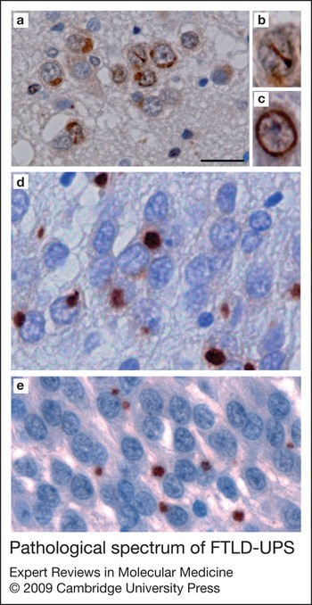
Although initial data suggested that all FTLD-U cases are characterised by TDP-43 pathology, recent follow-up studies have demonstrated that there are some cases in which the pathological inclusions are only detectable with markers of the ubiquitin proteasome system (FTLD-UPS), in which the pathological ubiquitinated protein(s) is still unknown (Refs Reference Cairns100, Reference Hatanpaa104, Reference Pikkarainen, Hartikainen and Alafuzoff142, Reference Josephs143). The most common subtype of FTLD-UPS, representing 10–20% of all FTLD-U cases, was recently described in detail (Refs Reference Mackenzie144, Reference Roeber145). The highly unusual and consistent clinical and pathological features of these cases suggest that they represent a newly recognised, discrete entity and the term atypical FTLD-U (aFTLD-U) was introduced. The clinical phenotype is sporadic, early-onset FTD with severe progressive behavioural and personality changes in the absence of aphasia or significant motor features. In addition to frontotemporal atrophy, severe degeneration of the anterior striatum and hippocampal sclerosis are common findings in aFTLD-U. TDP-43-negative, ubiquitin-positive and p62-positive NCIs are most abundant in the frontal and temporal neocortex and hippocampus (Fig. 4a). However, the most intriguing pathological feature is the presence of NIIs in the dentate granule cells and cortical pyramidal neurons with a unique morphology, where they appear as elongated straight or curved bars or thick twisted filaments (Fig. 4b, c). The immunohistochemical profile of these NIIs is also unusual and different from the NCIs, because they are reactive for ubiquitin but not for p62 (Fig. 4d).
Pathological spectrum of FTLD-UPS. An aFTLD-U case with ubiquitin-immunoreactive cytoplasmic (a) and intranuclear inclusions (b, c) in dentate granule cells. (d) The cytoplasmic inclusions are also immunoreactive for p62 but the intranuclear inclusions are not. (e) Familial FTD caused by CHMP2B mutations (FTD-3) also have ubiquitin-positive cytoplasmic inclusions in the dentate granule cells. a–c, e, ubiquitin immunohistochemistry; d, p62 immunohistochemistry. Scale bar: 20 µm (a, d, e); 10 µm (b, c).

Familial FTD linked to chromosome 3 (FTD-3), due to mutations in CHMP2B (Ref. Reference Skibinski17) has also recently been shown to have FTLD-UPS pathology (Ref. Reference Holm146). So far, four families have been identified with different CHMP2B mutations (AD&FTD Mutation database: http://www.molgen.ua.ac.be/FTDmutations). CHMP2B belongs to the chromatin-modifying protein/charged multivesicular body protein family and is a component of ESCRT-III (endosomal sorting complex required for transport III), a complex involved in the degradation of surface receptor proteins and the formation of endocytotic multivesicular bodies. Although previous reports failed to detect abnormal protein deposits in brains of patients with FTD-3, a recent study demonstrated the presence of granular NCIs that were immunoreactive for ubiquitin and p62, but negative for TDP-43 and tau, predominantly in the dentate granule cells of the hippocampus (Fig. 4e) (Ref. Reference Holm146).
FTLD-IF
Neuronal intermediate filament inclusion disease (NIFID) is an uncommon neurological disorder (Ref. Reference Uchikado147) with pathology characterised by neuronal inclusions that are immunoreactive for all of the Class IV intermediate filaments (IFs) [light, medium and heavy neurofilament (NF) subunits and α-internexin] (Refs Reference Cairns148, Reference Cairns149). The typical clinical presentation is early-onset sporadic FTD, associated with a pyramidal and/or extrapyramidal movement disorder (Refs Reference Cairns148, Reference Yokota150, Reference Roeber151, Reference Molina-Porcel152, Reference Mackenzie and Feldman153, Reference Josephs154, Reference Josephs155, Reference Bigio156, Reference Cairns157). Additional clinical manifestations that have been reported include falls, dystonia, myoclonus, ophthalmoplegia, memory deficits, seizures, eating disorders and psychiatric symptoms. There have been two cases published with possible childhood onset (Refs Reference Roeber151, Reference Mackenzie and Feldman153) and two with a questionable family history (Refs Reference Molina-Porcel152, Reference Mackenzie and Feldman153).
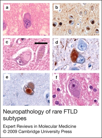
The neuropathological findings in NIFID are heterogeneous, but have a number of consistent features (Refs Reference Cairns148, Reference Yokota150, Reference Roeber151, Reference Molina-Porcel152, Reference Mackenzie and Feldman153, Reference Josephs154, Reference Josephs155, Reference Bigio156, Reference Cairns157). Chronic degenerative changes may affect a variety of cortical and subcortical regions, with the frontal and temporal lobes and caudate nucleus most consistently and severely involved. Several types of neuronal inclusions are found. The most common are small round Pick-body-like inclusions that are well-defined, slightly eosinophilic or basophilic and rarely argyrophilic (Fig. 5a). These are variably immunoreactive for ubiquitin, NF and α-internexin but negative for tau, α-synuclein and TDP-43 (Fig. 5b) (Ref. Reference Cairns149). The ultrastructure is a combination of electron-dense granular material and dispersed short filaments (Refs Reference Bigio156, Reference Mosaheb158, Reference Uchikado159). These are numerous in affected regions of cerebral neocortex, hippocampus and in select subcortical regions. Hyaline conglomerate inclusions are less frequent, and appear as irregular, multilobulated masses that often compress the nucleus (Fig. 5c). They are weakly eosinophilic, with a glassy, filamentous appearance and sometimes have a dense, brightly eosinophilic core. These stain intensely with the Bielschowsky silver method and are more consistently and intensely immunoreactive for IFs (Fig. 5d). The ultrastructure is compact, interlacing bundles of filaments (Ref. Reference Mosaheb158). NCI with other morphologies may also be present, and there are often IF-immunoreactive swollen axons. A less consistent feature is the presence of eosinophilic NIIs that are intensely immunoreactive for ubiquitin but generally not reactive for IFs (Fig. 5e) (Refs Reference Cairns149, Reference Mackenzie and Feldman153, Reference Josephs154, Reference Mosaheb158).
Neuropathology of rare FTLD subtypes. (a–d) Neuronal intermediate filament inclusion disease. The most common type of inclusion is Pick-body-like inclusions that are visible with haematoxylin and eosin stain (a) and immunoreactive for α-internexin (b). Hyaline conglomerate inclusions are also present (c) and are immunoreactive for phosphorylated neurofilament (d). (e) Neuronal intranuclear inclusions that are present in some cases of FTLD-IF are strongly immunoreactive for ubiquitin but do not stain for intermediate filaments. Note the presence of a Pick-body-like inclusion in the cytoplasm of the same neuron. (f) Typical inclusion of basophilic inclusion body disease. Scale bar: 20 µm (a, c–f); 40 µm (b).

Neuronal IFs include the triplet of light (68 kDa), medium (145 kDa) and heavy (200 kDa) NF subunits, α-internexin and peripherin (Ref. Reference Liu160). Based on sequence homology, the NF proteins and α-internexin have been grouped together as Class IV IFs (Ref. Reference Ching and Liem161). The expression of α-internexin precedes that of NFs during development and, in the adult brain, α-internexin is expressed at lower levels and in more restricted neuronal populations (Ref. Reference Fliegner162). The Class IV IFs can coassemble (Ref. Reference Ching and Liem163), and are part of the normal neuronal cytoskeleton, that is involved in maintaining cell structure and intracellular transport (Ref. Reference Lariviere and Julien164). The degree and pattern of phosphorylation of NFs is essential for normal intracellular localisation and function (Ref. Reference Liu160). The inclusions in NIFID are variably reactive with antibodies against phosphorylated and phosphorylation-independent epitopes of all three NF subunits; however, α-internexin immunohistochemistry is the most sensitive (Refs Reference Cairns148, Reference Cairns149). Although NF can be a minor component of the characteristic inclusion bodies of many different neurodegenerative conditions (including AD, PD, DLB and MND) (Ref. Reference Liu160), α-internexin immunoreactivity is relatively specific for NIFID (Ref. Reference Cairns165). Because the abnormal protein aggregates found in NIFID are immunoreactive for IFs but not for any other proteins currently known to be associated with neurodegenerative disease, the designation of NIFID or FTLD-IF is appropriate. However, the fact that IF proteins show no abnormal molecular modification in NIFID (Refs Reference Cairns149, Reference Mosaheb158, Reference Cairns165) and no pathogenic variants of the corresponding genes have been identified (Ref. Reference Momeni166), leaves open the possibility that some other protein might have a more central role in the pathogenesis of this condition (Refs Reference Yokota150, Reference Mackenzie and Feldman153).
BIBD
BIBD is a term that has been used for a small number of clinically and pathologically heterogeneous cases, in which the common finding is neuronal cytoplasmic inclusions that are basophilic with haematoxylin and eosin stain (Fig. 5f). The clinical phenotypes include sporadic ALS (Ref. Reference Kusaka, Matsumoto and Imai167), familial ALS (Ref. Reference Tsuchiya168), ALS with dementia (Refs Reference Yokota150, Reference Ishihara169, Reference Hamada170) and pure FTD (Refs Reference Yokota150, Reference Munoz-Garcia and Ludwin171). Although cases of BIBD with clinical FTD show chronic degeneration of the frontotemporal neocortex, the inclusions tend to be most numerous in subcortical regions, such as the basal ganglia and brainstem tegmentum (Refs Reference Yokota150, Reference Munoz-Garcia and Ludwin171). The inclusions are round, oval or crescentic, weakly argyrophilic and can be detected with histochemical stains for RNA (such as cresyl violet, methyl green pyronine and acridine orange). Immunohistochemical studies have found them to be negative for tau, α-synuclein, NF, α-internexin, tubulin, actin and TDP-43 (Refs Reference Cairns100, Reference Yokota150, Reference Ishihara169, Reference Munoz-Garcia and Ludwin171). Reports of ubiquitin immunoreactivity are inconsistent but some are positive for p62 (Ref. Reference Yokota150). The biochemical composition of the signature inclusions in BIBD is not presently known.
FTLD-ni
Until recently, most cases of tau-negative FTLD were thought to have no cellular inclusions and were classified as DLDH (Ref. Reference Knopman78). However, with the use of sensitive immunohistochemical techniques, most of these cases are found to have ubiquitin-immunoreactive pathology (previously designated FTLD-U) (Ref. Reference Mackenzie85) that is usually also reactive for TDP-43 (Ref. Reference Davidson99). Cases of true DLDH are now considered to be uncommon, and the existence of DLDH as a distinct entity has been questioned. The terminology that is now recommended for rare cases of FTLD in which no inclusions can be demonstrated with special histochemical stains or immunohistochemistry is FTLD-ni (no inclusions) (Ref. Reference Mackenzie22).
Other pathological causes of FTD
There are a number of rare neurodegenerative conditions, with unknown biochemical defects, that can present as FTD, such as hereditary diffuse leukoencephalopathy with spheroids (Ref. Reference Van Gerpen172) and neuronal intranuclear inclusion disease (Ref. Reference Munoz-Garcia and Ludwin173). In addition, most of the common neurodegenerative causes of dementia can occasionally fulfill clinical diagnostic criteria for FTD. Although the frequency of these ‘frontal variants’ and their overall contribution to FTD is difficult to determine, some studies have found more than 20% of clinical FTD to be associated with the pathology of AD, DLB, CJD or vascular disease (Refs Reference Kertesz174, Reference Forman175). The frontal variant of AD is best recognised and could account for 17% of all FTD (Refs Reference Kertesz174, Reference Forman175). Attempts to demonstrate a correlation between the regional severity of the AD pathology and the clinical phenotype have been inconsistent (Refs Reference Johnson56, Reference Taylor57, Reference Mesulam176). One recent study has described additional glial tau pathology in cases of AD with clinical PPA, suggesting a pathological overlap with FTLD-tau (Ref. Reference Munoz, Woulfe and Kertesz177). Another recent finding of interest is the presence of TDP-43 pathology in a significant proportion of AD and DLB cases (Refs Reference Uryu178, Reference Nakashima-Yasuda179, Reference Higashi180, Reference Arai181, Reference Amador-Ortiz182). Although this often has a restricted anatomical distribution, it is sometimes extensive and might closely resemble FTLD-TDP (Ref. Reference Arai181). Whether or not this additional pathology alters the clinical phenotype is yet to be determined.
Outstanding research questions
Despite the enormous gains made over the past few years in our understanding of the molecular basis of FTD, many fundamental issues are still unresolved. The FTLD gene on chromosome 9 remains to be identified and there is probably at least one other genetic cause of familial FTLD-tau. The normal functions of PGRN and TDP-43 in the nervous system need to be clarified, as do the mechanisms by which mutations in GRN, VCP and TARDBP lead to TDP-43 pathology. The possible role of GRN and TARDBP genetic variants in other (non-FTLD) neurodegenerative disease requires further investigation. The development of new animal models, particularly of FTLD-TDP, should help to answer these questions and will be crucial in the development of targeted therapies. Efforts to develop in vivo biomarkers that distinguish different subgroups of FTLD and aid in diagnosis and monitoring of disease are still preliminary (Refs Reference Ghidoni183, Reference Steinacker184, Reference Foulds185, Reference Tolnay186). Despite the enormous amount of work that lies ahead, recent discoveries have greatly improved our ability to offer meaningful genetic counselling for FTLD families and bring us much closer to developing useful diagnostic tests and rational therapies.
Acknowledgements and funding
This work was supported by funding from Stavros-Niarchos Foundation and the Synapsis Foundation (M.N.), the Swiss National Science Foundation (M.T.), the Canadian Institutes for Health Research and the Pacific Alzheimer Research Foundation (I.R.A.M.). The authors would like to thank the peer reviewers of this work for their constructive comments.