Non-technical Summary
Dasycladales are a group of green algae with a rich and varied fossil record, thanks to the mineralized aragonite skeleton. We present two new Dasycladales from the Eastern Carpathian (Romania) belt. The presence of several growth stages within the fossil sample allowed us to shed light on the poorly known growth of typical Dasycladales.
Introduction
Extant dasycladaleans follow growth stages related to the structure of the alga. Valet (Reference Valet1968) organized distinctive dasycladalean ontogenetic patterns into three groups: (1) uncalcified or poorly calcified plants with similar simple whorls (Dasycladus and Batophora); (2) calcified plants with whorls of a single type having secondary laterals forming a cortex (Neomeris, Bornetella, and Cymopolia); and (3) calcified plants with two types of whorls (Halicoryne and Acetabularia).
Some extant dasycladalean genera have representatives tracing back to the Cretaceous, and a “modern” type of reproduction (external gametophores) has been recorded since the Late Triassic (Barattolo et al., Reference Barattolo, Romano and Conrad2021b), so it is conceivable that extant species growth patterns can be confidently assigned to fossil Dasycladales, at least since the Mesozoic. Mineralization in fossil Dasycladales may cover almost the entire thallus or part of it (i.e., the vegetative or the fertile regions). Moreover, mineralization may appear either early or late during ontogeny, so that the fossilized calcareous skeletons reflect the onset and extent of mineralization of the alga during growth. The correct recognition of the preserved region, the occurrence of the apex and the bottom of the thallus may thus help interpreting the original morphology of the fossil algae. Herein, we describe two new Early Cretaceous dasycladalean species from the Rarău Syncline (Romania), which allow us to recognize and study key characters as they appear during ontogeny.
Geological setting
The Rarău Syncline is located in the northern part of the Eastern Carpathians and belongs to Median Dacides (Săndulescu, Reference Săndulescu1984, Reference Săndulescu2014) or Dacia Mega-unit (Schmid et al., Reference Schmid, Fügenschuh, Georgiev, Kounov and Mațenco2020, and references therein).
The Median Dacides sensu Săndulescu (Reference Săndulescu1984) or the informal “Crystalline Mesozoic Zone” roughly originated from European continental crust, which evolved during the Triassic–Cretaceous between the East Vardar Branch of the Neotethys (“Main Tethys Ocean Branch”) and the Alpine Tethys (“Ceahlău-Severin Ocean”). They are built by two kinds of nappes: one autochthonous (Bucovinian, Sub-Bucovinian, and Infra-Bucovinian nappes), and another allochthonous (Transylvanian nappes). The Bucovinian nappes are largely built by a basement of meso- and epimetamorphic rocks and sedimentary covers, deriving from European continental crust. In the Rarău Syncline, only the Bucovinian Nappe (Turculeț, Reference Turculeţ1971) is largely cropping out (Fig. 1), the Infra-Bucovian nappes are supposed to occur in the surroundings of Sadova (Săndulescu, Reference Săndulescu1976). The allochthonous Transylvanian nappes (Olt, Perşani, and Hăghimaş areas; Săndulescu, Reference Săndulescu1984) derive tectonically from the oceanic crust of the East Vardar Branch of the Neotethys and its sediments. They are scattered as various-sized sedimentary klippen and volcanic tectonic outliers embedded in the Lower Cretaceous wildflysch ending with Mesozoic sedimentation in the Bucovinian Nappe. The age of the klippen ranges from Triassic to Early Cretaceous (Turculeț, Reference Turculeţ2004a, Reference Turculeţb, and references therein).
Location of the studied material. (1), Location of the Rarău Mountains, Eastern Carpathians (Romania); (2), geological map of the environs of Câmpulung Moldovenesc (Suceava County, Romania). The study site is situated in the hanging wall of a NE-verging thrust forming part of a leading imbricate fan.

The sample with the dasycladal material presented here was collected at the front of a Lower Cretaceous klippe that is partially embedded in the wildflysch. It occurs on the Valea Seacă Stream, a tributary of the Moldova River (Fig. 1). The stream springs from the southeastern part of the Cretaceous wildflysch, obliquely crossing a large part of the wildflysch and the entire succession of the external flank syncline (e.g., Filipescu and Dragastan, Reference Filipescu and Dragastan1963; Popescu and Patrulius, Reference Popescu and Patrulius1964; Mutihac, Reference Mutihac1968; Turculeț, Reference Turculeţ1971; Săndulescu, Reference Săndulescu1976, etc.) (Fig. 1).
The Valea Seacă Stream is the locus-typicus of several new fossil taxa. From the various-sized blocks embedded in the wildflysch (klippen/olistoliths), Volz (Reference Volz1903) and Morycowa (Reference Morycowa1971) proposed several coral species and Patrulius (Reference Patrulius1965) proposed a new species of calcareous sponges. Also, the informal “Aptychus Beds” of the syncline flank provided the holotypes for several Late Jurassic Aptychus parataxa (e.g., Turculeț, 2004b, and references therein). Blocks from Valea Seacă-Rarău Peak were ascribed to the Barremian–Aptian (Popescu and Patrulius, Reference Popescu and Patrulius1964, and references therein) or recently to the late Barremian–early Aptian (Bucur et al., Reference Bucur, Kołodziej, Săsăran, Bucur and Filipescu2007) or early Aptian (Bedoulian) (Kołodziej et al., Reference Kołodziej, Golubic, Bucur, Radtke and Tribollet2012; Pleş et al., Reference Pleş, Bârtaş, Chelaru and Bucur2017) based on the presence of the orbitolinids Montseciella arabica (Henson, Reference Henson1948), Palorbitolina lenticularis (Blumenbach, Reference Blumenbach1805), and Rectodictyoconus giganteus Schroeder, Reference Schroeder1964.
The sample belongs to the wildflysh deposits near the contact with the so-called “Aptychus Beds” of the eastern syncline flank. There are many sedimentary blocks of various sizes in the riverbed or on the two slopes. Many blocks have metric dimensions, several being bigger (3–4 × 1–1.5 m and the largest ones reach more than 7 × 5.50 m). The blocks are massive and are composed of sandstone and limestone (mainly wackestone). Two types of Lower Cretaceous blocks previously described by Popescu and Patrulius (Reference Popescu and Patrulius1964) are frequently recognized: (1) “Reef limestone” or “Limestone with Chatetopsis zonata Patrulius, Reference Patrulius1965” and (2) “Limestone with rudists.” Other limestone types (mainly bioclastic grainstone) are also present as different remnants of a former carbonate platform.
Material and methods
The sample consists of a small boulder collected from the right side of Valea Seacă Stream in April 2013. The limestone block is a floatstone to rudstone with large fragments of rudists, sponges, and corals, rare echinoderms and gastropod fragments, Carpathocancer sp., calcareous algae, foraminifera, as well as many other small bioclasts. Micritic matrix is dominant, but an area with sparry calcite cement is also present. The large bioclasts are intensively bored, probably by clionid sponges. The low rate of sedimentation favored the growth of different crusts on the large bioclasts or on the substratum. The crusts consist of Coscinophragma or Bdelloidina foraminifera, Sporolithon red algae, different sponges, Koskinobulina socialis Cherchi and Schroeder, Reference Cherchi and Schroeder1979, Crescentiella sp., and microbial micrite. The assemblage of dasycladalean algae and foraminifera associated with the two new species described in this paper are represented by: Salpingoporella pygmaea (Gümbel, Reference Gümbel1891); Terquemella sp.; Triploporella? sp.; Protopeneroplis cf. P. ultragranulata (Gorbachik, Reference Gorbachik1971); Charentia cuvillieri Neumann, Reference Neumann1965; Nautiloculina bronnimanni Arnaud-Vanneau and Peybernès, Reference Arnaud-Vanneau and Peybernes1978; Melathrokerion praesigali (Banner, Reference Banner1966); Vercorsella scarsellai (De Castro, Reference De Castro1963); Pseudolituonella gavonensis Foury, Reference Foury1968; Derventina filipescui Neagu, Reference Neagu1968; Frentzenella odukpaniensis (Dessauvagie, Reference Dessauvagie1968); Ichnusella sp.; Neotrocholina sp.; Everticyclammina sp.; Ammobaculites sp.; Bolivinopsis sp.; Arenobulimina sp.; and Bullopora sp.
Most of the algal specimens described here occur in 58 thin sections of limestone, 42 labelled PSM-RT1 through PSM-RT42 and 16 labelled DiSTAR-BA.4283.1 through DiSTAR-BA.4283.16. A sample is also stored in Iaşi and labelled RS 8228 Museum of Paleontology, Original Collections (MP-UAIC). The algal specimens were observed under an optical stereomicroscope, then photographed and measured under a petrographic microscope.
Repositories and institutional abbreviations
PSM, Palaeontology-Stratigraphy Museum in the Geology Department of the Babeş-Bolyai University, Cluj-Napoca (Romania); DiSTAR, Department of Earth Sciences Environment and Resources, University of Naples Federico II; MP-UAIC, Museum of Palaeontology, Department of Geology, University Alexandru Ioan Cuza, Iaşi, Romania.
Morphogenesis of living Dasycladaleans and implications for interpreting growth stages of Suppiluliumaella schlagintweitii new species and Triploporella loducai new species
Morphogenesis of dasycladaleans follows steps related to the structure of the alga, which Valet (Reference Valet1968) assigned to three different groups: (1) uncalcified or poorly calcified plants with single type of simple whorls (Dasycladus and Batophora); (2) calcified plants with a single type of whorls having secondary laterals forming the cortex (Neomeris, Bornetella, and Cymopolia); and (3) calcified plants with two types of whorls (Halicoryne and Acetabularia). Group “1” is expected to occur in fossil Dasycladales-like thyrsoporellids (Radoičić and Özgen-Erdem, Reference Radoičić and Özgen-Erdem2011) and triploporellid-like Selliporella Sartoni and Crescenti, Reference Sartoni and Crescenti1963 (Barattolo et al., Reference Barattolo, Amodio, Bucur and Martino2021a). Growth type “3” pertains to fossil Acetabularia Lamouroux, Reference Lamouroux1812 (Barattolo et al., Reference Barattolo, Ionesi and Ţibuleac2019), and other genera such as Clypeina Michelin, Reference Michelin1845, and Aloisalthella Granier in Granier and Lethiers, Reference Granier and Lethiers2019 (Granier and Lethiers, Reference Granier and Lethiers2019). In contrast, the structure of the vegetative portions we found in Suppiluliumaella schlagintweitii n. sp. and Triploporella loducai n. sp. are best ascribed to group “2” growth type (i.e., plants forming whorls having secondary laterals forming cortex, as found in Neomeris and Bornetella).
Valet (Reference Valet1968) observed that the growth of club-shaped Bornetella is very close to that of Neomeris, as it is characterized by a non-branched simple thallus. Conversely, species of Bornetella displaying spherical thalli, follow a different type of growth. The morphogenesis in Neomeris has been described by several botanists (Cramer, Reference Cramer1887, Reference Cramer1890; Solms-Laubach, Reference Solms-Laubach1893; Church, Reference Church1895; Svedelius, Reference Svedelius1923; Egerod, Reference Egerod1952; and Valet, Reference Valet1968). Following Church (Reference Church1895), we summarize the growth of Neomeris and club-shaped Bornetella as passing through five consecutive stages: (1) an erect coenocytic filament rises up from the substratum (Fig. 2.1), then whorls of dichotomously branched laterals originate at its apex and delimit from the main axis by perforated septa (Fig. 2.2); a cap protects each incipient apical whorl (Mantelkappe by Cramer, Reference Cramer1890, p. 37–39; velum pectique by Valet, Reference Valet1968, p. 34–35). (2) The first-order lateral begins to be subdivided into an inner and an outer tract by means of a typical perforated septum formed as a ring-wall (Fig. 2.3, from the fifth whorl upward). (3) The inner tract starts to dilate, forming a pseudo-cortex (Fig. 2.3, from seventh whorl upward). (4) The fourth stage involves the development of an elongated first-order lateral bearing secondary laterals. The inner portions of these secondary laterals are separated from the outer portions by a perforated septum and then become swollen vesicles and repeat the cortex formation (Fig. 2.4, from seventh whorl upward). This stage represents the most important vegetative phase of the plant. (5) The fifth stage (not represented in Fig. 2) is characterized by the appearance of reproductive organs (distal gametophores).
Early developmental stages of extant calcified, not moniliform Dasycladales with similar whorls having secondary laterals forming cortex (Neomeris, Bornetella). (1) An erect coenocytic filament rises up from the substratum; (2) whorls of dichotomously branched laterals originate at its apex, delimited from the main axis by perforated septa; (3) first-order lateral begins to be subdivided into an inner and outer tract by means of a typical perforated septum formed as a ring-wall (from fifth whorl upward); the inner tract exhibits a tendency to dilate, thus forming a pseudo-cortex (from seventh whorl upward); (4) development of an elongated first-order lateral bearing secondary laterals (from seventh whorl upward). The inner portions of these secondary laterals are separated from the outer portions by a perforated septum and then become swollen vesicles and repeat the cortex formation. This fourth stage represents the most important vegetative phase of the plant. (5) After the coenocytic filament is formed, a first whorl of squat primary laterals appears; (6) the alga first produces a few simple whorls of primary laterals, then others with two orders of laterals; (7) soon primary laterals of new whorls lose secondary laterals and inflate, thus producing a pseudo-cortex (sixth to eighth whorl upward); then the alga produces whorls where secondary laterals swell and form a true cortex (from ninth whorl onwards). Sketches (1–4) based on Church (Reference Church1895), (5–7) based on Valet (Reference Valet1968, pl. 9). Scale bar for (1–4) represents 2.0 mm, scale bar for (5–7) represents 0.5 mm.

According to Valet (Reference Valet1968), who first described the growth of Bornetella, spherical thalli grow differently: (1) incipient whorls are not protected by a cap; (2) whorls consist of laterals that are not ramified over the second order; and (3) the growth unfolds faster. Morphogenesis of spherical Bornetella can be sketched-out as follows: (1) formation of an erect coenocytic filament, as in the previous scheme (Fig. 2.1). (2) A first whorl of squat primary laterals appears (Fig. 2.5). (3) The alga first produces a few simple whorls of primary laterals, then others with two orders of laterals (Fig. 2.6). (4) Soon the primary laterals of new whorls lose secondary laterals and inflate, thus producing a pseudo-cortex (Fig. 2.7, sixth to eighth whorls upward). (5) The alga produces whorls where secondary laterals become swollen and form a true cortex (Fig. 2.7, from nineth whorl onwards). (6) Growing upward the alga generates other whorls whose primary laterals greatly elongate to form a spherical thallus (not represented in Fig. 2).
Ontogeny and mineralization
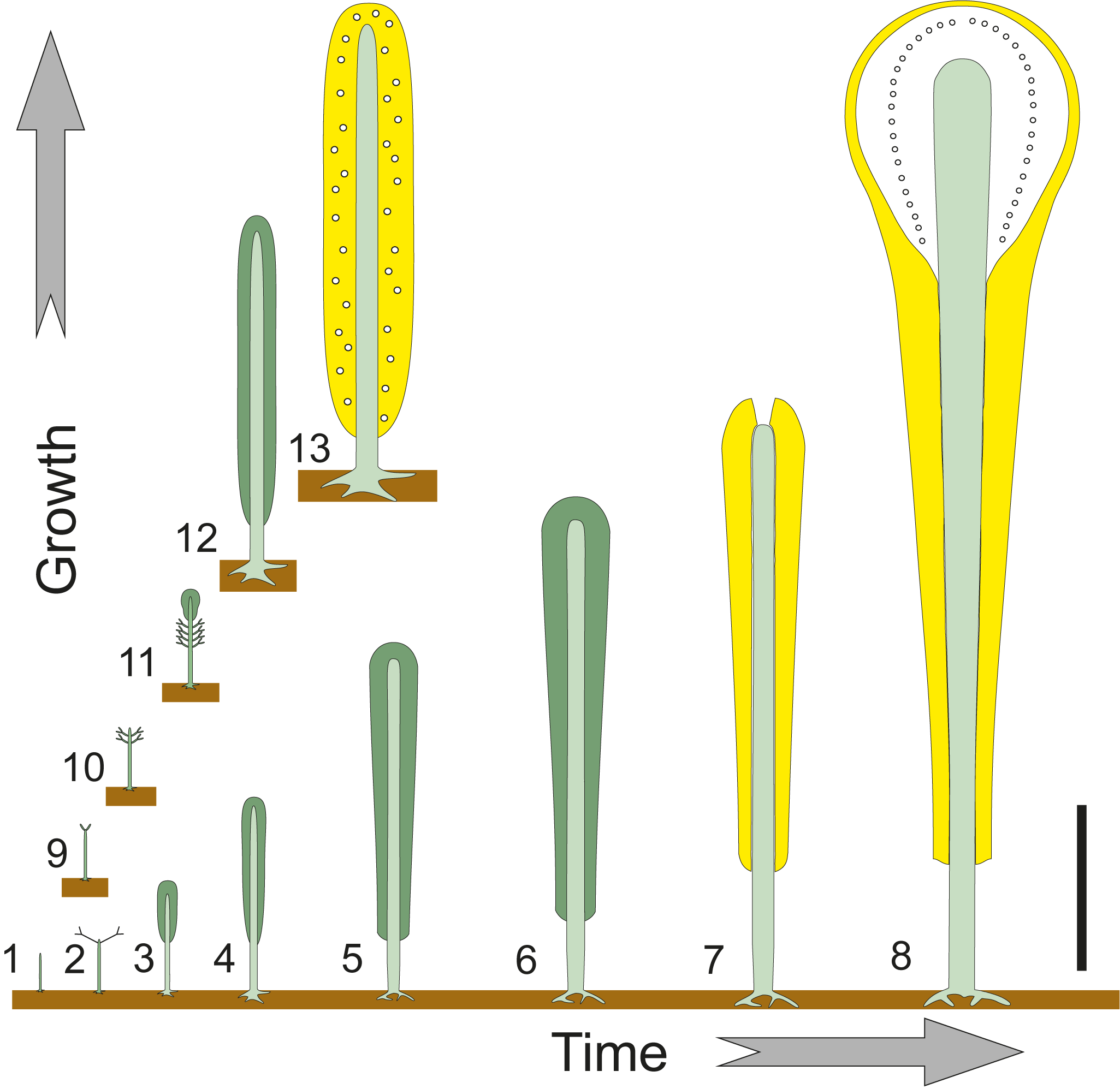
The fertile region of Suppiluliumaella schlagintweitii n. sp. can be estimated as one-third to one-fourth of the whole calcified thallus. Conversely, Triploporella loducai n. sp. displays a thallus quite completely represented by fertile whorls, like in mature thalli of spherical Bornetella where all whorls are fertile. Therefore, we can assume that the two species attained different rates of growth, faster for T. loducai n. sp. (Fig. 3.1, 3.9–3.13) and slower for S. schlagintweitii n. sp. (Fig. 3.1–3.8).
Development and mineralization. (1–8) Suppiluliumaella schlagintweitii n. sp., possible young uncalcified growth stages (1–6), calcified immature stage (7), and calcified mature stage (8); uncalcified reproductive organs are depicted with white circles, laterals are omitted. (1, 9–13) Triploporella loducai n. sp., possible young uncalcified growth stages (1, 9–12) and calcified mature stage (13); uncalcified reproductive organs are depicted with white circles, laterals are omitted. The growth rate of Triploporella loducai n. sp. was probably faster than that of Suppiluliumaella schlagintweitii n. sp. Scale bar represents 2.0 mm.

As for the time and location of mineralization, the two species behave differently. Suppiluliumaella schlagintweitii n. sp. displays specimens with a bud-like, Neomeris-type apex (immature specimens) and others with a “head” (mature specimens). This allows us to infer that mineralization enveloped the thallus well before the alga reached its fertile stage. The development of Suppiluliumaella schlagintweitii n. sp. can be reconstructed with several uncalcified vegetative stages (Fig. 3.1–3.6), as in Neomeris, followed by others calcified until maturation (Fig. 3.7 and 3.8). All specimens of Triploporella loducai n. sp. exhibit large, swollen primary laterals except at the ends. This suggests the specimens were mature, therefore mineralization occurred only in this phase. The transition from the early stages of growth (Fig. 3.1, 3.9–3.11) to the mature stage (Fig. 3.13) could have followed two different trajectories: (1) either with whorls rapidly appearing with large primary laterals followed by whorls added until the thallus is adult, so that the reproductive organs take place within the large laterals; or (b) simple whorls of two orders are formed until the thallus is adult, then primary laterals swell to host reproductive organs. Figure 3.12 depicts this second hypothesis.
Systematic paleontology
Order Dasycladales Pascher, Reference Pascher1931
Family Triploporellaceae (Pia, Reference Pia1920) Berger and Kaever, Reference Berger and Kaever1992
Tribe Triploporelleae (Pia, Reference Pia1920) Bassoullet et al., Reference Bassoullet, Bernier, Deloffre, Génot, Jaffrezo and Vachard1979
Genus Suppiluliumaella Elliott, Reference Elliott1968
Type species
Suppiluliumaella polyreme Elliott, Reference Elliott1968; Lower Cretaceous, 16 km SSE of Amasya (Black Sea Region, Turkey).
Other species
Suppiluliumaella elliotti Bakalova, Reference Bakalova1971; S. praebalcanica Bakalova, Reference Bakalova1971; S. alimani (Dragastan, Reference Dragastan1978) Dragastan, Reference Dragastan1989; S. tuberifera (Sokač and Nikler) in Granier and Deloffre, Reference Granier and Deloffre1993; S. erikfluegeli Dragastan, Reference Dragastan1989; and S. schroederi Barattolo, Reference Barattolo1984.
Diagnosis
According to Elliott (Reference Elliott1968) the alga is characterized by a cylindrical thallus bearing whorls of thin primary laterals, each terminating with a large conspicuous swelling. Short phloiophorous secondary laterals. Strong calcareous skeleton.
Remarks
Sokač and Nikler (Reference Sokač and Nikler1973) described three new species assigned to a new genus, Montenegrella. The authors considered as the main criterion for separating Montenegrella from Suppiluliumaella Elliott, Reference Elliott1968, the presence of secondary laterals that start approximately from a common point and that present an obvious narrowing at the contact with the primary lateral. Barattolo (Reference Barattolo1985) showed that such characteristics can derive from differences in the calcification process of the thallus, which calls into question their systematic value. As such, Montenegrella may represent a junior synonym of Suppiluliumaella. Since the description of Suppiluliumaella polyreme in 1968, 15 more species belonging to Suppiluliumaella/Montenegrella have been described: Suppiluliumaella praebalcanica Bakalova, Reference Bakalova1971 (Barremian–lower Aptian); S. elliotti Bakalova, Reference Bakalova1971 (Barremian–lower Aptian); S. gocanini Radoičić, Reference Radoičić1972 (lower Valanginian); Montenegrella tuberifera Sokač and Nikler, Reference Sokač and Nikler1973 (Barremian–Aptian); M. verae Sokač and Nikler, Reference Sokač and Nikler1973 (Barremian–Aptian); M. fustiformis Sokač and Nikler, Reference Sokač and Nikler1973 (Barremian–Aptian); M. corbarica Jaffrezo, Reference Jaffrezo1975 (Huterivian?–Barremian); M. florifera Bernier, Reference Bernier1978 (upper Valanginian); Suppiluliumaella erikfluegeli Dragastan, Reference Dragastan1978 (upper Valanginian); Montenegrella transdanubica Dragastan, Reference Dragastan1978 (upper Valanginian); M. alimani Dragastan, Reference Dragastan1978 (upper Valanginian); Suppiluliumaella schroederi Barattolo, Reference Barattolo1984 (Cenomanian); S. leini Dragastan, Reference Dragastan1989 (upper Oxfordian–Kimmeridgian); S. riedeli Dragastan, Reference Dragastan1989 (upper Oxfordian–Kimmeridgian), and Montenegrella? gracilis Sokač and Grgasović, Reference Sokač and Grgasović2015 (upper Barremian).
Suppiluliumaella gocanini was transferred by Radoičić (Reference Radoičić2005) to the new genus Zujovicella. Of the three species initially described by Dragastan in 1978 (incorrectly designated holotypes), Suppiluliumaella erikfluegeli and Montenegrella alimani have been validated by the author (Dragastan, Reference Dragastan1989) by designating a lectotype. Dragastan, Reference Dragastan1989, transferred M. alimani to Suppiluliumaella (Dragastan, Reference Dragastan1978). The third species, Montenegrella transdanubica, was considered by Dragastan (Reference Dragastan1989) a junior synonym of Suppiluliumaella erikfluegeli. Barattolo (Reference Barattolo1985) proposed that all three taxa described by Dragastan (Reference Dragastan1978) possibly pertained to a single species.
Suppiluliumaella praebalcanica Bakalova, Reference Bakalova1971, was transferred by Dragastan (Reference Dragastan1978) to the genus Trinocladus since it has three orders of laterals. Bucur (Reference Bucur1994, p. 155) showed that (translated from French): “The transfer to the genus Trinocladus starting from this characteristic (Dragastan, Reference Dragastan1978, Reference Dragastan1989) does not seem necessary to me, the shape of the thallus and the structure of the ramifications of Trinocladus being different (see Bassoullet et al., Reference Bassoullet, Bernier, Conrad, Deloffre and Jaffrezo1978, p. 281). On the other hand, the emendation of the generic diagnosis suggested by the same authors (Bassoullet et al., Reference Bassoullet, Bernier, Conrad, Deloffre and Jaffrezo1978) is necessary to include the existence of third-order ramifications.”
Montenegrella corbarica Jaffrezo, Reference Jaffrezo1975, was excluded by Barattolo (Reference Barattolo1985) from the genus Suppiluliumaella, because it lacks the primary laterals differentiated into a pedunculate proximal portion and a widened distal one.
Montenegrella florifera Bernier, Reference Bernier1978, has primary laterals whose shape is almost unknown, so its assignment to Suppiluliumaella was considered by Barattolo (Reference Barattolo1985) as uncertain; therefore, M. florifera is not considered here within Suppiluliumaella.
Suppiluliumaella leini Dragastan, Reference Dragastan1989, and Suppiluliumaella riedeli Dragastan, Reference Dragastan1989 (Dragastan, Reference Dragastan1989), were described on extremely poor material, with a high degree of recrystallization and with morphological features indicating the genus Salpingoporella (see also Carras et al., Reference Carras, Conrad and Radoičić2006). Moreover, the reconstruction of the laterals sketched by Dragastan (Reference Dragastan1989, pl. 9.6 and 9.9) does not correspond to the genus Suppiluliumaella, but rather to Steinmanniporella.
Suppiluliumaella minuta Masse, Arias, and Vilas, Reference Masse, Arias and Vilas1993, does not show secondary laterals, and, therefore, cannot be ascribed to the genus Suppiluliumaella, the taxon is tentatively assigned to Holosporella Pia, Reference Pia1930.
The primary laterals in the species Montenegrella? gracilis Sokač and Grgasović, Reference Sokač and Grgasović2015, display that the swollen distal part is not well differentiated from the inner thinner part, which excludes the species from the genus Suppiluliumaella.
Suppiluliumaella schlagintweitii new species
Possible growth of Suppiluliumaella schlagintweitii n. sp. (1–4) Early stages like in Neomeris and club-shaped Bornetella. (5) Reconstruction of a juvenile immature thallus. (6) Reconstruction of the adult thallus. Scale bar for (1–4) represents 1.0 mm like scale bar for (5, 6).

Suppiluliumaella schlagintweitii n. sp., upper Barremian, Valea Seacă, 3 km SSW of Câmpulung Moldovenesc (Suceava County, Romania). (1) Oblique section of a small-sized specimen (lower part of the thallus), DiSTAR-BA.4283.4, paratype; (2) oblique section of a small-sized specimen, DiSTAR-BA.4283.16, paratype, distal part of primary laterals is not well differentiated; (3) oblique section, PSM-RT10, paratype; (4) transversal section, PSM-RT25, paratype; (5) oblique section, DiSTAR-BA.4283.4, paratype; (6) oblique section, PSM-RT34, paratype; (7) oblique section, PSM-RT34, paratype; (8) oblique section, DiSTAR-BA.4283.12, paratype; (9) longitudinal section through the top of the juvenile thallus, DiSTAR-BA.4283.10, paratype, notice akrophorous primary laterals verging upwards near the top; (10) oblique section, PSM-RT23, paratype. Scale bar represents 0.5 mm for all figures.

Type material
Holotype: oblique section (Fig. 7.4), PSM-RT28. Paratypes: specimens contained in the thin sections, PSM-RT1 through PSM-RT42 and DiSTAR-BA.4283.1 through DiSTAR-BA.4283.16. The outcrop is located in the left slope of the Valea Seacă Stream, about 3 km SSW of Câmpulung Moldovenesc, coordinates 47°30′25″N, 25°32′33″E (Fig. 1).
Diagnosis
Simple, differentiated and club-shaped thallus. The primary laterals are moderately elongated and phloiophorous. Each primary lateral can be subdivided into two parts: (1) thin inner tract, ∼60–70% of the total length, nearly tubular, and (2) a swollen outer tract, globular. Small-sized specimens, probably corresponding to the lower vegetative part, either display tubular or modestly swollen outer primary laterals. Primary laterals are arranged in close whorls (euspondyl), 14–21 per whorl, and alternating in position between subsequent whorls. The inner tract is verging upright (20–30°) whereas the outer tract unfolds nearly orthogonally (70–80°). Phloiophorous secondary laterals are roughly orthogonal, rather thin but flaring outward to make a cortex distally. The upper fertile part of the thallus widens to form a subspherical “head” with several, thin phloiophorous laterals, probably of the third order. Reproductive organs are not observed. Most likely they were not mineralized and located in the subspherical part of the thallus (the “head”), probably in the swollen tract of primary laterals (cladosporous type). The mineralized skeleton extends from next to the central stem to the distal outer part of the secondary laterals before the cortical widening. Mineralization of the “head” is reduced to a thin wall around the outer part of tertiary laterals. Biometric data are shown in Table 1.
Suppiluliumaella schlagintweitii n. sp. Table of main biometrical parameters. d = inner diameter; D = outer diameter; d/D = ratio between inner and outer diameter; l = length of the primary lateral; p = width of the primary lateral; l′ = length of the secondary lateral; p′ = width of the secondary lateral; h = height between whorls; w = number of primary laterals per tuft; w′ = number of secondary laterals per tuft. Values of h and w from oblique sections are mathematically inferred. All dimensions are in millimeters

Occurrence
Type locality and horizon only.
Description
The calcareous sleeve is simple, not articulated, club-shaped and differentiated in a long conical lower part (“stalk”) and a rounded subspherical top (“head”). Although a complete longitudinal section has not been observed, it is likely that the lower part was rather long and moderately increasing in size upwards as evinced by the eccentric inner ellipse in oblique sections (Fig. 5.1 and 5.10). The inner surface is rarely regular and sharp (Fig. 5.4), commonly slightly irregular (Fig. 5.6 and 5.7), less frequently very irregular (Fig. 6.3 and 6.7). This variety of shapes can be interpreted with a mineralization occasionally in contact with the central stem, but usually next to the main axis. Calcification occupies the area between laterals (external mineralization), from the inner part (Fig. 5.6 and 5.10) to the distal end of the primary laterals and the greatest part of secondary laterals just before the cortical swelling (Fig. 6.1 and 6.7). Sometimes mineralization encrusts the secondary laterals for a small tract (Fig. 5.5 and 5.10). Therefore, the outer surface appears more or less regular depending on the extent of mineralization. In any case, the outer surface lacks a regular ornamentation. The upper subspherical part of the thallus (the “head”) displays very delicate, thin-walled calcification (Fig. 7.2, 7.3, 7.7) crossed by thin tubular pores flaring outwards, thus forming the “empty room” (Fig. 7.3; sensu Barattolo, Reference Barattolo2019). The transition between the stalk and the head is obtained by a mineralization tapering upward and outwards (Fig. 7.2; i.e., a “fitting sleeve” sensu Barattolo (Reference Barattolo2019, p. 3, fig. 6).
Suppiluliumaella schlagintweitii n. sp., upper Barremian, Valea Seacă, 3 km SSW of Câmpulung Moldovenesc (Suceava County, Romania). (1) Transversal-oblique section, DiSTAR-BA.4283.5, paratype; (2) transversal section, DiSTAR-BA.4283.6, paratype; (3) oblique section, DiSTAR-BA.4283.5, paratype, annular, uncalcified channel as in the specimen in Figure 8.5; (4) oblique section, DiSTAR-BA.4283.12, paratype, calcareous skeleton reaches to the cortex, outer tract aligned along the lateral axis; (5) oblique section, DiSTAR-BA.4283.15, paratype; (6) detail of a tangential section showing secondary laterals distally packed, DiSTAR-BA.4283.1, paratype; (7) transversal-oblique section of a large specimen (upper part of the vegetative region), DiSTAR-BA.4283.11, paratype; (8) oblique section of a large specimen (upper part of the vegetative region), DiSTAR-BA.4283.1, paratype. Scale bar represents 0.5 mm for all figures.

Suppiluliumaella schlagintweitii n. sp., upper Barremian, Valea Seacă, 3 km SSW of Câmpulung Moldovenesc (Suceava County, Romania). (1) Tangential-oblique section cutting the lower-middle part of the vegetative region, DiSTAR-BA.4283.3, paratype; in the upper part primary laterals are swollen distally, conversely in the lower part they are not well differentiated; (2) longitudinal section through the upper part of the thallus, PSM-RT37, paratype, showing the thin-walled “head”; (3) oblique section though top of the thallus (“head” or fertile region), below the vegetative region cut tangentially, DiSTAR-BA.4283.2, paratype; (4) oblique section though the upper part of vegetative region, PSM-RT28, holotype; (5) tangential section, PSM-RT24, paratype; (6) tangential section, PSM-RT37, paratype; (7) detail of an oblique section of the fertile region (“head”), PSM-RT27, paratype; pores through the thin wall seemingly represent tertiary laterals; (8) detail of an oblique section though the upper part of vegetative region, DiSTAR-BA.4283.1, paratype. Scale bar A represents 0.5 mm (7, 8), scale bar B represents 1.0 mm (1–6).

The primary laterals are set in close whorls (Fig. 7.4 and 7.5), with 14–21 per whorl. The distance between whorls is relatively low (0.20 mm). The arrangement is alternated in two subsequent whorls (Figs. 6.4, 6.5, 6.7, 6.8, 7.2). Phloiophorous primary laterals consist of a thin, long inner tract, tubular, followed by a swollen, globular outer tract. However, the shape and inclination of primary laterals change according to the specimen size. Small-sized specimens, probably corresponding to the lowermost vegetative part, display tubular primary laterals or with a modest swollen outer part, inclined at ~45° (Fig. 5.1 and 5.2). Primary laterals from medium- to large-sized specimens are more verging upright in their inner tract (20–30°) and nearly orthogonal in their outer tract (70–80°). Their initial part is not preserved because it is uncalcified, but the proximal part of the inner tract looks slightly larger than the middle part (Figs. 6.4 and 7.8). The ovoidal outer tract is sometimes not well developed (e.g., Figs. 6.8 and 7.8); other times better differentiated from the inner tract (Figs. 6.5 and 7.4). In this latter case, primary laterals are distally close packed sideways with a polygonal cross section (Fig. 7.4). Each primary lateral distally holds a tuft of 4–5 secondary laterals arranged in two rows (Fig. 6.6). Secondary laterals are moderately long (0.1–0.25 mm) and tubular in their inner part, then flaring outwards to make an external cortex (Fig. 6.1); they are rather thin (Figs. 5.5, 5.6, 6.1) to particularly strong (Figs. 5.4 and 6.3). Secondary laterals near the transition to upper part of the thallus (the “head”) appear to get longer (Fig. 7.2 and 7.8). Only the longitudinal section seen in Figure 7.2 is potentially useful to determine the order of ramification of pores crossing the thin calcified wall of the “head,” but its state of preservation of the key region (fitting sleeve; see Barattolo, Reference Barattolo2019, p. 3) does not allow us to understand whether they correspond to second- or third-order laterals. Nevertheless, we suspect they are third-order laterals (see discussion in supplemental data; Barattolo et al., Reference Barattolo, Bucur and Marian2021c). Such laterals appear to flare out distally and seemingly formed a cortex beyond the calcified wall.
No evidence of reproductive organs has been observed. However, the differentiated club shape of the thallus allows us to recognize the “head” as the fertile region; thus, all elements contained in the empty room (laterals and gametophores) were not mineralized. However, the globular outer tract of primary laterals was probably also present in those of the “head” and produced gametophores within them (cladosporous type).
Etymology
Dedicated to colleague Felix Schlagintweit for his contributions to the study of fossil calcareous algae.
Remarks
The new species reveals some aspects of morphogenesis in fossil Dasycladales, which are difficult to detect in fossil specimens. Characters of the calcareous skeleton allow reconstruction of a reliable thallus.
Growth stages in Suppiluliumaella schlagintweitii n. sp
Early stages of growth in Neomeris and Bornetella are not calcified, and seemingly the same happened in Suppiluliumaella schlagintweitii n. sp. Therefore, discussion about the early stages in fossil dasycladeans is somewhat speculative. However, the club shape of the thallus in S. schlagintweitii n. sp., the very elongated vegetative part below the fertile zone (“head”), and the occurrence of a cortex of secondary laterals all point to a Neomeris growth scheme (Fig. 4.1–4.4). The long calcified vegetative part records the changes of laterals from the base to the top (Fig. 4.6). The adult, fertile thallus ends with a swollen “head,” although a specimen ending without a “head” was found (drawing of Fig. 4.5, corresponding to photograph of Fig. 5.9), which is a very unusual occurrence in fossil material. This specimen may hardly represent a different species. Rather, it testifies to the occurrence, within the same population, of an immature stage of growth preceding the formation of the fertile “head.” Pores at the top of the head appear to be comparable to the rest of the head. This means that the apical whorls were fully developed, like in club-shaped Bornetella, and not close to form a sort of bud like in Neomeris. Fragmentation of the calcareous skeleton and random sectioning (transversal and oblique sections) do not allow us to assign the stalk either to mature or immature specimens if the apex of the thallus is missing.
Thallus reconstruction
The mineralized skeleton covers most of the thallus. The lower and middle tract of the calcareous sleeve is strongly calcified (vegetative region). Mineralization is close to the main axis, so that the reconstruction does not need a high degree of inference. The reconstruction of these parts is essentially based on some longitudinal and oblique sections (e.g., Figs. 5.1, 7.1, 7.2, 7.4). The reconstruction of the juvenile stage is based on Figure 5.9. Conversely, the fertile region is poorly calcified (calcareous shield). The inference of soft parts existing within the empty room is very high. The reconstruction, based on Figure 7.2, 7.3, and 7.8, therefore followed several assumptions: (1) the main axis enlarged moderately, as in the genus Bornetella (Berger and Kaever, 1991, Berger Reference Berger2006); (2) the number of primary laterals per whorl does not increase consistently and number of secondary laterals remains constant; (3) no new structures (e.g., external gametophores) appear; and (4) laterals grow more in length than in size. Accordingly, a possible reconstruction of the adult stage of the alga is shown in Figure 9.
Suppiluliumaella schlagintweitii n. sp. Reconstruction of the juvenile thallus (upper part, based on Fig. 5.9). (1) Reconstruction of the alga in axial section; yellow = calcified skeleton; gray = soft parts. (2) Axial view of the vegetative parts (green). Scale bar represents 0.5 mm.

Suppiluliumaella schlagintweitii n. sp. Reconstruction of the alga; yellow = extracellular calcification, gray = soft parts, green = vegetative parts. (1) Reconstruction of the alga in axial section. (2) Axial view of the vegetative parts. (3) Sector of a whorl projected on a transversal plane across the lower part of the thallus. (4) Upper view of whorl in the lower part of the thallus. (5) Sector of a whorl projected on a transversal plane across the middle part of the thallus. (6) Upper view of whorl in the middle part of the thallus. (7) Sector of a whorl projected on a transversal plane across the upper part of the thallus, the “head.” (8) Upper view of whorl in the upper part of the thallus, the “head.” Scale bar represents 1.0 mm.

Comparison
Considering the features described above, it follows that the genus Suppiluliumaella is represented by eight species. Sketches of all species based on the relevant holotype/lectotype are supplied in Figure 10. The species listed are Suppiluliumaella polyreme Elliott, Reference Elliott1968 (Fig. 10.13 and 10.14), which is the type species of the genus, S. elliotti Bakalova, Reference Bakalova1971 (Fig. 10.1 and 10.2), S. praebalcanica Bakalova, Reference Bakalova1971 (Fig. 10.3 and 10.4), S. alimani (Dragastan, Reference Dragastan1978) Dragastan, Reference Dragastan1989 (Fig. 10.11 and 10.12), S. tuberifera Sokač and Nikler in Granier and Deloffre, Reference Granier and Deloffre1993 (Fig. 10.15 and 10.16), S. erikfluegeli Dragastan, Reference Dragastan1989 (Fig. 10.5 and 10.6), S. schroederi Barattolo, Reference Barattolo1984 (Fig. 10.7 and 10.8), and S. schlagintweitii n. sp. (Figs. 10.9 and 10.10). It is noteworthy that except for S. tuberifera and S. schroederi, the other species were described based on relatively poor material (e.g., Bakalova, Reference Bakalova1971; Dragastan, Reference Dragastan1978, Reference Dragastan1989) or with minimal illustration (e.g., Elliott, Reference Elliott1968).
Species of the genus Suppiluliumaella Elliott, Reference Elliott1968, sketched for comparison. (1, 2) axial and verticillar views of Suppiluliumaella elliotti Bakalova, Reference Bakalova1971. (3, 4) Axial and verticillar views of Suppiluliumaella praebalcanica Bakalova, Reference Bakalova1971. (5, 6) Axial and verticillar views of Suppiluliumaella erikfluegeli Dragastan, Reference Dragastan1989. (7, 8) Axial and verticillar views of Suppiluliumaella schroederi Barattolo, Reference Barattolo1984. (9, 10) Axial and verticillar views of Suppiluliumaella schlagintweitii n. sp. (11, 12) Axial and verticillar views of Suppiluliumaella alimani (Dragastan, Reference Dragastan1978) Dragastan, Reference Dragastan1989. (13, 14) Axial and verticillar views of Suppiluliumaella polyreme Elliott, Reference Elliott1968. (15, 16) Axial and verticillar views of Suppiluliumaella tuberifera (Sokač and Nikler, Reference Sokač and Nikler1973). Scale bar represents 1.0 mm (1–14) and 2.0 mm (15, 16).

Table 2 shows the main dimensional parameters of the different species of Suppiluliumaella compared to the new species from Rarău. The primary laterals of the new species display the typical shape characterizing the genus Suppiluliumaella (compare species in Fig. 10), but it is the only species preserving a spherical fertile region and laterals of the third order, a pattern well known in the genus Petrascula Pia, Reference Pia1920 (Bucur and Săsăran, Reference Bucur and Săsăran2012). From a morphological point of view, Suppiluliumaella schlagintweitii n. sp. (Fig. 10.9 and 10.10) looks like S. alimani (Dragastan, Reference Dragastan1978) Dragastan, Reference Dragastan1989, but in this latter species the primary laterals and especially secondary laterals are stronger.
Biometrical dimensions compared. L = Maximum observed length of thallus; d = inner diameter; D = outer diameter; l = length of the primary lateral; p = width of the primary lateral; l′ = length of the secondary lateral; p′ = width of the secondary lateral; h = height between whorls; w = number of primary laterals per tuft; w′ = number of secondary laterals per tuft; ɑ = angle of inclination of branches. Values in parentheses are estimated from original photographs. All dimensions are in millimeters

Genus Triploporella Steinmann, Reference Steinmann1880
Type species
Triploporella fraasi Steinmann, Reference Steinmann1880; Albian, Damur Valley near Abeih (Lebanon).
Other species
Triploporella apenninica Baretti, Reference Baretti1922; T.? atlasica Granier, Ait Sliman, and Fedan, Reference Granier, Ait Sliman and Fedan1997 (validated in Granier et al., Reference Granier, Ait Sliman, Fedan, Granier, Ait Sliman and Fedan2002); T. carpatica Bucur, Reference Bucur1993; T. decastroi Barattolo, Reference Barattolo1980; T. duplicata (Sokač and Nikler, Reference Sokač and Nikler1973); T. friulana Schindler and Conrad, Reference Schindler and Conrad1994; T. imecikae (Jaffrezo, Poisson, and Akbulut, Reference Jaffrezo, Poisson and Akbulut1980); T. issaensis Sokač, and Nikler, Reference Sokač and Nikler1975; T. marsicana Praturlon, Reference Praturlon1964; T. minor (Steinmann, Reference Steinmann1899) Barattolo, Reference Barattolo1981; T. praturlonii Barattolo, Reference Barattolo1982b; T. remesi (Steinmann, Reference Steinmann1903); T.? sarda Jaffrezo et al., Reference Jaffrezo, Bassoullet, Chabrier and Fourcade1978; T. steinmannii Barattolo, Reference Barattolo1982c; and T.? uragielliformis Conrad and Peybernès, Reference Conrad and Peybernès1976.
Diagnosis
According to the emended diagnosis by Barattolo (Reference Barattolo1981), the genus is characterized by a simple thallus, cylindrical to club-shaped. Primary laterals very stout that, at least in their verticillar section, widen outwards, arranged in verticils close together. Phloiophorous secondary laterals generally in tufts of three or four, at the end of the primary laterals. Reproductive organs formed by cyst-containers calcified or not, placed in the primary laterals (cladosporate type); the cyst-containers may in part come out from the laterals and set secondarily among the laterals.
Remarks
The genus Triploporella Steinmann, Reference Steinmann1880, is fundamentally characterized by two orders of laterals where the euspondyl primary laterals are strong and considered fertile, but the occurrence of reproductive organs inside primary laterals, already demonstrated by Steinmann (Reference Steinmann1899), is not proved in all species, included the type species. Barattolo (Reference Barattolo1981, p. 36) clustered the species of Triploporella in three groups according to (1) well-calcified cystophores (T. steinmannii group), (2) only their external wall preserved (T. remesi group), and (3) cystophores not preserved (uncalcified, T. fraasi group). Successively, Barattolo et al. (Reference Barattolo, Bucur, Kołodziej, Hoffmann and Skupien2013) showed cystophores also in T. remesi.
Triploporella bacilliformis Sokač, Reference Sokač1985, shows relatively narrow and long primary laterals, morphologically comparable to those of Acroporella (Praturlon, Reference Praturlon1964) (Praturlon and Radoičić, Reference Praturlon and Radoičić1974). Thus, as proposed by Bucur et al. (Reference Bucur, Bruchental, Cociuba, Granier, Hebriştean, Lazar, Marian and Săsăran2013), this species is attributed to Acroporella (Praturlon, Reference Praturlon1964). As to Triploporella? karabiensis Maslov, Reference Maslov1958, Bucur et al. (Reference Bucur, Bruchental, Cociuba, Granier, Hebriştean, Lazar, Marian and Săsăran2013) suggested an attribution to the genus Suppiluliumaella Elliott, Reference Elliott1968, or Steinmanniporella Bucur et al., 2010.
Triploporella loducai new species
Figures 3.1, 3.9–3.13, 11, 12–14
Possible growth of Triploporella loducai n. sp. (1–4) Early stages like in spherical Bornetella. (5) Reconstruction of the adult thallus. Scale bar for (1–4) represents 0.5 mm, scale bar for (5) represents 1.0 mm.

Type material
Holotype: oblique section (Fig. 12.3), DiSTAR-BA.4283.8. Paratypes: specimens contained in the thin sections, PSM-RT1 through PSM-RT42 and DiSTAR-BA.4283.1 through DiSTAR-BA.4283.16. The outcrop is located in the left slope of Valea Seacă Stream, about 3 km SSW of Câmpulung Moldovenesc, coordinates 47°30′25″N, 25°32′33″E (Fig. 1).
Triploporella loducai n. sp., upper Barremian, Valea Seacă, 3 km SSW of Câmpulung Moldovenesc (Suceava County, Romania). (1) Oblique section, PSM-RT29, paratype; (2) oblique section, PSM-RT24, paratype; close packed secondary laterals are visible at the top; (3) oblique section, DiSTAR-BA.4283.8, holotype; a tuft of five secondary laterals is visible at the top; (4) tangential section, PSM-RT19, paratype. Scale bar represents 0.5 mm for all figures.

Diagnosis
Simple, cylindrical thallus. The primary laterals are rather strong and phloiophorous. The initial part is very thin and short, ∼10% of the total length, set at the base of the whorl so that the swollen outer part widens upward. Primary laterals are arranged in close whorls (euspondyl), number 12–16 per whorl, and are aligned between subsequent whorls. The primary laterals are orthogonal to the axis throughout the thallus, also in the short, tapered lower end, but set upright at the top. Phloiophorous secondary laterals, rather thin but flaring outward to make a cortex distally. Reproductive organs not observed. Most likely they were not mineralized and located in the swollen part of primary (cladosporous type). The mineralized skeleton extends from the central stem to the distal outer part of the secondary laterals before the cortical widening. However, mineralization is missing between primary laterals of the same whorl along their swollen part. Biometric data are shown in Table 3.
Triploporella loducai new species. Table of main biometrical parameters. d = inner diameter; D = outer diameter; d/D = ratio between inner and outer diameter; l = length of the primary lateral; p = width of the primary lateral; l′ = length of the secondary lateral; p′ = width of the secondary lateral; h = height between whorls; w = number of primary laterals per tuft; w′ = number of secondary laterals per tuft. Values of h and w from oblique sections are mathematically inferred. All dimensions are in millimeters

Occurrence
Type locality and horizon only.
Description
The calcareous sleeve is simple, unarticulated, closed at the top and rounded at the lower end. The shape is cylindrical. No complete axial section has been observed, but it was over four times longer than wide. The lower part is tapered, with few whorls decreasing in size downwards (Fig. 13.7). The inner surface is always sharp, indicating that mineralization was in contact with the central stem; therefore, the inner cavity exactly reflects the size of the main axis. The outer surface is regular, without ornamentation. Calcification encrusts the space between laterals (external mineralization), from the initial to the distal end (Fig. 13.3 and 13.8) and the greatest part of secondary laterals just before the cortical swelling (Figs. 12.1, 12.2, 13.3). Calcification is lacking at the level of whorls along the swollen part of primary laterals. As a result, adjacent pores of the same whorl coalesce together, thus forming an annular channel (Fig. 13.6 and 13.10). In sub-transversal sections through two subsequent whorls the annular channel appears interrupted by the interverticillar calcified wall (Fig. 13.1 and 13.11). This wall is regularly present, but sometimes some vertical channels connecting two subsequent whorls are observed (Fig. 12.1 and 12.3).
Triploporella loducai n. sp., upper Barremian, Valea Seacă, 3 km SSW of Câmpulung Moldovenesc (Suceava County, Romania). (1) Transversal-oblique section, DiSTAR-BA.4283.5, paratype; (2) oblique section cutting the top of the thallus, DiSTAR-BA.4283.13, paratype; (3) oblique section, DiSTAR-BA.4283.1, paratype; (4) transversal section, DiSTAR-BA.4283.6, paratype; (5) oblique section across the tapered top of the thallus, DiSTAR-BA.4283.5, paratype; (6) transverse section, DiSTAR-BA.4283.1, paratype; notice the annular channel produced by swollen part of primary laterals; (7) oblique section across the lower end of the calcareous skeleton, PSM-RT4, paratype; notice the tapered bottom and the laterals decreasing in size; (8) oblique section, PSM-RT21, paratype; (9) oblique section, DiSTAR-BA.4283.9, paratype; (10) transversal section across a whorl section, PSM-RT24, paratype; notice the annular channel and the thin calcified wall around the main axis; (11) transversal-oblique section cutting two subsequent whorls, PSM-RT32, paratype. Scale bar represents 0.5 mm for all figures.

The primary laterals are set in close whorls (Fig. 12.1–12.4), with 12–16 per whorl. The distance between whorls is relatively low (0.14–0.20 mm) but sufficiently high to separate two subsequent whorls so that in the swollen part primary laterals appear only horizontally packed (e.g., Fig. 12.1). The arrangement is aligned in two subsequent whorls (Figs. 12.3 and 13.7), but this organization is not well respected distally (Fig. 12.4). Throughout the thallus primary laterals are orthogonal (Figs. 12.3 and 13.7), but gradually bend upward towards the top where they are arranged vertically (Fig. 13.2). The primary laterals are rather strong and phloiophorous. Their initial part is very small (0.025–0.035 mm wide and 0.030–0.090 mm long), slightly flaring outwards, representing ∼10% of total length. This initial part is set at the base of the whorl so that it appears shifted downwards as to the vertical section of the whole primary lateral. The swollen part of the primary lateral appears akrophorous in vertical section (Fig. 12.3) and phloiophorous in verticillar section (Fig. 13.4); therefore, its inner transversal section appears like a laterally compressed ellipse (Fig. 12.3), whereas the outer transversal section is like a circle (Fig. 12.4). Each primary lateral distally holds a tuft of four to six secondary laterals (Fig. 12.2–12.4). The inner part of each secondary lateral is thin and long, slightly flaring outwards. The secondary lateral distally tends to flare out abruptly to make an external cortex, mostly uncalcified (Fig. 12.2 and 12.3). At times secondary laterals appear very thin (Figs. 13.8 and 13.10), other times rather strong (Fig.13.3–13.5). No evidence of reproductive organs has been observed; most likely gametophores were not mineralized. However, the primary laterals with their bulkiness make us think they were adapted to reproduction and produced gametophores within them (cladosporous type).
Etymology
Dedicated to colleague Steven LoDuca for his contributions to the study of fossil dasycladalean algae.
Remarks
The new species displays a very short vegetative part that is recorded only in one other species, Triploporella apenninica Baretti, Reference Baretti1922. As for Suppiluliumaella schlagintweitii n. sp., different characters of the thallus and of the type of growth, are here analyzed. In addition, we made a key of interpretation to compare species of the genus Triploporella with Triploporella loducai n. sp. as a function of thallus growth and calcification.
Growth stages in Triploporella loducai n. sp
The cylindrical thallus and the occurrence of secondary phloiophorous laterals making a cortex allow us to recognize that Triploporella loducai n. sp. grew like Neomeris or Bornetella (Fig. 11.5). In order to distinguish between the two, we took into account the general shape of the thallus, the type of apex, and the structure of the lower part of the thallus. Triploporella loducai n. sp. clearly shows a cylindrical thallus, although rather short. This suggests early growth close to that of Neomeris and club-shaped Bornetella. The same indication derives from the apex, which is morphologically close to that of club-shaped Bornetella. And yet, these elements reflect the late growth of the thallus. In contrast, the sharp tapering rounded lower end of Triploporella loducai n. sp. (Fig. 11.5) most likely suggests that the transition between early stages of growth and the fertile region (vegetative stage) was very short, like in spherical Bornetella. Therefore, these early stages are proposed here for Triploporella loducai n. sp. (Fig. 11.1–11.4).
Thallus reconstruction
The thallus is widely mineralized and mineralization coats from the main axis up to secondary laterals on this side of the cortical layer, often with an intermediate empty space (Barattolo, Reference Barattolo2019) at the level of the primary laterals. Such empty space does not limit the possibility of reconstructing the alga with good confidence (Fig. 14). The general framework of the alga (Fig. 17.2) has been made possible thanks to the occurrence of specimens showing the apex (Fig. 13.2) and the base (Fig. 13.7) besides longitudinal and oblique sections (Fig. 12.1 and 12.3).
Triploporella loducai n. sp. Reconstruction of the alga; yellow = extracellular calcification, gray = soft parts, green = vegetative parts. (1) Axial section; (2) axial view of the vegetative parts, below, the cortical layer; (3) sector of a whorl in transverse section; black = calcified skeleton; (4) sector of a whorl in transverse section; (5) upper view of whorl. Scale bar represents 0.5 mm.

Comparison
The genus Triploporella Steinmann, Reference Steinmann1880, apparently displays great diversity in the shape of the thallus. Sketches of all species based on the relative holotype/lectotype is supplied in Figures 15, 16, 18. Many species, which we indicate as belonging to the Triploporella steinmannii group, have short cylindrical to ovoid thalli showing a mineralized apex, and gametophores (cystophores) within primary laterals, including Triploporella steinmannii Barattolo, Reference Barattolo1982c (Fig. 16.1 and 16.2); Triploporella decastroi Barattolo, Reference Barattolo1980 (Fig. 16.3 and 16.4); Triploporella duplicata (Sokač and Nikler, Reference Sokač and Nikler1973) (Fig. 16.5 and 16.6); Triploporella praturlonii Barattolo, Reference Barattolo1982b (Fig. 16.7 and 16.8); and Triploporella remesi (Steinmann, Reference Steinmann1903) (Fig. 15.1 and 15.2). Triploporella carpatica Bucur, Reference Bucur1993 (Fig. 15.3 and 15.4) and Triploporella minor (Steinmann, Reference Steinmann1899) Barattolo, Reference Barattolo1981 (Fig. 15.5 and 15.6) fit well in the Triploporella steinmannii group, but the absence of mineralized secondary laterals makes the attribution to the genus dubious. Conversely, Triploporella friulana Schindler and Conrad, Reference Schindler and Conrad1994 (Fig. 15.7 and 15.8) and Triploporella imecikae (Jaffrezo et al., Reference Jaffrezo, Poisson and Akbulut1980; Fig. 15.9 and 15.10) can be properly assigned to this group but lack mineralized gametophores. Several other species that we indicate as belonging to the Triploporella fraasi group, namely Triploporella fraasi Steinmann, Reference Steinmann1880 (Fig. 18.5 and 18.6), Triploporella issaensis Sokač and Nikler, Reference Sokač and Nikler1975 (Fig. 18.3 and 18.4), and Triploporella marsicana Praturlon, Reference Praturlon1964 (Fig. 18.1 and 18.2) exhibit elongated club-shaped thalli.
Species of the Triploporella steinmannii group (1–10) and Triploporella apenninica group (11–14) sketched for comparison; yellow = extracellular calcification, gray = soft parts, green = vegetative parts. (1, 2) Axial and verticillar views of Triploporella remesi (Steinmann, Reference Steinmann1903). (3, 4) Axial and verticillar views of Triploporella carpatica Bucur, Reference Bucur1993. (5, 6) Axial and verticillar views of Triploporella minor (Steinmann, Reference Steinmann1899) Barattolo, Reference Barattolo1981. (7, 8) Axial and verticillar views of Triploporella friulana Schindler and Conrad, Reference Schindler and Conrad1994. (9, 10) Axial and verticillar views of Triploporella imecikae (Jaffrezo, Poisson, and Akbulut, Reference Jaffrezo, Poisson and Akbulut1980) Granier and Deloffre, Reference Granier and Deloffre1993. (11, 12) Axial and verticillar views of Triploporella apenninica Baretti Reference Baretti1922. (13, 14) Axial and verticillar views of Triploporella loducai n. sp. Species depicted in (1–10) fit with a mineralized fertile region (Triploporella steinmannii group, see Fig. 15.3), (7–10) do not display mineralized reproductive organs. Species depicted in (11–14) fit with a thallus displaying a reduced vegetative region (Triploporella apenninica group, see Fig. 16.2). Scale bar represents 2.5 mm (1–4), 2.0 mm (5–12), and 1.0 mm (13, 14).

Species of the Triploporella steinmannii group sketched for comparison; yellow = extracellular calcification, gray = soft parts, green = vegetative parts. (1, 2) Axial and verticillar views of Triploporella steinmannii Barattolo, Reference Barattolo1982c; (3, 4) axial and verticillar views of Triploporella decastroi Barattolo, Reference Barattolo1980; (5, 6), axial and verticillar views of Triploporella duplicata (Sokač, and Nikler, Reference Sokač and Nikler1973) Barattolo, Reference Barattolo1980; (7, 8),\ axial and verticillar views of Triploporella praturlonii Barattolo, Reference Barattolo1982b. Scale bar represents 2.0 mm for all figures.

Possible structure of the thalli in the genus Triploporella Steinmann, Reference Steinmann1880. (1) Club-shaped thallus with a long vegetative part (e.g., Triploporella marsicana Praturlon, Reference Praturlon1964). (2) Cylindrical thallus with a short vegetative part (e.g., Triploporella loducai n. sp.). Black circles represent calcified cystophores. Scale bar represents 4.0 mm (1) and 1.0 mm (2).

Triploporella apenninica Baretti, Reference Baretti1922 (Fig. 15.11 and 15.12) is the only species with a moderately short cylindrical thallus showing a mineralized apex, and calcified gametophores (cystophores) within primary laterals. The lower part constantly appears very short and rounded (Barattolo, Reference Barattolo1982a, pls. VI.2, VII.1, VII.2). Triploporella loducai n. sp. (Fig. 15.13 and 15.14) is similar to this latter species but has no evidence of mineralized gametophores. Both species are included in the Triploporella apenninica group.
Triploporella? atlasica Granier et al., Reference Granier, Ait Sliman and Fedan1997 (Fig. 18.7 and 18.8), Triploporella? sarda Jaffrezo et al., Reference Jaffrezo, Bassoullet, Chabrier and Fourcade1978 (Fig. 18.9 and 18.10), and Triploporella? uragielliformis Conrad and Peybernès, Reference Conrad and Peybernès1976 (Fig. 18.11 and 18.12) are species of uncertain attribution to a specific group because the top or bottom of the thallus is not documented; in addition, no gametophores are recorded inside primary laterals.
Species of the Triploporella fraasi group (1–6) and those of uncertain position (7–12) sketched for comparison; yellow = extracellular calcification, gray = soft parts, green = vegetative parts. (1, 2) Axial and verticillar views of Triploporella marsicana Praturlon, Reference Praturlon1964. (3, 4) Axial and verticillar views of Triploporella issaensis Sokač, and Nikler, Reference Sokač and Nikler1975. (5, 6) Axial and verticillar views of Triploporella fraasi Steinmann, Reference Steinmann1880. (7, 8) Axial and verticillar views of Triploporella? atlasica Granier, Ait Sliman, and Fedan, Reference Granier, Ait Sliman and Fedan1997. (9, 10) Axial and verticillar views of Triploporella? sarda Jaffrezo et al., Reference Jaffrezo, Bassoullet, Chabrier and Fourcade1978. (11, 12) Axial and verticillar views of Triploporella? uragielliformis Conrad and Peybernès, Reference Conrad and Peybernès1976. Scale bar represents 2.0 mm (1–10), and 1.0 mm (11, 12).

Among the species listed above, Triploporella loducai n. sp. appears to be the smallest, at the most comparable with Triploporella? uragielliformis Conrad and Peybernès, Reference Conrad and Peybernès1976, but primary laterals look stronger and straighter towards the main axis in the new species.
Key of interpretation
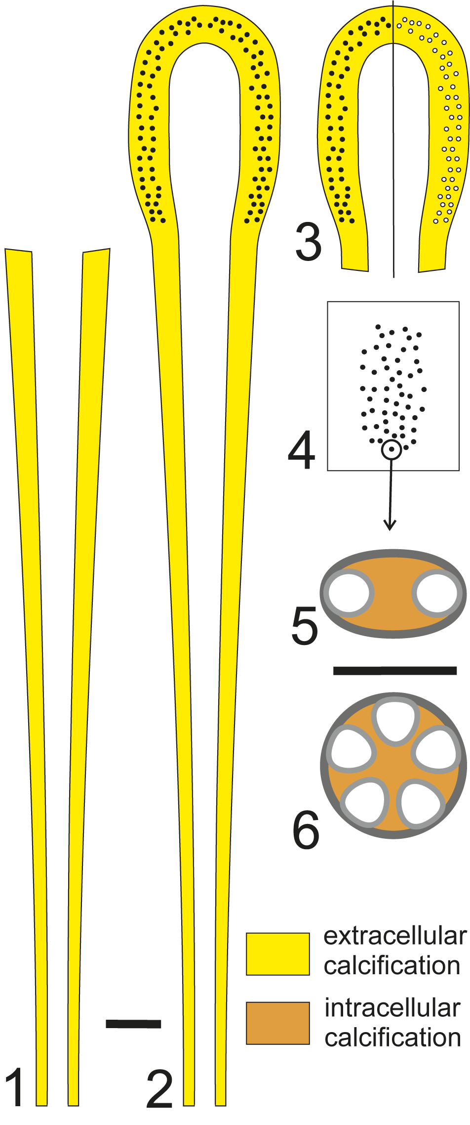
Despite abundant material (Barattolo, Reference Barattolo1981), Triploporella fraasi Steinmann, Reference Steinmann1880, displays a long club-shaped calcareous skeleton but never reveals its apical region or calcified gametophores inside the primary laterals (see Fig. 19.1). Conversely, Triploporella marsicana Praturlon, Reference Praturlon1964 (Barattolo, personal data) mainly displays a calcareous skeleton like that of Triploporella fraasi, but the top is preserved and looks like that of the Triploporella steinmannii species group (see Fig. 19.2). On the other hand, species of the latter, such as Triploporella praturlonii Barattolo, Reference Barattolo1982b (Barattolo Reference Barattolo1982b, pl. III.1; Barattolo et al., Reference Barattolo, Bucur, Kołodziej, Hoffmann and Skupien2013, fig. 5.e), Triploporella remesi (Steinmann, Reference Steinmann1903) (Barattolo et al., Reference Barattolo, Bucur, Kołodziej, Hoffmann and Skupien2013, fig. 4.c), and most probably also Triploporella duplicata (Sokač and Nikler, Reference Sokač and Nikler1973) (Sokač and Nikler Reference Sokač and Nikler1973, pl XI.1), usually retain sterile laterals at the bottom of the stocky calcareous skeleton. However, even if the sterile laterals are missing, the bottom of such calcareous skeletons is always irregular and truncated. In conclusion, the calcareous skeleton in the Triploporella fraasi group and Triploporella steinmannii group may represent the vegetative region (Fig. 19.1), nearly the complete thallus (Fig. 19.2), or the fertile region (Fig. 19.3). Moreover, it is very likely that in some species only the gametophores were mineralized and are sparse in the sediment (Fig. 19.4). Even if not specific to the genus Triploporella Steinmann, Reference Steinmann1880, Russoella Barattolo, Reference Barattolo1984, represents such reproductive organs.
Mineralized parts in the genus Triploporella with an extended vegetative part. (1) Only vegetative part mineralized (e.g., Triploporella fraasi Steinmann, Reference Steinmann1880). (2) Vegetative and fertile parts mineralized (e.g., Triploporella marsicana Praturlon, Reference Praturlon1964). (3) Mainly fertile part mineralized with calcified cystophores (left, e.g., Triploporella steinmannii Barattolo, Reference Barattolo1981) or uncalcified (right, e.g., Triploporella friulana Schindler and Conrad, Reference Schindler and Conrad1994). (4) Only cystophores mineralized (e.g., Russoella Barattolo, Reference Barattolo1984). (5, 6) Structure of cystophores in axial and transversal section, respectively. Scale bar represents 2.0 mm (1–4) and 0.1 mm (5, 6).

On comparing all species attributed to the genus Triploporella Steinmann, Reference Steinmann1880, it is conceivable to identify two kinds of growth (Fig. 17). The first (Triploporella fraasi group and Triploporella steinmannii group; Fig. 17.1) envisages a simple club-shaped thallus with a long vegetative part followed by a variously differentiated upper fertile region. The second (Triploporella apenninica group; Fig. 17.2), with only Triploporella apenninica Baretti, Reference Baretti1922, and Triploporella loducai n. sp., represents a rather short cylindrical thallus, totally fertile, with a reduced vegetative lower part.
Species of the Triploporella fraasi group and Suppiluliumaella schlagintweitii n. sp. share a similar kind of growth (see Possible different growth patterns and stages of growth; Fig. 3). Therefore, in the same genus Triploporella it is possible to infer two growth patterns, that of Neomeris and club-shaped Bornetella for the Triploporella fraasi group and Triploporella steinmannii group (Fig. 20.1–20.8) and that of spherical Bornetella for the Triploporella apenninica group (Fig. 20.1, 20.9–20.13).
Development and mineralization in the genus Triploporella Steinmann. (1–8) Development of species with an extended vegetative part (Triploporella marsicana group); yellow = mineralization; black circles = mineralized reproductive organs, laterals are omitted. (1, 9–13) Development of species with a reduced vegetative part (Triploporella apenninica group); yellow = mineralization; black circles = mineralized reproductive organs, laterals are omitted. Scale bar represents 5.0 mm.

For most species of Triploporella Steinmann, Reference Steinmann1880 (the growth pattern depicted in Fig. 17.1), we can expect that the vegetative region of some species was mineralized before maturity (Fig. 20.7), namely before the fertile region was formed. This fact could be evinced by the occurrence of a mineralized apex confidently referable to the sterile region, as in Suppiluliumaella schlagintweitii n. sp. (Fig. 8).
Sokač and Nikler (Reference Sokač and Nikler1975, pl IV.4) provided an oblique section of Triploporella issaensis Sokač and Nikler, Reference Sokač and Nikler1975, that most likely corresponds to the apical part cut longitudinally. The specimen as well as its primary laterals are of moderate size, which suggests that it belonged to the vegetative part.
Acknowledgments
We thank B. Granier and an anonymous reviewer for their useful suggestions that improved the manuscript.
Competing interests
We the authors have no competing interest to disclose.
Data availability statement
Data available from the Dryad Digital Repository https://doi.org/10.5061/dryad.n5tb2rc51.