Introduction
Brain functional changes are well documented in bipolar disorder, and while the findings have been heterogeneous, there is an emerging consensus that the overall pattern is one of reduced activity in prefrontal and some other cortical regions coupled with overactivity in subcortical structures such as the amygdala, hippocampus and basal ganglia (Strakowski et al. Reference Strakowski, Delbello and Adler2005, Reference Strakowski, Adler, Almeida, Altshuler, Blumberg, Chang, DelBello, Frangou, McIntosh, Phillips, Sussman and Townsend2012; Bellack et al. Reference Bellack, Green, Cook, Fenton, Harvey, Heaton, Laughren, Leon, Mayo, Patrick, Patterson, Rose, Stover and Wykes2007; Savitz & Drevets, Reference Savitz and Drevets2009). Most studies have examined patients in the manic or depressed phase, but changes have also been found in euthymia. Thus, a meta-analysis of positron emission tomography, single-photon emission computed tomography and functional magnetic resonance (fMRI) studies (Kupferschmidt & Zakzanis, Reference Kupferschmidt and Zakzanis2011) found support for cognitive task-related hypoactivations affecting the inferior and middle lateral frontal cortex in euthymic patients, plus hyperactivations in the superior temporal gyrus and ventrolateral prefrontal cortex. On the other hand, in another meta-analysis (Chen et al. Reference Chen, Suckling, Lennox, Ooi and Bullmore2011), this time restricted to voxel-based fMRI studies, changes in euthymia were seen only in the lingual gyrus.
Since bipolar disorder has a hereditary component (McGuffin et al. Reference McGuffin, Rijsdijk, Andrew, Sham, Katz and Cardno2003; Craddock & Sklar, Reference Craddock and Sklar2013), the question arises of whether brain functional changes, perhaps similar to those seen in euthymic patients, might also be seen in their first-degree relatives. Studies to date, however, have been relatively few and have mixed findings. Drapier et al. (Reference Drapier, Surguladze, Marshall, Schulze, Fern, Hall, Walshe, Murray and McDonald2008) found increased activation in the left orbitofrontal cortex extending to the frontopolar and ventrolateral prefrontal cortex in 20 unaffected first-degree relatives of bipolar patients compared with 20 healthy controls during performance of the n-back working memory task. Thermenos et al. (Reference Thermenos, Goldstein, Milanovic, Whitfield-Gabrieli, Makris, Laviolette, Koch, Faraone, Tsuang, Buka and Seidman2010) also used the n-back task and found a pattern of increased activation in the left frontopolar cortex, the anterior insula and the right parietal lobe in 18 first-degree relatives compared with 19 controls. Pompei et al. (Reference Pompei, Jogia, Tatarelli, Girardi, Rubia, Kumari and Frangou2011) examined 25 relatives and 48 controls using the Stroop task and found reduced activation affecting the superior and inferior parietal cortex. In contrast, four studies failed to find any cortical activation differences between relatives and controls (Allin et al. Reference Allin, Marshall, Schulze, Walshe, Hall, Picchioni, Murray and McDonald2010; Whalley et al. Reference Whalley, Sussmann, Chakirova, Mukerjee, Peel, McKirdy, Hall, Johnstone, Lawrie and McIntosh2011; Sepede et al. Reference Sepede, De Berardis, Campanella, Perrucci, Ferretti, Serroni, Moschetta, Del Gratta, Salerno, Ferro, Di Giannantonio, Onofrj, Romani and Gambi2012; Roberts et al. Reference Roberts, Green, Breakspear, McCormack, Frankland, Wright, Levy, Lenroot, Chan and Mitchell2013), although one of them (Whalley et al. Reference Whalley, Sussmann, Chakirova, Mukerjee, Peel, McKirdy, Hall, Johnstone, Lawrie and McIntosh2011) found greater activation in a subcortical structure, the left amygdala, in a secondary analysis taking into account task difficulty (Whalley et al. Reference Whalley, Sussmann, Chakirova, Mukerjee, Peel, McKirdy, Hall, Johnstone, Lawrie and McIntosh2011). Findings in studies using emotional tasks (typically facial expression recognition) rather than cognitive tasks have been similarly heterogeneous (Surguladze et al. Reference Surguladze, Marshall, Schulze, Hall, Walshe, Bramon, Phillips, Murray and McDonald2010; Linke et al. Reference Linke, King, Rietschel, Strohmaier, Hennerici, Gass, Meyer-Lindenberg and Wessa2012; Kanske et al. Reference Kanske, Heissler, Schonfelder, Forneck and Wessa2013; Roberts et al. Reference Roberts, Green, Breakspear, McCormack, Frankland, Wright, Levy, Lenroot, Chan and Mitchell2013).
Since 2001, the existence has been recognized of brain regions which, rather than activating, de-activate in response to performance of a wide range of attention-demanding tasks (Gusnard & Raichle, Reference Gusnard and Raichle2001; Raichle et al. Reference Raichle, MacLeod, Snyder, Powers, Gusnard and Shulman2001). These regions are jointly referred to as the default mode network, and include the medial prefrontal cortex, the posterior cingulate cortex/precuneus, parts of the parietal and temporal lobe cortex, and also the hippocampus (Buckner et al. Reference Buckner, Andrews-Hanna and Schacter2008). Failure of de-activation in parts of this network has been found in a range of psychiatric disorders, including schizophrenia, autism and attention-deficit/hyperactivity disorder (see Broyd et al. Reference Broyd, Demanuele, Debener, Helps, James and Sonuga-Barke2009). It is also increasingly well documented in major depression (e.g. Frodl et al. Reference Frodl, Scheuerecker, Albrecht, Kleemann, Muller-Schunk, Koutsouleris, Moller, Bruckmann, Wiesmann and Meisenzahl2009; Grimm et al. Reference Grimm, Boesiger, Beck, Schuepbach, Bermpohl, Walter, Ernst, Hell, Boeker and Northoff2009; Sheline et al. Reference Sheline, Barch, Price, Rundle, Vaishnavi, Snyder, Mintun, Wang, Coalson and Raichle2009) and bipolar disorder (Allin et al. Reference Allin, Marshall, Schulze, Walshe, Hall, Picchioni, Murray and McDonald2010; Fernandez-Corcuera et al. Reference Fernandez-Corcuera, Salvador, Monte, Salvador Sarro, Goikolea, Amann, Moro, Sans-Sansa, Ortiz-Gil, Vieta, Maristany, McKenna and Pomarol-Clotet2013; Pomarol-Clotet et al. Reference Pomarol-Clotet, Alonso-Lana, Moro, Sarro, Bonnin, Goikolea, Fernandez-Corcuera, Amann, Romaguera, Vieta, Blanch, McKenna and Salvador2015), where it has been found to affect particularly the anterior midline ‘node’ in the medial frontal cortex. Pomarol-Clotet et al. (Reference Pomarol-Clotet, Alonso-Lana, Moro, Sarro, Bonnin, Goikolea, Fernandez-Corcuera, Amann, Romaguera, Vieta, Blanch, McKenna and Salvador2015) found medial frontal failure of de-activation in euthymic bipolar patients, whereas two other studies (Allin et al. Reference Allin, Marshall, Schulze, Walshe, Hall, Picchioni, Murray and McDonald2010; Costafreda et al. Reference Costafreda, Fu, Picchioni, Toulopoulou, McDonald, Kravariti, Walshe, Prata, Murray and McGuire2011) found that the region affected was the posterior cingulate gyrus/precuneus, corresponding to the posterior midline node of the network.
Currently, findings concerning default mode network function in relatives of patients with bipolar disorder are few. Allin et al. (Reference Allin, Marshall, Schulze, Walshe, Hall, Picchioni, Murray and McDonald2010) found failure of de-activation in the posterior cingulate cortex/precuneus in 18 euthymic bipolar patients and in 19 of their unaffected first-degree relatives, in a study using a verbal fluency task. In contrast, Sepede et al. (Reference Sepede, De Berardis, Campanella, Perrucci, Ferretti, Serroni, Moschetta, Del Gratta, Salerno, Ferro, Di Giannantonio, Onofrj, Romani and Gambi2012), using the Continuous Performance Test, found increased de-activation in the posterior cingulate cortex in 22 relatives compared with 24 controls.
The aim of this study was to further examine the pattern of brain functional changes, including both activations and de-activations, in the unaffected siblings of bipolar patients. We used a cognitive task, the n-back task, which has regularly been found to produce activation changes in bipolar disorder (Townsend et al. Reference Townsend, Bookheimer, Foland-Ross, Sugar and Altshuler2010; Fernandez-Corcuera et al. Reference Fernandez-Corcuera, Salvador, Monte, Salvador Sarro, Goikolea, Amann, Moro, Sans-Sansa, Ortiz-Gil, Vieta, Maristany, McKenna and Pomarol-Clotet2013), including in euthymia (Cremaschi et al. Reference Cremaschi, Penzo, Palazzo, Dobrea, Cristoffanini, Dell'Osso and Altamura2013), and which, as an attention-demanding cognitive task, should also induce de-activation in the default mode network.
Method
Subjects
The patient samples consisted of 20 right-handed individuals with bipolar disorder (n = 16 type I, n = 4 type II) and 20 of their unaffected siblings. A total of 40 healthy controls, two for each patient–relative pair, were also recruited. This strategy was adopted in order to be able to match properly in those cases where the patient–relative pair consisted of a male and a female, and to allow for better age and pre-morbid intelligence quotient (IQ) matching.
The patients all met Diagnostic and Statistical Manual of Mental Disorders, 4th edition (DSM-IV) criteria for bipolar disorder, based on interview and review of case notes, and were euthymic at the time of scanning. Euthymia was defined on the basis of having experienced no episodes of illness for at least 3 months and having a score of ⩽8 on the 21-item Hamilton Rating Scale for Depression (Hamilton, Reference Hamilton1960), and ⩽8 on the Young Mania Rating Scale (Young et al. Reference Young, Biggs, Ziegler and Meyer1978). Patients were excluded: (a) if they were younger than 18 or older than 65 years; (b) if they had a history of brain trauma or neurological disease; (c) if they had shown alcohol/substance abuse within 12 months prior to participation; and (d) if they had undergone electroconvulsive therapy in the previous 12 months.
The unaffected siblings met the same exclusion criteria as the patients. They were also excluded if they reported a history of mental illness and/or treatment with psychotropic medication as assessed using the Computerized Diagnostic Interview Schedule for the DSM-IV (C DIS-IV; Robins et al. Reference Robins, Cottler, Bucholz, Compton, North and Rourke2000).
Healthy subjects were recruited via poster and web-based advertisement in the hospital and local community, plus word-of-mouth requests from staff in the research unit. They met the same exclusion criteria as the patients and, like the unaffected siblings, were excluded if they reported a history of mental illness and/or treatment with psychotropic medication, on interview with the C DIS-IV. They were also excluded if they had a first-degree relative with a major psychiatric disorder.
The three groups were matched for age, sex and pre-morbid IQ. This latter variable was estimated using the Word Accentuation Test (Test de Acentuación de Palabras; TAP) (Del Ser et al. Reference Del Ser, Gonzalez-Montalvo, Martinez-Espinosa, Delgado-Villapalos and Bermejo1997; Gomar et al. Reference Gomar, Ortiz-Gil, McKenna, Salvador, Sans-Sansa, Sarro, Guerrero and Pomarol-Clotet2011), a test that is conceptually similar to the National Adult Reading Test used in the UK (Nelson & Willison, Reference Nelson and Willison1991) and the Wide Range of Achievement Test in the USA (Jastak & Wilkinson, Reference Jastak and Wilkinson1984). Subjects have to pronounce low-frequency Spanish words whose accents have been removed.
All participants gave written informed consent and the study was approved by the hospital research ethics committee. All procedures were carried out according to the Declaration of Helsinki.
Procedure
While being scanned, participants performed a sequential-letter version of the n-back task (Gevins & Cutillo, Reference Gevins and Cutillo1993). Two levels of memory load (1-back and 2-back) were presented in a blocked design manner. Each block consisted of 24 letters that were shown every 2 s (1 s on, 1 s off) and all blocks contained five repetitions (1-back and 2-back depending on the block) located randomly within the blocks. Individuals had to indicate repetitions by pressing a button. Four 1-back and four 2-back blocks were presented in an interleaved way, and between them a baseline stimulus (an asterisk flashing with the same frequency as the letters) was presented for 16 s. To identify which task had to be performed, characters were shown in green in 1-back blocks and in red in the 2-back blocks. All participants first went through a training session outside the scanner.
The behavioural measure used was the signal detection theory index of sensitivity, d’ (Green & Swets, Reference Green and Swets1966). Higher values of d’ indicate better ability to discriminate between targets and distractors. If subjects showed negative d’ values in either or both of the 1-back and 2-back versions of the task, which suggests that they were not performing it, they were not included in the study.
In each individual scanning session 266 volumes were acquired from a 1.5-T GE Signa scanner. A gradient echo echo-planar imaging (EPI) sequence depicting the blood oxygenation level-dependent (BOLD) contrast was used. Each volume contained 16 axial planes acquired with the following parameters: repetition time = 2000 ms, echo time = 20 ms, flip angle = 70°, section thickness = 7 mm, section skip = 0.7 mm, in-plane resolution = 3r3 mm. The first 10 volumes are discarded to avoid T1 saturation effects.
fMRI image analyses were performed with the FEAT module included in FSL software (Smith et al. Reference Smith, Jenkinson, Woolrich, Beckmann, Behrens, Johansen-Berg, Bannister, De Luca, Drobnjak, Flitney, Niazy, Saunders, Vickers, Zhang, De Stefano, Brady and Matthews2004). At a first level, images were corrected for movement and then co-registered to a common stereotaxic space [Montreal Neurological Institute (MNI) template]. Before the group analyses, normalized images were spatially filtered with a Gaussian filter [full-width at half maximum (FWHM) = 5 mm]. To minimize unwanted movement-related effects, individuals with an estimated maximum absolute movement >3.0 mm or an average absolute movement >0.3 mm were excluded from the study. General linear models were fitted to generate individual activation maps for the 1-back v. baseline and 2-back v. baseline contrasts. Values for movement parameters were included as nuisance covariates in the fitting of individual linear models.
Data analysis
The FEAT module was used to fit a linear mixed-effects model including the baseline v. 1-back and the baseline v. 2-back activation images for the three groups. The groups were then compared using a whole-brain (voxel-level) analysis of variance (ANOVA). We did not do this simply by carrying out a one-way between-group ANOVA with three levels, because such an analysis assumes the complete independence of individuals (apart from the shared group effect). This was not the case in our study, where each bipolar patient was sampled together with his/her sibling, thus creating covariation between pairs. Therefore, a block design was employed to take into account the within-family covariability (in a block design, related individuals are included in the same block). Specifically, each set of patient–sibling–matched pair of controls was considered as a block, and a one-way blocked ANOVA was performed to detect statistical differences between groups. Statistical tests on these contrasts were carried out at the cluster level with a family-wise corrected p value of 0.05 using Gaussian random field methods. The default threshold of z = 2.3 was used to define the initial set of clusters.
Any clusters showing significant differences among the groups in the ANOVA were examined further in a region of interest (ROI) analysis. Specifically, for each individual, the mean value of each ROI was calculated. Then, these mean values were compared between pairs of groups by means of paired t tests. For the bipolar v. relative comparison, the 20 patients were compared with their 20 relatives. For the bipolar v. control comparison we compared the 20 patients with the 20 controls that had been matched for sex, age and pre-morbid IQ with the patients. For the relative v. control comparison we used the remaining 20 controls (who had been specifically matched with the group of relatives).
Results
Demographic and behavioural findings
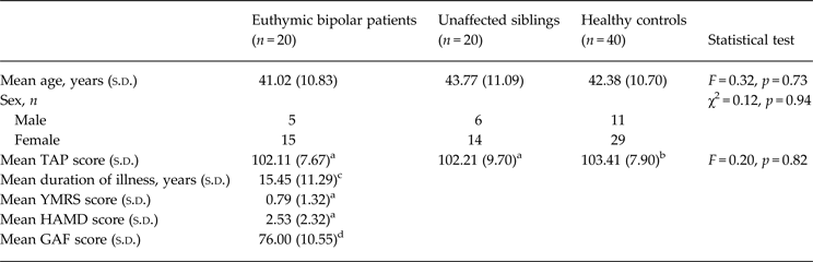
As shown in Table 1, the three groups were matched for age, sex and TAP-estimated IQ. None of the participants was excluded due to excessive head movement or because of lack of compliance with the task (i.e. negative d’ scores).
Demographic and clinical characteristics of the sample

s.d., Standard deviation; TAP, Word Accentuation Test (Test de Acentuación de Palabras); YMRS, Young Mania Rating Scale; HAMD, Hamilton Rating Scale for Depression; GAF, Global Assessment of Functioning.
a Data missing for one participant.
b Data missing for six participants.
c Data missing for two participants.
d Data missing for five participants.
All patients were taking mood stabilizers (lithium 11; other mood stabilizer 2; combinations 7). Six patients were taking antidepressants and 14 were also taking antipsychotics (second generation 13 and combined first and second generation 1). The mean daily dose (in chlorpromazine equivalents) in these patients was 268.37 (s.d. = 276.88) mg/day.
There were no differences on 1-back performance between the unaffected siblings and the healthy controls [d’ 4.39 (s.d. = 0.76) v. 4.24 (s.d. = 0.68); t = 0.58, p = 0.57], between the bipolar patients and the healthy controls [d’ 4.18 (s.d. = 0.81) v. 4.41 (s.d. = 0.63); t = −1.32, p = 0.20], and between the bipolar patients and their unaffected siblings [d’ 4.18 (s.d. = 0.81) v. 4.39 (s.d. = 0.76); t = −0.83, p = 0.42]. In the 2-back task, the bipolar patients performed more poorly than the healthy controls [d’ 2.68 (s.d. = 0.82) v. 3.58 (s.d. = 0.75); t = −3.54, p = 0.002]. There were no significant differences between the unaffected siblings and the healthy controls [d’ 2.91 (s.d. = 0.81) v. 3.40 (s.d. = 0.93); t = −1.76, p = 0.09] or between the bipolar patients and their unaffected siblings [mean d’ 2.68 (s.d. = 0.82) v. 2.91 (s.d. = 0.81); t = −0.86, p = 0.40].
Mean activations and de-activations in the three groups
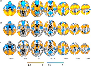
Mean activations in the 1-back v. baseline contrast were generally similar to but less marked than in the 2-back v. baseline contrast. Therefore, only the findings for the 2-back v. baseline contrast are described here. Clusters of activation for the three groups are shown in Fig. 1. At p < 0.05 corrected, the healthy controls showed bilateral clusters of activation in the anterior insula, the dorsolateral prefrontal cortex, the precentral gyrus, the supplementary motor area, the cerebellum, the thalamus, the basal ganglia, and parts of the temporal and parietal cortex. De-activations were seen bilaterally in the medial frontal cortex, the amygdala, the hippocampus and adjacent cortical regions, the medial parietal cortex extending to primary visual areas, the posterior insula and the lateral parietal cortex.
Brain regions showing a significant effect in the 2-back v. baseline contrast in healthy controls (a), unaffected siblings (b) and euthymic bipolar patients (c). Colour bars indicate z scores; red to yellow colours indicate significant activation and blue to cyan colours indicate regions with significant deactivation. Numbers refer to Montreal Neurological Institute (MNI) z coordinates of the slice shown. The right side of the image is the right side of the brain.

Activations and de-activations in the euthymic bipolar patients and their unaffected siblings followed a broadly similar pattern to those in the controls. Both the clusters of activation and de-activation appeared less extensive in the unaffected siblings and the euthymic bipolar patients, but it should be borne in mind that this may have reflected the fact that there were only half the number of subjects in these groups than in the healthy controls.
ANOVA comparing the three groups
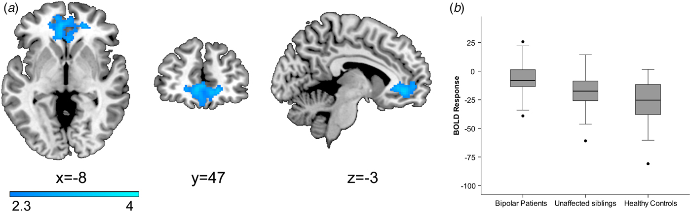
No clusters of significant difference emerged in the 1-back v. baseline contrast. In the 2-back v. baseline contrast there was a single cluster of significant difference in the medial and inferior frontal cortex bilaterally, also including portions of the orbitofrontal cortex [cluster size 1167 voxels, peak activation in MNI coordinates (2, 52, −14), z max = 3.66, p = 0.006] (see Fig. 2a ).
(a) Cluster of significant difference in the medial prefrontal cortex found comparing bipolar patients, unaffected siblings and healthy controls in the 2-back v. baseline contrast. Numbers refer to Montreal Neurological Institute (MNI) coordinates of the slice shown. Colour bar indicates z scores from the group-level analysis. The right side of the image is the right side of the brain. (b) Boxplot based on individual mean activation values from the region of interest extracted from this significant cluster. Centre lines show the medians; box limits indicate the 25th and 75th percentiles; whiskers extend 1.5 times the interquartile range from the 25th and 75th percentiles; and outliers are represented by dots. BOLD, Blood oxygen level dependent.

Mean activation values for this cluster in the three groups are shown in Fig. 2b . It can be seen that the euthymic bipolar patients showed significantly less de-activation than healthy controls (p = 0.009) and their unaffected siblings (p = 0.03). The unaffected siblings also showed significant failure of de-activation compared with healthy controls (p = 0.03).
No correlations were found between individual mean activation values and the behavioural performance (d’) for any of the groups: euthymic bipolar patients (r = −0.13, p = 0.59); unaffected siblings (r = 0.19, p = 0.43); and healthy controls (r = 0.03, p = 0.87).
Discussion
This study found that euthymic bipolar patients showed failure of de-activation in the medial frontal cortex, an area which constitutes the anterior midline node of the default mode network. This was not accompanied by any activation changes. A similar, though less marked failure of de-activation, again with no activation changes, was also seen in the unaffected siblings of patients with the disorder.
At first sight our failure to find activation changes in euthymic bipolar patients seems surprising, since the disorder is widely recognized as being associated with a pattern of task-related hypoactivations and hyperactivations (Strakowski et al. Reference Strakowski, Adler, Almeida, Altshuler, Blumberg, Chang, DelBello, Frangou, McIntosh, Phillips, Sussman and Townsend2012). However, it should be noted that these changes have mostly been documented in patients in the manic or depressed phase. In fact, in Chen et al. (Reference Chen, Suckling, Lennox, Ooi and Bullmore2011)'s meta-analysis of voxel-based fMRI studies the only area of hypoactivation found in euthymic patients was the lingual gyrus. This meta-analysis did find evidence of task-related hyperactivations in euthymia; here, however, the areas affected included the medial frontal cortex, the superior temporal gyrus, the parahippocampal gyrus and the cingulate cortex, and so it seems possible that at least some of the changes actually represented failure of de-activation rather than true hyperactivation (hyperactivation and failure of de-activation give the same appearance in functional imaging studies using subtraction analysis; for discussions, see Gusnard & Raichle, Reference Gusnard and Raichle2001; Pomarol-Clotet et al. Reference Pomarol-Clotet, Salvador, Sarro, Gomar, Vila, Martinez, Guerrero, Ortiz-Gil, Sans-Sansa, Capdevila, Cebamanos and McKenna2008).
Similarly, the relatives of the bipolar patients did not show activation changes. This finding goes against those of a number of studies that have found hypoactivations, and more consistently hyperactivations, in such individuals (Drapier et al. Reference Drapier, Surguladze, Marshall, Schulze, Fern, Hall, Walshe, Murray and McDonald2008; Thermenos et al. Reference Thermenos, Goldstein, Milanovic, Whitfield-Gabrieli, Makris, Laviolette, Koch, Faraone, Tsuang, Buka and Seidman2010; Pompei et al. Reference Pompei, Jogia, Tatarelli, Girardi, Rubia, Kumari and Frangou2011). However, as noted in the Introduction, other studies have failed to find any evidence of activation differences (Allin et al. Reference Allin, Marshall, Schulze, Walshe, Hall, Picchioni, Murray and McDonald2010; Sepede et al. Reference Sepede, De Berardis, Campanella, Perrucci, Ferretti, Serroni, Moschetta, Del Gratta, Salerno, Ferro, Di Giannantonio, Onofrj, Romani and Gambi2012; Roberts et al. Reference Roberts, Green, Breakspear, McCormack, Frankland, Wright, Levy, Lenroot, Chan and Mitchell2013), or have found evidence of subcortical changes only (Whalley et al. Reference Whalley, Sussmann, Chakirova, Mukerjee, Peel, McKirdy, Hall, Johnstone, Lawrie and McIntosh2011).
As noted in the Introduction, failure of de-activation is now a relatively robust finding in bipolar disorder. It has been documented in both manic (Fernandez-Corcuera et al. Reference Fernandez-Corcuera, Salvador, Monte, Salvador Sarro, Goikolea, Amann, Moro, Sans-Sansa, Ortiz-Gil, Vieta, Maristany, McKenna and Pomarol-Clotet2013) and depressed (Fernandez-Corcuera et al. Reference Fernandez-Corcuera, Salvador, Monte, Salvador Sarro, Goikolea, Amann, Moro, Sans-Sansa, Ortiz-Gil, Vieta, Maristany, McKenna and Pomarol-Clotet2013) patients, and also in patients unselected for phase of illness (Calhoun et al. Reference Calhoun, Maciejewski, Pearlson and Kiehl2008), where it affects particularly the medial frontal cortex. We (Pomarol-Clotet et al. Reference Pomarol-Clotet, Alonso-Lana, Moro, Sarro, Bonnin, Goikolea, Fernandez-Corcuera, Amann, Romaguera, Vieta, Blanch, McKenna and Salvador2015) also found failure of de-activation in the same location in euthymic patients, and two other studies (Allin et al. Reference Allin, Marshall, Schulze, Walshe, Hall, Picchioni, Murray and McDonald2010; Costafreda et al. Reference Costafreda, Fu, Picchioni, Toulopoulou, McDonald, Kravariti, Walshe, Prata, Murray and McGuire2011) found that the region affected was the posterior cingulate gyrus/precuneus. The present study found that the relatives of bipolar patients also showed failure of de-activation, which was located in the medial frontal cortex and was less marked than that seen in the patients. Previous studies have also found de-activation changes in relatives: Allin et al. (Reference Allin, Marshall, Schulze, Walshe, Hall, Picchioni, Murray and McDonald2010) found failure of de-activation, although this was in the posterior cingulate cortex/precuneus rather than in the medial prefrontal cortex as in our study. In contrast, Sepede et al. (Reference Sepede, De Berardis, Campanella, Perrucci, Ferretti, Serroni, Moschetta, Del Gratta, Salerno, Ferro, Di Giannantonio, Onofrj, Romani and Gambi2012) found exaggerated de-activation in the same area. Also relevant here is the study of Thermenos et al. (Reference Thermenos, Goldstein, Milanovic, Whitfield-Gabrieli, Makris, Laviolette, Koch, Faraone, Tsuang, Buka and Seidman2010) which found four areas of what the authors considered to be increased activation in relatives of bipolar patients; however, in two of these regions, the left orbitofrontal cortex and the parietal cortex, plots of mean activations revealed that this actually represented failure of de-activation.
The question arises of what medial frontal failure of de-activation in relatives of bipolar patients might mean. The fact that it was also present in euthymic patients suggests that one is dealing with a trait abnormality. Beyond this, evidence from studies using a small number of tasks that have been found to activate parts of the default mode network rather than de-activate it suggests that it has roles as diverse as autobiographical recall, thinking about the future, theory of mind, moral decision-making and making judgements about characteristics that apply to oneself v. others (Buckner et al. Reference Buckner, Andrews-Hanna and Schacter2008; Whitfield-Gabrieli et al. Reference Whitfield-Gabrieli, Moran, Nieto-Castanon, Triantafyllou, Saxe and Gabrieli2011). The medial frontal cortex, in particular, may also have a role in emotion: Price & Drevets (Reference Price and Drevets2012) have pointed out that this region has close connections with the amygdala and both structures form part of a wider network that includes the ventral striatum, the medial thalamus, the hypothalamus and the brainstem. Data from animals suggest that this system is involved in forebrain modulation of visceral function in response to sensory or emotive stimuli.
In conclusion, this study provides evidence that bipolar disorder in euthymia is characterized by a failure to de-activate the medial prefrontal cortex, and that this change is present to a lesser extent in the unaffected first-degree relatives of patients. Taken together these findings suggest that default mode network dysfunction might represent a trait abnormality and possibly even an endophenotype for the disorder. Limitations of the study include the relatively small sample sizes for the patients and their relatives, and it is possible that a larger sample would have revealed changes in activation as well as in de-activation. As in most studies, the bipolar patients were on medication; however, current evidence suggests that the confounding effects of this are relatively limited (Hafeman et al. Reference Hafeman, Chang, Garrett, Sanders and Phillips2012). Finally, the finding that the relatives showed an intermediate level of medial frontal de-activation between the bipolar patients and the controls depended on ROI analysis. When reported clusters are large (which was not the case in our study) they may contain functionally and anatomically heterogeneous ensembles of voxels, which therefore may not be well characterized by a single ROI (Poldrack, Reference Poldrack2007).
Acknowledgements
This work was supported by the Catalonian Government (2014-SGR-1573 to FIDMAG and 2014-SGR-398 to the Bipolar Disorders Group) and several grants from the Plan Nacional de I+D+i 2008–2011 and 2013–2013 and co-funded by the Instituto de Salud Carlos III-Subdirección General de Evaluación y Fomento de la Investigación and the European Regional Development Fund (FEDER): Miguel Servet Research Contracts (CP10/00596 to E.P.-C. and CPII13/00018 to R.S.), Stabilization Contract grant (CES12/024 to B.L.A.), Rio Hortega Contract (CM14/00048 to A.R.), Predoctoral grant (FI11/00221 to S.A.-L.) and Research Projects (PI10/01058 and PI14/01148 to E.P.-C. and PI14/01151 to R.S.). The funding organizations played no role in the study design, data collection and analysis, or manuscript approval.
Declaration of Interest
None.