Echocardiographic assessment of single right ventricular function using standard echocardiographic techniques remains problematic. Recently, newer echocardiographic techniques have shown promise in overcoming certain limitations, the latest being two-dimensional speckle tracking.Reference Amundsen, Helle-Valle and Edvardsen1 This technique has particular appeal because it is independent of the angle of insonation as well as ventricular geometry and thus theoretically superior when assessing right ventricular function.Reference Teske, De Boeck, Olimulder, Prakken, Doevendans and Cramer2 Speckle tracking is validated for the right ventricle against tissue DopplerReference Teske, De Boeck, Olimulder, Prakken, Doevendans and Cramer2 and cardiac magnetic resonance imagingReference Bansal, Cho, Chan, Leano, Haluska and Marwick3 for strain calculation. Strain and strain rate analysis have proven useful to assess left ventricularReference Pettersen, Fredriksen and Urheim4 as well as subpulmonaryReference Knirsch, Dodge-Khatami and Kadner5 and systemicReference Kalogeropoulos, Georgiopoulou and Giamouzis6, Reference Moiduddin, Texter and Zaidi7 right ventricular function in patients with congenital heart disease.
There is evidence that the presence of a significant “piggyback” left ventricle in hypoplastic left heart syndrome may influence systemic right ventricular functionReference Wisler, Khoury and Kimball8 and outcome.Reference Walsh, McCrindle and Dipchand9 Patients with mitral and aortic atresia typically have a negligible left ventricle and are shown to have better actuarial survival than other anatomical subtypes with larger left ventricles,Reference Furck, Uebing and Hansen10 but whether this survival benefit truly relates to the size of the left ventricle, right ventricular function, or other factors like, for example, electromechanical dyssynchrony remains unclear.
We, therefore, aimed to investigate potential differences in global and regional systemic right ventricular deformation and dyssynchrony between hypoplastic left heart syndrome patients with mitral and aortic atresia – the anatomic variant with the smallest left ventricle – and the other anatomical subtypes after Fontan surgery using two-dimensional speckle tracking.
Materials and methods
Patients
A database search identified 29 children with hypoplastic left heart syndrome after Fontan completion in whom echocardiographic data suitable for speckle tracking analysis of the systemic right ventricle were available.
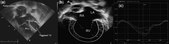
On the basis of echocardiography performed before the Norwood operation, two senior investigators allocated patients to two different groups. The first group consisted of 15 patients with mitral and aortic valve atresia. In these cases, neither colour Doppler nor spectral Doppler flow could be demonstrated across either valve at birth. The second group comprised 14 patients with the remaining anatomical subgroups – mitral/aortic stenosis (Fig 1a), mitral atresia/aortic stenosis, and mitral stenosis/aortic atresia. All patients had a Norwood operation with modified Blalock–Taussig shunt as stage 1, a superior cavo-atrio-pulmonary anastomosis (Hemi Fontan) as stage 2, and an intra-atrial lateral tunnel modification of the Fontan operation as stage 3 of their palliation. All patients except three had all steps of their surgeries performed at our centre by a single surgeon (J.S.). Informed consent for anonymised data analysis was obtained for every patient. Ethics committee approval was waived due to the retrospective nature of the study.
An example of a patient with hypoplastic left heart syndrome and mitral and aortic stenosis and a significant left ventricle is shown in Figure 1a. Figure 1b shows a patient with hypoplastic left heart syndrome and mitral and aortic atresia with the six right ventricular segments. Figure 1c demonstrates an example of six segmental strain tracings. Of note, peak strain values for the cardiac cycle do not always occur before aortic valve closure and therefore may be different from peak systolic strain values.

Two-dimensional Speckle tracking
All echocardiographic recordings used for two-dimensional speckle tracking were obtained using a GE Vingmed Vivid 7 scanner (GE Healthcare, Wauwatosa, United States of America) by trained physicians during routine outpatient visits and stored digitally for offline analysis. In addition to the standard echocardiographic study, grey-scale images optimised for two-dimensional speckle tracking analysis were acquired from the apical four-chamber view between 79 and 92 frames per second. An experienced echocardiographer, blinded to the anatomical subgroup (C.P.), retrospectively analysed the data offline using dedicated software (EchoPac, GE Healthcare). Manual tracing of the endocardial border started from the septal tricuspid valve annular hinge point, and commenced along the apex to the lateral tricuspid valve annular hinge point. The region of interest was adjusted according to myocardial thickness. The software divided the right ventricular myocardium automatically into six segments according to the terminology originally intended for the left ventricle of hearts with normal anatomy: basal septal, mid-septal, apical septal, apical lateral, mid-lateral, and basal lateral (Fig 1b). Of note, this segmental allocation for the right ventricle in hypoplastic left heart syndrome does not always correlate to the anatomy. For instance, in the absence of a left ventricle, there may not be a true apical septal segment because there is no identifiable septum. Aortic valve closure was determined from pulsed Doppler tracings of the neo-aortic outflow. Peak systolic longitudinal strain, percent change in segment length from end-diastole, and peak systolic longitudinal strain rate – representing the rate of deformation during systole – were recorded.
From the regional segmental data, the global systolic strain and strain rate were calculated by the software.
Dyssynchrony
We recorded the time intervals from the beginning of the QRS interval to the peak strain for each segment. Of note, peak strain may be reached after aortic valve closure (Fig 1c). We calculated the wall-to-wall delay of the time from the onset of electrical activation (beginning of the Q wave) to peak strain by subtracting the time interval of the basal septal segment from that of the basal lateral segment.
Standard echocardiography parameters
On the same echocardiogram used for the speckle tracking analysis, the end diastolic area of the left ventricle was obtained by manual tracing from the apical four-chamber imaging plane in end-diastole. We averaged three measurements.
Assessment of right ventricular function was graded semi-quantitatively as 1 = normal, 2 = mildly depressed, 3 = moderately depressed, and 4 = severely depressed.
The severity of tricuspid valve regurgitation was graded accordingly from 0 to 4 (0 = none, 1 = trivial, 2 = mild, 3 = moderate, and 4 = severe) taking into account the vena contracta width and area of the colour Doppler jet in relation to size of the right ventricle and atrium.
The presence of fenestrations was assessed by colour Doppler imaging of the intra-atrial tunnel.
The diameter of the ascending aorta at birth was measured from parasternal long-axis imaging on the same echocardiogram used to determine the anatomical subtype and to allocate patients to groups.
Additional data
On the day of echocardiography, we recorded the QRS duration from a 12-lead surface electrocardiogram, peak oxygen uptake as a measure of exercise capacity from a same day exercise test and the New York Heart Association functional class (1–4) as a measure of clinical functional status. Exercise data were available for 23 patients – 10 patients with mitral and aortic atresia and 13 patients with other anatomical subtypes; the remaining six patients were too young to perform an exercise test. The method of cardiopulmonary exercise testing was previously described.Reference Moller, Weitz and Jensen11
Intraobserver variability
To assess intraobserver variability, the same observer made two measurements at different points in time in 20 randomly selected patients. Duplicate measurements were made for the global longitudinal strain and strain rate as well as regional strain, and the strain rate in the six segments and the coefficient of variation were calculated.
Statistical methods
Data analysis was performed using SPSS 15.0 for Windows software (SPSS Inc., Chicago, Illinois, United States of America).
Continuous data are displayed as the mean, standard deviation. Testing for normality was performed with the Kolmogorov–Smirnov test. Comparisons between groups were made with a two-sided Student's t-test for independent samples or the Wilcoxon rank sum test as appropriate.
Dichotomous data were examined using the chi-square test. Correlation of parameters was assessed using Pearson's correlation coefficient for normally distributed data and Spearman's rank correlation coefficient for not normally distributed data. For all calculations, a p-value of less than 0.05 was considered statistically significant.
Results
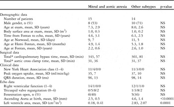
Patient data, including demographic, surgical, functional, and echo data, are presented in Table 1. Gender, age at study, age at the different steps of surgical palliation, total – all three steps combined – time of cardiopulmonary bypass, and aortic cross-clamp times as well as body surface area at the time of the study were similar between both groups. QRS duration, New York Heart Association functional class, and peak oxygen uptake were not different between groups. All patients had New York Heart Association functional class 1 or 2. The diameters of the ascending aorta at birth and the left ventricle area were significantly smaller in patients with mitral and aortic atresia. We found no difference in the amount of tricuspid valve regurgitation, right ventricular function assessment, and number of patients with fenestrations.
Patient data.

*Total refers to combined values for stages 1, 2, and 3
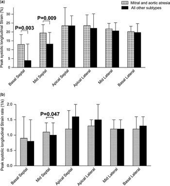
Global strain (−19.5, 2.8% versus −17.4, 3.9%, p = 0.11) as well as global strain rate (−1.0, 0.2 per second versus −0.9, 0.3 per second, p = 0.51) was similar between groups.
Figure 2 shows the regional results for the peak systolic longitudinal strain and strain rate in the six segments of the right ventricle. The basal septal and mid-septal segments showed a higher longitudinal strain (−13.0, 5.0% versus −3.9, 9.3%, p = 0.003 and −19.4, 4.7% versus −13.2, 6.5%, p = 0.009, respectively) and strain rate (−1.14, 0.3 per second versus −0.95, 0.4 per second, p = 0.047) in the mid-septal segment in patients with mitral and aortic atresia.
Shows the comparison of strain in percent (Fig 2a) and the strain rate in per second (Fig 2b) in the six right ventricular segments of patients with mitral and aortic atresia (grey columns) and the three other subtypes (black columns). The columns indicate the mean and the error bars the standard deviation. Significant differences between groups are shown as p-values above the column.

A total of 10 – five apical septal and five apical lateral – out of 176 segments (6%) could not be analysed.
Intraobserver variability was 4.4% for global strain and 8.9% for global strain rate, and was in the range of 9.7–16.6% for the regional strain and strain rate.
The basal septal to basal lateral wall-to-wall delay of the time interval from the onset of the Q wave to peak strain was significantly shorter in patients with mitral and aortic atresia (81, 57 milliseconds versus 38, 29 milliseconds, p = 0.02). No correlation was found between these two parameters and QRS duration.
Discussion
Using two-dimensional speckle tracking echocardiography, this study shows that children with hypoplastic left heart syndrome and mitral and aortic atresia have distinct differences in regional deformation parameters and intraventricular dyssynchrony when compared to other anatomical subtypes of hypoplastic left heart syndrome. These differences do not compromise global ventricular function or clinical functional status in our cohort, although relatively early after Fontan surgery.
Despite the fact that speckle tracking echocardiography was originally designed and validated for left ventricular function analysis,Reference Amundsen, Helle-Valle and Edvardsen1, Reference Bansal, Cho, Chan, Leano, Haluska and Marwick3, Reference Perk, Tunick and Kronzon12 it was recently shown to be also useful for right ventricular strain and strain rate calculation.Reference Teske, De Boeck, Olimulder, Prakken, Doevendans and Cramer2 To date, there are only a few clinical studies using this method in patients with congenital heart disease.Reference Pettersen, Fredriksen and Urheim4, Reference Knirsch, Dodge-Khatami and Kadner5 However, a recent study by Moiduddin et alReference Moiduddin, Texter and Zaidi7 has successfully applied the use of two-dimensional speckle tracking to show decreased deformation and increased intraventricular dyssynchrony of the systemic right ventricle after Fontan when compared to subpulmonary right ventricles of normal children. Our study is the first to use two-dimensional speckle tracking to compare differences in global and regional right ventricular deformation and dyssynchrony in a group of children with different anatomical subtypes of hypoplastic left heart syndrome after Fontan surgery.
There is much debate about the different outcome of hypoplastic left heart syndrome based on the anatomical subtype. Jonas et alReference Jonas, Hansen, Cook and Wessel13 showed in an early study that 3-year survival was lowest in the aortic and mitral atresia group and that mitral and aortic stenosis patients had better outcome. This is in contrast to the recently published data from our group, where mitral and aortic atresia patients had the best actuarial survival.Reference Furck, Uebing and Hansen10
Morphology and size of the left ventricle are reported to influence outcome in hypoplastic left heart syndrome. There is an undeniable interaction between left ventricular size and anatomic subtype in hypoplastic left heart syndrome. Thus, patients with mitral/aortic atresia have smaller, often negligible left ventricles, whereas the other subtypes typically have larger left ventricles. In a study that correlated angiographic and pathologic findings, Sugiyama et alReference Sugiyama, Yutani, Iida, Arakaki, Yamada and Kamiya14 postulated that a larger muscle bulk of the left ventricle, as found in mitral stenosis and aortic atresia, could impair right ventricular function when compared to mitral and aortic atresia with less prominent left ventricles. Walsh et alReference Walsh, McCrindle and Dipchand9 showed in a retrospective echocardiographic study that a thicker interventricular septum – independent of anatomic subtype – at birth predicted worse survival up to stage II palliation.
It is unclear at this point how global and particularly regional right ventricular function is affected by the left ventricle and how right ventricular function itself affects outcome in different anatomical subtypes of hypoplastic left heart syndrome. Wisler et alReference Wisler, Khoury and Kimball8 found in a retrospective study that left ventricular size did not impact on right ventricular fractional area change and myocardial performance index at different stages of palliation. Our own group recently published no differences in multiple two-dimensional echocardiographic parameters as well as tissue Doppler between Fontan patients with mitral atresia and aortic atresia and small left ventricles and other subtypes of hypoplastic left heart syndrome.Reference Petko, Moller, Hoffmann, Kramer and Uebing15
This study shows that distinct differences in regional deformation exist depending on anatomy. The mitral atresia and aortic atresia subtype typically has no or little left ventricular cavity, and therefore the leftward and posterior free wall of the right ventricle starts right beneath the tricuspid valve. According to our study, the nomenclature of segmental allocation includes the “basal septal” and “mid-septal” segments in patients with mitral and aortic atresia. In patients with other anatomical subtypes, however, these two segments are comprised more or less of the interventricular septum, depending on the actual size of the left ventricle. The septum, if present, is often fibrotic and less contractile. We showed that strain and strain rate, and thus deformation or regional function, are compromised in this region when there is a more prominent left ventricle. Despite the differences in regional deformation, we found no statistically significant differences in global strain or the strain rate. When interpreting these results, one might argue that while septal deformation may be compromised in the presence of a significant left ventricle, the rest of the myocardium compensates effectively in preserving pump function up to this point in time in our cohort. It has to be taken into account that our follow-up is relatively early after Fontan surgery. It is conceivable, however, that the subtle differences in regional right ventricular function presented here progress to discernible differences in global right ventricular function on further follow-up. Adding to this notion is the fact that right ventricular dyssynchrony was greater in the group with the left ventricles. This finding is most likely related to the differences in deformation characteristics of the septal region between groups. Whether these differences will lead to a more rapid deterioration of global right ventricular function as time progresses for patients with larger left ventricles is unclear. Incorporating two-dimensional speckle tracking into longitudinal follow-up to assess global and regional deformation and the synchronicity of electromechanical coupling could provide an answer.
Our study is limited by its retrospective design. The size of our study sample may have limited power to detect certain differences, and due to imaging quality, we did not have data available for each segment in each patient. The apical segments especially are sometimes difficult to track appropriately in older patients with hypoplastic left heart syndrome.
There is a natural selection bias in our study, because we could only include those patients who survived Fontan surgery and were doing well as outpatients.
It should also be noted that the measurement of the left ventricle area by two-dimensional echocardiography has limited accuracy, but this is unlikely to have affected the findings or main message of the data presented here.
The echocardiography software was designed for the left ventricle of normal hearts, and therefore the allocation and terminology of the six right ventricular segments by the software does not always correlate with the anatomy of patients with hypoplastic left heart syndrome, particularly in the absence of a left ventricle.
Variability of regional strain and strain rate data in our study is not negligible but in a similar range as found by other investigators using the same technology.Reference Singh, Cupps, Pasque, Woodard, Holland and Ludomirsky16
We did not recruit our own age-matched reference population for two reasons: first, lower deformation and higher dyssynchrony of the systemic single right ventricle after Fontan surgery compared to the normal subpulmonary right ventricle has been demonstrated;Reference Moiduddin, Texter and Zaidi7 second, the intention of our study was to investigate two distinct groups of patients with hypoplastic left heart syndrome and not to compare the systemic right ventricle with the subpulmonary right ventricle of healthy children.
Assessment of right ventricular dyssynchrony in hypoplastic left heart syndrome has not yet been standardised and the parameter used in this study has been applied extensively in patients with normal anatomy but may need further investigation in this population.
In conclusion, we found that hypoplastic left heart syndrome patients with mitral and aortic atresia and small left ventricles early after Fontan have similar global right ventricular function, clinical functional status, and exercise capacity when compared with other anatomical subtypes with larger left ventricles. Differences in regional deformation and dyssynchrony exist relatively early after Fontan surgery. Whether these differences progress over time and impact on global function and outcome is unclear. We therefore advocate that longitudinal follow-up of hypoplastic left heart syndrome patients should be on a regular basis and include newer echocardiographic techniques like two-dimensional speckle tracking to assess regional and global systemic right ventricular deformation as well as dyssynchrony.
Acknowledgements
This work was supported by the non-profit organisation “Kinderherzen wollen leben e.V.”. None of the authors have any conflict of interest to declare.