Introduction
A major challenge to the field of neuropsychology has been to identify tests that are specific to frontal lobe function. Executive function tests have long been assumed to measure frontal lobe function; in fact, these tests are often referred to as “frontal-executive tests.” However, there is increasing evidence that the total achievement scores derived from commonly used executive function tests, including the Stroop Test, the Trail Making Test, the Wisconsin Card Sorting Test, and fluency tests, rely on the integrity of posterior cortex in addition to frontal regions (Heflin et al., Reference Heflin, Laluz, Jang, Ketelle, Miller and Kramer2011; Kramer et al., Reference Kramer, Quitania, Dean, Neuhaus, Rosen, Halabi and Millwe2007; Nyhus & Barcelo, Reference Nyhus and Barcelo2009; Pa et al., Reference Pa, Possin, Wilson, Quintania, Kramer, Boxer and Johnson2010; Porter, Collins, Muetzel, Lim, & Luciana, Reference Porter, Collins, Muetzel, Lim and Luciana2011). For example, design fluency total correct scores have been shown to correlate with gray matter volume in the frontal, parietal, and temporal lobes (Kramer et al., Reference Kramer, Quitania, Dean, Neuhaus, Rosen, Halabi and Millwe2007). Consequently, there may not be a strong empirical basis for making inferences about frontal lobe function from these total achievement scores.
In contrast to total achievement scores on tests of executive function, recent evidence indicates that certain error scores may have greater frontal-anatomic specificity. For example, we recently demonstrated that rule violations (sometimes called set-loss errors) made across several tests of executive function in patients with neurodegenerative disease were associated with cortical volumes in right lateral prefrontal cortex but not in any posterior cortex regions (Possin et al., Reference Possin, Brambati, Rosen, Johnson, Pa, Weiner and Kramer2009). Similarly, Carey and colleagues (2008) demonstrated that rule violation errors made on the Delis-Kaplan Executive Function System (D-KEFS) (Delis, Kaplan, & Kramer, Reference Delis, Kaplan and Kramer2001) Tower Test were specifically associated with frontal volumes, whereas the total achievement scores correlated with both frontal and parietal volumes. Like rule violation errors, repetition errors (often called perseverations) are also frequently made by patients on certain executive function tests, such as design fluency, verbal fluency, and word list learning. The underlying cognitive mechanisms and neuroanatomical bases of repetition errors are not well understood. Theoretical models of repetition errors point primarily to anatomical substrates in frontal systems (Goldberg, Reference Goldberg1986; Luria, Reference Luria1965; Sandson & Albert, Reference Sandson and Albert1984), and patients with frontal system dysfunction due to subcortical ischemic vascular dementia have been shown to make more repetition errors on graphomotor tests than patients with Alzheimer's disease (Lamar et al., Reference Lamar, Podell, Carew, Cloud, Resh, Kennedy and Libon1997). Posterior dysfunction has also been implicated. For example, in Sandson and Albert's model of perseveration subtypes, “recurrent perseverations,” which are the unintentional repetition, after cessation, of a previously emitted response and would include fluency repetition errors, were postulated to arise from posterior left hemisphere damage. It should be noted, however, that Sandson and Albert focused primarily on studies of verbal repetitions when making this connection.
The present investigation was inspired by clinical observation that patients with frontal dysfunction frequently show elevated repetitions on tests of design fluency. In addition, we were motivated to investigate design fluency repetitions in particular because these errors have been less studied than verbal repetitions. Repetition errors were tabulated on the D-KEFS Design Fluency Test, which requires subjects to create designs in a series of identical dot arrays using four lines connecting dots. Compared to previous design fluency formats, this format provides for precise scoring of responses, ensures the same complexity of designs across examinees, and captures error rates (Delis et al., Reference Delis, Kaplan and Kramer2001). The D-KEFS exemplifies the Boston Process Approach to executive function assessment, pioneered by Edith Kaplan, Ph.D., in that it provides standard methods for characterizing a patient's behavior en route to a solution (Kaplan, Reference Kaplan1988).
The first purpose of this study was to identify the neuroanatomical correlates of the D-KEFS Design Fluency Test repetition errors in a large cohort of patients diagnosed with dementia, mild cognitive impairment, or as neurologically healthy. We hypothesized that the frontal lobes would be critical for preventing these errors more so than the parietal and temporal lobes and that the repetition errors would show greater frontal-anatomic specificity than total achievement scores. In addition, we sought to determine which frontal subregions were most crucial.
The second purpose of this study was to determine the clinical utility of design fluency repetition errors for patients with neurodegenerative disease. Current methods for neurocognitive evaluation of behavioral variant frontotemporal dementia (bvFTD) are limited by the lack of measures that show greater impairment in bvFTD than in patients with other neurodegenerative diseases, including Alzheimer's disease (Wittenberg et al., Reference Wittenberg, Possin, Rascovsky, Rankin, Miller and Kramer2008). In fact, despite the widespread frontal atrophy and behavioral disturbances that characterize bvFTD, these patients often show no greater impairment on executive function tests than patients with AD (Hutchinson & Mathias, Reference Hutchinson and Mathias2007). This problem may be due to the lack of frontal-anatomic specificity of executive function test total achievement scores, discussed above. However, bvFTD patients have shown greater impairment than AD on certain error scores including rule violation errors on the Tower Test (Carey et al., Reference Carey, Woods, Damon, Halabi, Dean, Delis and Kramer2008), a composite error score comprised of both rule violations and repetitions (Kramer et al., Reference Kramer, Jurik, Sha, Rankin, Rosen, Johnson and Miller2003), perseverative strokes on a figure copy test, and perseverations on a confrontation naming test (Thompson, Stopford, Snowden, & Neary, Reference Thompson, Stopford, Snowden and Neary2005). These patients have also been shown to perform more quickly on the Trail Making Test than patients with AD or semantic dementia, but make an elevated number of errors, suggesting they do not slow their performance in an attempt to avoid errors (Libon et al., Reference Libon, Xie, Moore, Farmer, Antani, McCawley and Grossman2007).
Despite their potential clinical importance, the diagnostic utility of design fluency repetition errors in bvFTD has not been empirically examined. We hypothesized that design fluency repetition errors, but not design fluency total correct, would distinguish bvFTD patients from patients with other types of neurodegenerative disease.
Method
Subjects
We searched the University of California, San Francisco Memory and Aging Center (UCSF MAC) database for all patients who received a 1.5 T high-definition MR anatomical scan within 90 days of design fluency assessment and scored at least 15 on the Mini Mental State Examination (MMSE) (Folstein, Folstein, & McHugh, Reference Folstein, Folstein and McHugh1975). When there was more than one visit when the patients met these criteria, the first visit was selected. We included subjects who received a research diagnosis of mild cognitive impairment (MCI; n = 53), probable behavioral variant frontotemporal dementia (bvFTD; n = 32), probable Alzheimer's disease (AD; n = 32), semantic dementia (SD; n = 25), progressive nonfluent aphasia (n = 6), progressive supranuclear palsy (n = 10), corticobasal syndrome (CBS; n = 5), and neurologically healthy control (n = 37). These diagnoses were derived based upon a comprehensive evaluation including neurological history and examination, a caregiver interview, and a brief neuropsychological assessment that included tests of memory, executive function, language, visual spatial skills, and mood using a previously described standard protocol (Kramer et al., Reference Kramer, Jurik, Sha, Rankin, Rosen, Johnson and Miller2003). Diagnosis of MCI was based on Winblad criteria (Winblad et al., Reference Winblad, Palmer, Kivipelto, Jelic, Fratiglioni, Wahlund and Petersen2004) and required: (1) complaint in one or more cognitive domains (memory, executive function, visuospatial, or language) reported by the subject, informant, or clinician; (2) report of a meaningful decline in one or more cognitive domains over a period of at least 1 year; (3) report of difficulty in the cognitive domain compared to age- and education-matched peers; (4) absence of dementia (APA, 1994); and (5) the absence of other factors that could account for cognitive decline (e.g., major depression, substance abuse, hypothyroid). Our MCI patients were further classified as dysexecutive (N = 28), amnestic (N = 21), or other (N = 4) subtype (Pa et al., Reference Pa, Boxer, Chao, Gazzaley, Freeman, Kramer and Johnson2009). All other diagnoses were derived using published criteria (Gorno-Tempini et al., Reference Gorno-Tempini, Hillis, Weintraub, Kertesz, Mendez, Cappa and Grossman2011; Lee et al., Reference Lee, Rabinovici, Mayo, Wilson, Seeley, Dearmond and Miller2011; Litvan et al., Reference Litvan, Agid, Calne, Campbell, Dubois, Duvoisin and Zee1996; McKhann et al., Reference McKhann, Drachman, Folstein, Katzman, Price and Stadlan1984; Neary et al., Reference Neary, Snowden, Gustafson, Passant, Stuss, Black and Benson1998). The prevalence of atypical dementia syndromes and the dysexecutive subtype of MCI are higher in our sample than in the general population with neurodegenerative syndromes, a diversity that reflects the research foci of the Memory and Aging Center. By including this wide range of neurodegenerative disease diagnoses in our sample we maximized the cognitive and anatomic variability and thus the power of the correlation analyses and the validity of our statistical models. Furthermore, by including a wide range of neurodegenerative disease diagnoses we were able to investigate more comprehensively the clinical utility of the cognitive scores and in particular their specificity to bvFTD.
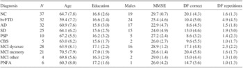
Exclusionary criteria included the presence of another neurologic condition, severe metabolic disorder, or other severe medical illness that was suspected to have a significant impact on behavior or cognition. Subjects were also excluded if they had a longstanding Axis I psychiatric disorder, major organ dysfunction, alcohol abuse or dependence within 5 years, head trauma with loss of consciousness greater than 30 minutes, deteriorating cardiovascular disease, or prominent white matter disease. Demographic and clinical variables are reported in Table 1 and were compared by group using analysis of variance with Tukey post hoc with α = .05. The controls and the patients diagnosed with the MCI (amnestic or dysexecutive subtype) scored higher on the MMSE than patients diagnosed with AD, bvFTD, or SD. In addition, the AD patients scored lower than patients with PSP or MCI other subtype. The MCI memory patients were older than the AD and the bvFTD patients. There were no other group differences in MMSE scores or age, and there were no group differences in education or gender. The study was approved by the UCSF committee on human research. All subjects provided written informed consent before participating.
Demographic characteristics and design fluency scores by diagnostic group

Note. Values represent mean (SD).
MMSE = Mini Mental State Examination; DF = design fluency; NC = normal control; bvFTD = behavioral variant frontotemporal dementia; AD = Alzheimer's disease; SD = semantic dementia; PSP/CBS = progressive supranuclear palsy or corticobasal degeneration; MCI = mild cognitive impairment; PNFA = progressive nonfluent aphasia.
Design Fluency Assessment
Subjects were administered the D-KEFS Design Fluency Test Conditions 2 and 3 by a research associate who was trained and supervised by a neuropsychologist. During each condition, subjects were presented with rows of squares containing an array of dots and are asked to draw as many different designs as possible in 60 s using only four lines connecting dots. The subject could view all previous designs drawn throughout the condition. Each condition began with a practice session during which the subject generated three designs and the examiner explained and corrected any errors. The requirement to make every design different was emphasized both during the practice trials and at the beginning of each condition. During each condition, the first time an examinee made two consecutive repetition errors, the examiner would say “make every design different.” Self-corrections were not allowed. Repetition errors and total correct were tabulated and summed across the conditions, as per standard D-KEFS scoring procedures. A repetition error is the generation of a previously emitted correct design.
Inter-rater reliability was examined in a subset of 14 randomly selected protocols. For total correct, r = .94, and for total repetitions, r = .84. To ensure scoring accuracy, all tests were scored by two raters.
Neuroimaging Data
MRI scans were obtained on a 1.5T Magnetom VISION system (Siemens, Iselin, NJ) at the San Francisco Veteran's Administration Hospital. A volumetric magnetization prepared rapid gradient-echo MRI (MPRG, TR/TE/TI = 10/4/300 ms) was used to obtain T1-weighted (MP-RAGE) images of the entire brain, 15-degree flip angle, coronal orientation perpendicular to the double spin-echo sequence, 1.0 × 1.0 mm in-plane resolution and 1.5 mm slab thickness.
Freesurfer Software Package
The T1 MPRAGE structural MR images were analyzed using Freesurfer version 4.0.2, which is documented and freely available online (http://surfer.nmr.mgh.harvard.edu). Previous publications have detailed and validated the software (Dale, Fischl, & Sereno, Reference Dale, Fischl and Sereno1999; Fischl, Liu, & Dale, Reference Fischl, Liu and Dale2001; Segonne et al., Reference Segonne, Dale, Busa, Glessner, Salat, Hahn and Fischl2004). Cortical regions were demarcated as described in Desikan et al. (Reference Desikan, Segonne, Fischl, Quinn, Dickerson, Blacker and Kiliany2006) and the thalamus and striatum (caudate + putamen) as described in Fischl et al. (Reference Fischl, Salat, Busa, Albert, Dieterich, Haselgrove and Dale2002). Figure 1 was rendered using the tool “tksurfer” and template brain included in the Freesurfer package using the default parcellation (Desikan et al., Reference Desikan, Segonne, Fischl, Quinn, Dickerson, Blacker and Kiliany2006); subcortical regions are not rendered.
Gray matter correlates of Design Fluency Total Correct and Repetitions, controlling for intracranial volume and Mini Mental State Exam scores.

Statistical Analyses
Statistical analyses were performed with PASW 17.0 for Windows (SPSS Inc., Chicago, IL). For all correlation and regression analyses with the gray matter volumes, ICV and MMSE were included as covariates. A square root transformation was performed on the repetition error scores to reduce positive skew.
To examine the neuroanatomical correlates of design fluency repetition errors and total correct designs, partial correlations were performed with all 56 regions in the frontal, parietal, and temporal cortex and with the four subcortical regions: the right and left thalamus and striatum. To control Type I error rate, a Bonferroni correction was applied to the alpha levels separately for the repetition error and for the total correct analyses by dividing .05 by 60; p values < .00083 were considered significant. To determine which regions contributed uniquely to repetition errors and to total correct designs, stepwise regression analyses with backward elimination were performed, including all regions that correlated significantly with these performance measures as predictors in the model. Regions were eliminated if they did not significantly predict the errors, α = .05, consecutively and starting with the weakest predictors. These analyses were performed separately for the right and left hemispheres due to high collinearity between corresponding regions (e.g., the right and left lateral OFC volumes correlate .77 in our sample). To facilitate interpretation of unstandardized regression coefficients for the regression analyses, gray matter volumes and ICV are expressed as cubic mm/10,000.
To assess diagnostic group differences in design fluency repetition errors and total correct designs, an analysis of variance with Tukey post hoc was conducted for each of these measures, comparing the performance of patient groups with sample size greater than 10. Specifically, we examined group differences in patients with a diagnosis of the following neurodegenerative disease syndromes: AD, bvFTD, SD, or PSP/CBS, as well as the two subtypes of MCI: dysexecutive subtype and amnestic subtype, and neurologically healthy controls. PSP and CBS were combined because these disorders share many clinical and pathological features and because clinical diagnostic separation of these syndromes may be unreliable (Josephs, Reference Josephs2008; Lee et al., Reference Lee, Rabinovici, Mayo, Wilson, Seeley, Dearmond and Miller2011).
Results
Neuroanatomical Correlates of Design Fluency Test Performance
Repetition errors
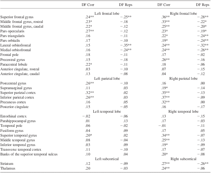
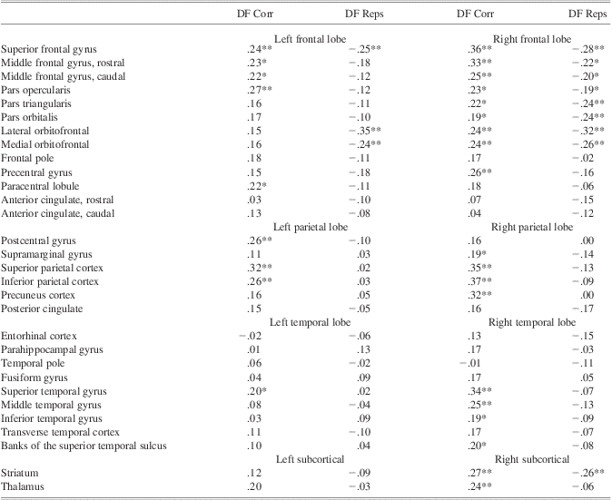
As reported in Table 2 and depicted in Figure 1, in the frontal lobes, a propensity to make repetition errors was significantly associated with reduced gray matter in all orbitofrontal cortex (OFC) regions: right lateral OFC, r = −.32, left lateral OFC, r = −.35, right medial OFC, r = −.26, left medial OFC, r = −.24, all ps < .00083. Repetition errors were also associated with reduced gray matter in three right lateral prefrontal cortex regions: the right superior frontal gyrus: r = −.28, the right pars triangularis, r = −.24, and the right pars orbitalis, r = −.24, and one left lateral prefrontal region, the left superior frontal gyrus: r = −.24, all ps < .00083. Repetition errors were not significantly associated with the other frontal regions including other lateral prefrontal cortex regions, the anterior cingulate regions, the precentral gyrus, or the paracentral lobule (all ps > .00083). No regions in the parietal or temporal lobes correlated significantly with repetition errors at the corrected threshold or at a more lenient exploratory threshold of p < .01 uncorrected. Of the subcortical volumes, the only significant correlation was with the right striatum, r = −.26.
Correlations of design fluency total correct and repetition errors with gray matter regions
Note. All correlations control for Mini Mental State Exam scores and intracranial volume.
DF corr = design fluency total correct designs; DF reps = design fluency repetition errors.
**p < .00083.
*p < .01, uncorrected.
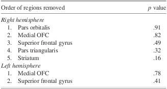
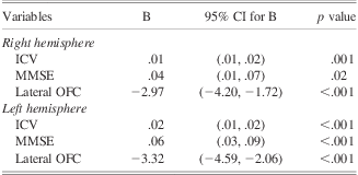
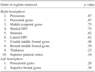
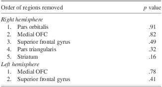
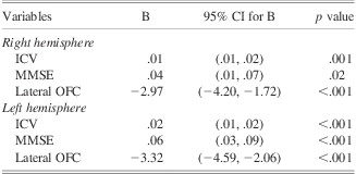
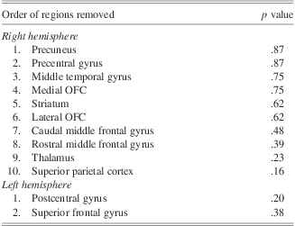
Stepwise regression with backward elimination was performed to determine which of the regions that correlated significantly with design fluency repetition errors predicted a significant amount of unique variance after controlling for ICV and MMSE scores. Regions were eliminated from the final model if they did not significantly predict the errors, α = .05, consecutively and starting with the weakest predictors (Table 3). In the final model for each hemisphere, only the lateral OFC was retained as a significant predictor (Table 4).
Summary of backward elimination for regions predicting repetition errors
Final model predicting design fluency repetitions, adjusting for Mini Mental State Exam (MMSE) scores and total intracranial volume (ICV)
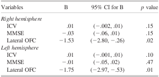
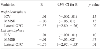
We explored whether similar neuroanatomical correlates for repetition errors would be observed in our sample of subjects who showed some propensity to make repetition errors (>1) after removing the subjects with a bvFTD diagnosis (N = 69). The purpose of these analyses was to explore whether the pattern of results discussed above are specific to bvFTD or if they may generalize to other patients who tend to make these errors. No partial correlations were significant with the Bonferroni correction in this smaller sample. When we removed the multiple comparison correction on a strictly exploratory basis, a similar pattern of associations emerged as were present in the full sample. Repetition errors correlated with the right lateral OFC, r = −.29, p = .02, the left lateral OFC, r = −.34, p = .01, left medial OFC, r = −.27, p = .03, and there were trends with the right medial OFC, r = −.21, p = .09, and the right superior frontal gyrus, r = −.22, p = .08. The correlations with the right pars triangularis, r = .01, p = .97, right pars orbitalis, r = −.05, p = .67, the left superior frontal gyrus, r = −.18, p = .15, and the right striatum, r = −.13, p = .30, however, were not significant. A significant correlation was also observed with the right posterior cingulate at the uncorrected threshold, r = −.27, p = .03. Significant correlations were not observed with any other regions in the frontal, parietal, or temporal cortex or the subcortical regions, even at this lenient exploratory threshold (all ps > .05, uncorrected). Stepwise regression analyses with backward elimination were performed including the same regions as were included in the regressions for the full sample and also including the right posterior cingulate. For the right hemisphere analysis, regions were eliminated in the following order: Step 1: medial OFC, p = .93; Step 2: the superior frontal gyrus, p = .85; Step 3: the striatum, p = .55; Step 4: the pars orbitalis, p = .24; Step 5: the pars triangularis p = .19; Step 6: the posterior cingulate, p = .08. In the final model, only the lateral OFC was retained, B = −1.53, p = .02. For the left hemisphere analysis, the medial OFC was removed, p = .59, and the lateral OFC was retained, B = −8.57, p = .002 (Table 5).
Final model predicting design fluency repetitions in the sample without behavioral variant frontotemporal dementia, adjusting for Mini Mental State Exam (MMSE) scores and total intracranial volume (ICV)
Total correct designs
Partial correlations coefficients of total correct with all cortical and subcortical regions are reported in Table 2 and Figure 1. Significant correlations were found with several regions in the right and left frontal, right and left parietal, and right temporal lobes, and also with the right striatum and the right thalamus.
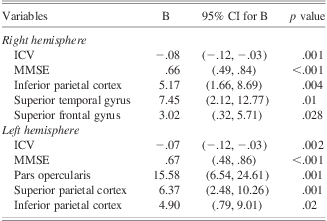
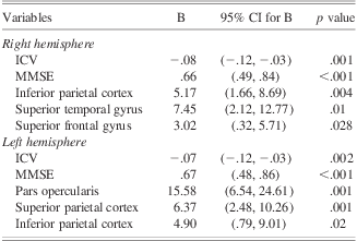
Stepwise regression analyses with backward elimination were performed to determine which of the regions that correlated significantly with design fluency total correct predicted a significant amount of unique variance in design fluency total correct, controlling for ICV and MMSE scores. Regions were eliminated from the final model if they did not significantly predict total correct scores, α = .05 (Table 6). In the final right hemisphere model, the inferior parietal cortex, superior temporal gyrus, and the superior frontal gyrus were retained. In the final left hemisphere model, the pars opercularis, superior parietal cortex, and the inferior parietal cortex were retained (Table 7).
Summary of backward elimination for regions predicting total correct designs
Final model predicting design fluency total correct designs, adjusting for Mini Mental State Exam (MMSE) scores and total intracranial volume (ICV)
Diagnostic Group Differences in Design Fluency Test Performance
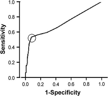
Analysis of repetition errors revealed a significant effect of diagnostic group, F(6,183) = 6.77, p < .001. BvFTD patients made more repetition errors than controls and all other patient groups (all ps < .01). The other patient groups and controls did not differ from each other in the number of repetition errors (Table 1). Effect size differences in repetition errors were large between the bvFTD patients and the healthy controls, d = 1.00, the bvFTD patients and all the other patients combined, d = .92. Whereas 53% of the bvFTD patients made more than four repetition errors, 0% of the healthy controls and 8% of all non-bvFTD patients combined made more than four errors. The non-bvFTD patients who made more than four errors were diagnosed with SD (3), MCI-dysexecutive (3), AD (2), PSP (1), and MCI-amnestic (1). Using an optimal cut point of >4, repetition errors demonstrated excellent specificity (.92) with lower sensitivity (.53) for bvFTD versus other patient diagnosis. This cut point was considered optimal because, although lower cut points increased sensitivity, the increases were offset by larger decreases in specificity; for example, a cut point of two repetition errors demonstrated a slightly increased sensitivity of 60% but a much lower specificity of 72%. See Figure 2 for the receiver operating characteristics (ROC) curve.
Receiver operating characteristics (ROC) curve depicting sensitivity and 1-specificity values for separating behavioral variant frontotemporal dementia (bvFTD) patients from the other patients. The optimal cutoff of more than four repetition errors is circled.

The analysis of variance with total correct designs, F(6,183) = 31.56, p < .001, indicated that all four groups of dementia patients generated fewer correct designs than controls or patients with MCI amnestic subtype or MCI dyexecutive subtype. The MCI groups did not differ from controls or from each other. Among the patients diagnosed with the four types of dementia, the only significant difference was that SD patients generated more designs than patients with AD; otherwise the groups performed similarly.
Discussion
The purpose of this study was to investigate the neural substrates and clinical utility of design fluency repetition errors for patients with neurodegenerative disease. A high propensity to make repetition errors on design fluency was associated with atrophy in several regions in the frontal lobes and with the right striatum. The errors did not correlate with any regions in the parietal or temporal lobes even at a lenient uncorrected significance threshold. Multiple regression analyses indicated that the right and left lateral OFC uniquely predicted error propensity. In contrast, the total number of correct designs, which is the score typically used from this test, showed more widespread cortical correlates that included regions in the right and left frontal lobes, the right and left parietal lobes, the right temporal lobe, and the right striatum and thalamus. Multiple regression analyses indicated that frontal (the right superior frontal gyrus and the left pars opercularis), parietal (the right and left inferior parietal cortex and the left superior parietal cortex), right temporal (the right superior temporal gyrus) regions uniquely predicted design fluency total correct. Taken together, design fluency repetition errors may be a more useful measure for making inferences about frontal system integrity, whereas a low number of total correct designs could signal frontal, parietal, and/or right temporal dysfunction.
The study results also highlight the utility of design fluency repetition errors for assisting in the clinical diagnosis of bvFTD, and indicate that total correct designs is less useful for this purpose. Whereas making an elevated number of repetition errors (>4) was common for patients diagnosed with bvFTD (53%), it was rare for patients diagnosed other types of neurodegenerative disease or mild cognitive impairment (8%), and was not observed in any of our controls, indicating excellent diagnostic specificity for bvFTD. In contrast, the different dementia diagnostic groups achieved similar numbers of total correct designs, with the exception that the AD patients made fewer than the SD patients.
Design fluency total correct was historically assumed to reflect frontal lobe integrity (Baldo, Shimamura, Delis, Kramer, & Kaplan, Reference Baldo, Shimamura, Delis, Kramer and Kaplan2001; Elfgren & Risberg, Reference Elfgren and Risberg1998). This test relies on multiple cognitive processes including processing speed, motor planning, visual scanning, and cognitive flexibility. Perhaps because of its multi-factorial nature, there is growing evidence that design fluency total correct reflects the integrity of a diffuse network of cortical regions (Kramer et al., Reference Kramer, Quitania, Dean, Neuhaus, Rosen, Halabi and Millwe2007; Pa et al., Reference Pa, Possin, Wilson, Quintania, Kramer, Boxer and Johnson2010). In this study, we identified several correlates throughout the right and left frontal and parietal lobes and the right temporal lobe, and with the right striatum and the right thalamus. In contrast, whereas repetition errors correlated with volumes of several frontal regions, the errors did not correlate significantly with any parietal or temporal lobe subregion volumes, even when a lenient significance level without multiple comparisons correction was applied on an exploratory basis.
The present study's findings highlight the frontal-anatomic specificity of design fluency repetitions errors and raise the question of why some patients show a propensity to make these errors from a neuropsychological standpoint. On design fluency, patients are instructed to make as many designs as quickly as possible while also trying to minimize repetitions. Good performance, therefore, requires a subject to shift their attentional resources between response generation and response monitoring. These two task demands are associated with conflicting contingencies: to direct attentional resources toward response monitoring, one may need to sacrifice generation speed. The motivation to shift between task goals is fueled by social drives, for example, to please the examiner. Consistent with this conceptualization, the OFC is thought to be critical for self-monitoring and subsequent regulation based on rewarding and punishing contingencies (e.g., in this case, producing a high number of designs is rewarding and repeating designs is punishing, in terms of the patient's experience of success) (Bechara, Reference Bechara2004; Viskontas, Possin, & Miller, Reference Viskontas, Possin and Miller2007). The lateral OFC in particular is thought to be critical for using punishing cues to modulate behavior (Kringelbach & Rolls, Reference Kringelbach and Rolls2004; Liu et al., Reference Liu, Powell, Wang, Gold, Corbly and Joseph2007; O'Doherty, Kringelbach, Rolls, Hornak, & Andrews, Reference O'Doherty, Kringelbach, Rolls, Hornak and Andrews2001), particularly in response to social cues (Liu et al., Reference Liu, Powell, Wang, Gold, Corbly and Joseph2007).
The right and left lateral OFC appear to be crucial for preventing the repetition errors because they predicted unique variance in error propensity even when the other regions that correlated with repetition errors were included in the regression models. In contrast, although the right and left medial OFC, the right and left superior frontal gyrus, the right pars triangularis, the right pars orbitalis, and the right striatum correlated robustly with these errors even after multiple comparison correction, they did not contribute significant unique variance when lateral OFC was included in the model; thus, the critical importance of these regions in preventing these errors is less clear. Nevertheless, it is well-established that the superior and inferior prefrontal cortex play important roles in working memory and cognitive flexibility (D'Esposito, Reference D'Esposito2007; Manes et al., Reference Manes, Sahakian, Clark, Rogers, Antoun, Aitken and Robbins2002; Muller & Knight, Reference Muller and Knight2006; Robbins, Reference Robbins1996; Wager & Smith, Reference Wager and Smith2003), and it may be that these lateral frontal regions help patients to keep track of and continuously update which designs they have already generated, and to shift between representations of possible designs. The ventral portion of the striatum supports behavior regulation via its massive corticostriatal connections from limbic regions, including the orbitofrontal cortex, and the anterodorsal striatum supports executive function via its corticostriatal connections from dorsolateral prefrontal cortex (Heimer & Van Hoesen, Reference Heimer and Van Hoesen2006; Schmahmann & Pandya, Reference Schmahmann and Pandya2008; Yeterian & Pandya, Reference Yeterian and Pandya1991). Of note, more correlations with the lateral prefrontal cortex and with the striatum were observed in the right hemisphere, which supports the view that right frontal systems may be particularly important for cognitive monitoring (Possin et al., Reference Possin, Brambati, Rosen, Johnson, Pa, Weiner and Kramer2009; Stuss, Reference Stuss2011).
An important question is whether these findings would generalize to repetition errors made on other tests. In particular, repetition errors are frequently observed on tests of verbal fluency and verbal list learning, but few studies have examined the neural bases of repetition errors on these tests. Some evidence suggests that verbal fluency repetitions could arise from left temporal lobe atrophy or brain injury and that they are not specifically frontal (Hotz & Helm-Estabrooks, Reference Hotz and Helm-Estabrooks1995; Possin et al., Reference Possin, Brambati, Rosen, Johnson, Pa, Weiner and Kramer2009); in other words, verbal repetitions may arise from damage to verbal processing areas. In contrast, the present study suggests that design fluency repetitions are not significantly predicted by damage to visual processing areas, but rather arise from frontal regions important for executive control. In a study of verbal and design fluency repetitions in aging, making repetitions on verbal fluency tasks did not increase the probability of making repetitions on a design fluency task (Foldi, Helm-Estabrooks, Redfield, & Nickel, Reference Foldi, Helm-Estabrooks, Redfield and Nickel2010). Furthermore, while design fluency repetitions increased as a function of age, a similar effect was not found for letter or category fluency. Verbal fluency and memory repetitions have been shown not to correlate significantly with a large number of traditional neuropsychological test indices including measures of executive function, processing speed, language, and memory (Possin et al., Reference Possin, Filoteo, Roesch, Zizak, Rilling and Davis2005) and have been shown not to be elevated in patients with executive function deficits (Brooks, Weaver, & Scialfa, Reference Brooks, Weaver and Scialfa2006). Although more work is needed to understand the neural underpinnings, underlying cognitive processes, and possible clinical significance of verbal repetition errors, they appear to be measuring something different from design fluency repetitions. Modality distinctions in perseveration, which have been only minimally emphasized by leading classification schemes, may be important for making inferences about etiology.
With the exception of the bvFTD patients, our subjects made very few design fluency repetition errors; in fact, only 10 of the 168 participants without a bvFTD diagnosis made more than four repetition errors. This specificity highlights the utility of this measure for the identification of bvFTD, discussed below, but it made it difficult to determine whether the neuroanatomical correlates of these errors would be similar in other patients who also show a propensity to make these errors. Nevertheless, when we explored the neuroanatomical correlates of repetition errors in the non-bvFTD subjects with at least a mild propensity to make these errors (>1), a frontally-specific pattern was again identified and the regression analyses indicated that the right and left lateral OFC significantly predicted repetition propensity. Based on these findings, the frontal-specificity of design fluency repetition errors and the importance of the lateral OFC in preventing these errors do appear to generalize to patients without bvFTD.
In our sample of neurodegenerative disease and MCI patients, the bvFTD patients were unique in their tendency to make an elevated number of repetition errors; in fact, they made more repetition errors than patients diagnosed with AD, SD, PSP/CBS, or MCI. The specificity of repetition errors for bvFTD versus other diagnosis was very high (92%), whereas the sensitivity was lower (53%). Similarly, rule violation errors quantified on a test of spatial planning showed good specificity (80%) and lower sensitivity (50%) for bvFTD versus AD diagnosis (Carey et al., Reference Carey, Woods, Damon, Halabi, Dean, Delis and Kramer2008). These findings suggest that when incorporating analysis of rule violation errors or design fluency repetition errors into a neurodegenerative disease evaluation, the presence of a high number of these errors supports the diagnosis of bvFTD, whereas the absence of these errors has less clinical utility.
Misdiagnosis rates of bvFTD are high (Rascovsky et al., Reference Rascovsky, Hodges, Kipps, Johnson, Seeley, Mendez and Miller2007; Woolley, Khan, Murthy, Miller, & Rankin, Reference Woolley, Khan, Murthy, Miller and Rankin2011) and a major challenge of neuropsychology has been to identify standardized tests that differentiate bvFTD from other neurodegenerative diseases or normal controls. BvFTD patients typically show relative preservation of visuospatial skills and memory in comparison to patients with AD (Hodges et al., Reference Hodges, Patterson, Ward, Garrard, Bak, Perry and Gregory1999; Kramer et al., Reference Kramer, Jurik, Sha, Rankin, Rosen, Johnson and Miller2003; Rascovsky et al., Reference Rascovsky, Salmon, Ho, Galasko, Peavy, Hansen and Thal2002). The identification of measures that show greater impairment in bvFTD compared to other diagnostic groups has been less successful. In early bvFTD, patients often perform normally on traditional executive function tests, perhaps because at this stage the severe effects of the disease are restricted to orbital and medial aspects of prefrontal cortex and do not substantially involve the lateral prefrontal cortex (Perry et al., Reference Perry, Graham, Williams, Rosen, Erzinclioglu, Weiner and Hodges2006; Seeley et al., Reference Seeley, Crawford, Rascovsky, Kramer, Weiner, Miller and Gorno-Tempini2008). As the disease progresses, impairments on traditional executive function tests usually emerge, but these impairments often do not reliably differentiate bvFTD from AD (Giovagnoli, Erbetta, Reati, & Bugiani, Reference Giovagnoli, Erbetta, Reati and Bugiani2008; Jenner, Reali, Puopolo, & Silveri, Reference Jenner, Reali, Puopolo and Silveri2006), consistent with the results from this study that patients with bvFTD did not differ from patients with AD, SD, or PSP/CBS on design fluency total correct. Changes in behavior and personality are the hallmark early features of bvFTD, and methods of quantifying these changes have shown promise for supporting diagnosis (Levy, Miller, Cummings, Fairbanks, & Craig, Reference Levy, Miller, Cummings, Fairbanks and Craig1996; Rankin, Kramer, & Miller, Reference Rankin, Kramer and Miller2005; Rankin et al., Reference Rankin, Santos-Modesitt, Kramer, Pavlic, Beckman and Miller2008; Salmon et al., Reference Salmon, Perani, Collette, Feyers, Kalbe, Holthoff and Herholz2008). In addition, the results of this study augment a growing body of literature that the analysis of certain cognitive error types can also help identify bvFTD (Carey et al., Reference Carey, Woods, Damon, Halabi, Dean, Delis and Kramer2008; Kramer et al., Reference Kramer, Jurik, Sha, Rankin, Rosen, Johnson and Miller2003; Libon et al., Reference Libon, Xie, Moore, Farmer, Antani, McCawley and Grossman2007; Thompson et al., Reference Thompson, Stopford, Snowden and Neary2005).
In conclusion, the results of this study indicate that an elevated propensity to make design fluency repetition errors is associated with frontal atrophy but not with atrophy in the parietal or temporal lobes in patients with neurodegenerative disease. Within the frontal lobes, the errors were associated with reduced volumes in the right and left OFC, the right superior and inferior frontal gyri, and the left superior frontal gyrus, and the errors were also associated with smaller right striatal volumes. The lateral OFC volumes predicted unique variance in repetition errors, suggesting a particularly important role of this region in preventing these errors. In contrast to repetition errors, design fluency total correct correlated with volumes in a diffuse network of bilateral frontal, bilateral parietal, right temporal, and right subcortical regions and thus lack frontal-anatomic specificity. The tendency to make an elevated number of repetition errors was much more common in patients with bvFTD than in patients with other types of dementia or mild cognitive impairment, highlighting their clinical utility.
Acknowledgments
This work was supported by the Larry L. Hillblom Foundation [2008-A-020-FEL and 2007/2I], and the National Institute on Aging [5 P01 AG019724, 3 P50 AG023501, 1K23AG037566-01A1, 1 R01 AG022983-01, 1 R01 AG032289]. The authors have no conflicts of interest to report. This work has not been published previously either electronically or in print.