Case report/brief report
Our patient was 42-year-old woman with tricuspid atresia, who had undergone previous atriopulmonary Fontan. She was symptomatic with abdominal swelling, shortness of breath, and palpitations. By echocardiogram, her right atrium was massively dilated, and her single ventricle ejection fraction was 40%. Cardiac catheterisation showed a right atrial pressure of 20 and a pulmonary capillary wedge pressure of 13. She was referred for Fontan conversion secondary to intractable arrhythmias – atrial fibrillation and flutter – despite three previous ablations and sotalol therapy. Further work-up by CT scan and liver biopsy showed enlargement of the liver and focal peri-central sinusoidal fibrosis, both suggestive of a chronic, passive congestive hepatopathy. Liver ultrasound revealed a dilated inferior caval vein and flow reversal during systole. Unfortunately, social behaviour and non-compliance issues would likely preclude her from future heart or heart/liver transplantation. We wanted to perform a Fontan conversion to eliminate the right atrium from the Fontan pathway and reduce her arrhythmia burden, but also wanted to protect her liver from further decompensation and the potential need for liver transplantation. Thus, we performed a Fontan conversion with hepatic vein exclusion.
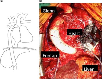
Midline sternotomy and laparotomy incisions were made, as well as a left groin incision. The necessary dissection was performed, and she was cannulated for cardiopulmonary bypass. The common femoral vein, the supra-hepatic inferior caval vein, and the innominate vein were cannulated for venous drainage; the common femoral via an 8 mm end-to-side graft was cannulated for arterial return. Her operation consisted of the following components: takedown of the atriopulmonary connection; superior and inferior cavopulmonary anastomoses; atrial septectomy, bi-atrial maze procedure, and right atrial plication; and dual chamber epicardial pacemaker implantation. All were performed in the standard manner, except for the Glenn and the extra-cardiac Fontan conduit. Owing to the location of the superior caval vein and the main pulmonary artery, the Glenn connection was performed using a short inter-position graft. For the extra-cardiac Fontan, a 22-mm ringed Gore-Tex tube (W. L. Gore and Associates, Flagstaff, Arizona, United States of America) was passed through a small diaphragmatic defect and behind the left lobe of the liver. The superior aspect of the conduit was anastomosed to the pulmonary artery, and the inferior aspect of the conduit was anastomosed to the infra-hepatic inferior caval vein, rather than the supra-hepatic caval vein. The infra-hepatic inferior caval vein was then ligated just above the conduit insertion site (Fig 1).
Operative images. ( a ) Schematic showing Fontan with hepatic vein exclusion. ( b ) Intra-operative view showing Glenn and extra-cardiac Fontan conduit. The intra-abdominal portion is hidden behind the left lobe of the liver. The nodular appearance of the liver can be appreciated.

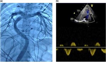
Her postoperative haemodynamics were favourable. Pressures within the Fontan pathway were mildly elevated at 15 mmHg. Common atrial pressure, and thus pressure transmitted to the splanchnic venous system, was low at 8 mmHg. Systemic oxygen saturations were 83%. Pre-discharge cardiac catheterisation demonstrated a widely patent extra-cardiac conduit (Fig 2). She was discharged on warfarin with a goal INR between 2 and 3.
Postoperative imaging. ( a ) Postoperative cardiac catheterisation shows a patent extra-cardiac Fontan conduit from the intra-hepatic inferior caval vein to the pulmonary artery. ( b ) Postoperative hepatic venous Dopplers are normal with minimal flow reversal during late systole.

At the most recent follow-up at 18 months after surgery, she was asymptomatic with a normal daily routine. Her abdominal fullness and fatigue were much improved. She has had no documented atrial arrhythmias on sotalol. Her oxygen saturations were typically 85%. Her follow-up hepatic venous Doppler studies were normal, suggesting decompression of the liver and intestines (Fig 2).
Comments
Patients with single ventricle heart disease often undergo staged surgical palliation resulting in Fontan anatomy and physiology. Although operative survival is reported to be 95% or greater, the 10-year survival rate ranges from 60 to 81%.Reference Mahle, Spray, Wernovsky, Gaynor and Clark 1 , Reference Lee, Choi, Kang, Bae, Kim and Rho 2 The reduction in long-term survival is due to ventricular dysfunction, increased pulmonary vascular resistance, arrhythmias, protein-losing enteropathy, and hepatic complications.Reference de Laval and Deanfield 3 – Reference Ghaferi and Hutchins 6
Of specific interest regarding this operation are the debilitating effects of Fontan circulation on the liver and intestines. The increase in hepatic venous pressure likely contributes to chronic passive congestion, hepatic cirrhosis, hepatic adenoma, hepatocellular carcinoma, ascites, and formation of arteriovenous malformations. The increase in mesenteric venous pressure likely contributes to varices and protein-losing enteropathy.
Attempts have been made previously to exclude the hepatic venous return from the Fontan pathway;Reference Reed, Leonard, Zellers and Nikaidoh 7 , Reference Schneider, Banerjee, Mendelsohn and Norwood 8 however, the majority of attempts were made at the supra-hepatic level or with partial hepatic vein exclusion, resulting in the formation of intense intra-hepatic collaterals between the high-pressure and low-pressure systems.Reference Reed, Leonard, Zellers and Nikaidoh 7 , Reference Schneider, Banerjee, Mendelsohn and Norwood 8 A previous animal study used a model similar to ours with favourable results: few veno-venous collaterals formed in the presence of favourable haemodynamics, and, when they did, the location was consistent and potentially amenable to catheter-based occlusion.Reference Brizard, Goussef, Chachques and Carpentier 9
The proposed novel Fontan operation may relieve the elevated Fontan pressure on the liver and intestines; however, the obvious concern is the development of pulmonary arteriovenous malformations and worsening cyanosis from the lack of an unidentified “hepatic factor” in the lungs. Should this occur, additional strategies could be used to overcome this obstacle. First, a systemic-to-pulmonary artery shunt, upper extremity arteriovenous fistula, or neck artrerio-venous fistula could be performed. This would augment pulmonary blood flow and allow aortic blood with “hepatic factor” to be diverted to the pulmonary vasculature.Reference Garg, Talwar, Kothari, Rajashekar, Saxena and Airan 10 Second, the hepatic veins could be subsequently re-incorporated into the Fontan circuit if necessary. Although this would again expose the liver and intestines to elevated pressures, the onset of harmful sequelae could be delayed.
Ongoing research is needed to improve the lives of patients with Fontan-type physiology, and this modification may play a role in the staged palliative approach in these patients. At present, we would consider this particular configuration in adults with atriopulmonary Fontan anatomy and physiology who are good candidates for Fontan conversion. As in our patient, we hope that this modification will improve clinical symptoms, arrhythmia burden, and visceral health. We also hope that this modification will reduce the overall morbidity and mortality associated with liver and intestinal complications inherent to Fontan anatomy and physiology. Ultimately, the reconstruction set forth by this model may also play a role in children undergoing initial staged palliation and in adults with total cavopulmonary Fontan anatomy and physiology.
Acknowledgement
None.
Financial Support
This research received no specific grant from any funding agency, commercial, or not-for-profit sectors.
Conflicts of Interest
None.
Ethical Standards
The Institutional Review Board was queried and approval was granted to perform this operation and case review. Consent was waived.