Introduction
The subperiosteal pocket technique, which has become popular over the last decade, is a surgical technique used in cochlear implantation. Unlike the standard technique, with this technique there is no need to drill a bone bed in the skull.
Implant migration is one of the major concerns of cochlear implant surgery. To avoid this complication, in the standard technique, a bone bed is created for the internal receiver-stimulator, and various fixation techniques are used. However, by using the subperiosteal pocket technique in paediatric revision cases, we have demonstrated spontaneous bone bed formation, at least six months after surgery.Reference Orhan, Polat, Enver, Celik, Güldiken and Değer1 In terms of receiver-stimulator stabilisation, there is no significant difference between spontaneous and created bone bed formation. However, the risk of migration may increase, especially for patients for whom the subperiosteal pocket is not sufficiently tight to entrap the receiver-stimulator, in the early post-operative period before formation of the spontaneous bone bed.
We observed anterior implant migration resulting in exposure of the receiver-stimulator in the early post-operative period in two patients for whom the subperiosteal pocket technique with standard Palva flap was performed. In these patients, we performed re-implantation by drilling the calvarium, as described for the standard technique. Thus, we considered that we needed to modify our technique.
In this retrospective clinical study, a new periosteal incision is described for placing the receiver and avoiding migration in the early post-operative period.
Materials and methods
This study comprised 280 patients (aged between 11 months and 72 years; mean ± standard deviation (SD) = 74 ± 134 months) who underwent cochlear implantation with the subperiosteal technique between June 2008 and January 2013. All implantations were performed by two surgeons (YG and KSO), who used the same surgical technique.
Surgical technique
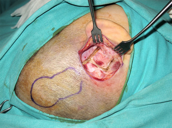
The probable implant area was marked on the skin and a small C-shaped retroauricular skin incision was made. Following elevation of the retroauricular skin, the periosteum of the temporal bone was exposed. The temporal lines were determined by palpation. Along this line, a 2 cm horizontal periosteal incision was made from the zygomatic root towards the posterior using electrocautery. A second periosteal incision was made parallel to the first incision at the mastoid tip. The two periosteal incisions were connected by a posterior vertical incision, and an anterior-based quadrangular Palva flap was created. Additionally, at the posterosuperior corner of the flap, we extended the superior incision by 2 cm posteriorly. This extended incision provides easy access for subperiosteal pocket preparation and placement of the receiver-stimulator. We have called this modified incision a ‘tailed Palva flap’ (Figure 1).
Periosteal incisions.

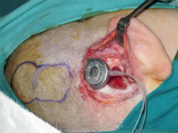
A subperiosteal pocket was created using a Freer elevator (Karl Storz, Tuttlingen, Germany). Subsequently, we performed standard mastoidectomy, posterior tympanotomy and cochleostomy. The implant and reference electrode, if present, were placed in the subperiosteal pocket (Figure 2).
Receiver-stimulator is placed into subperiosteal pocket.

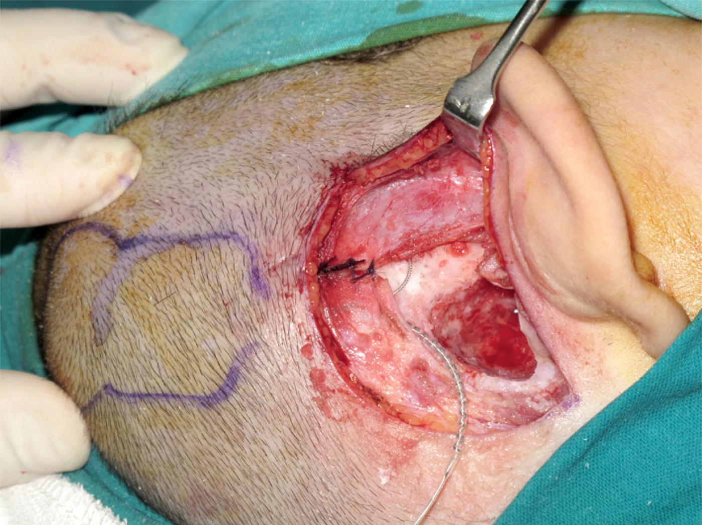
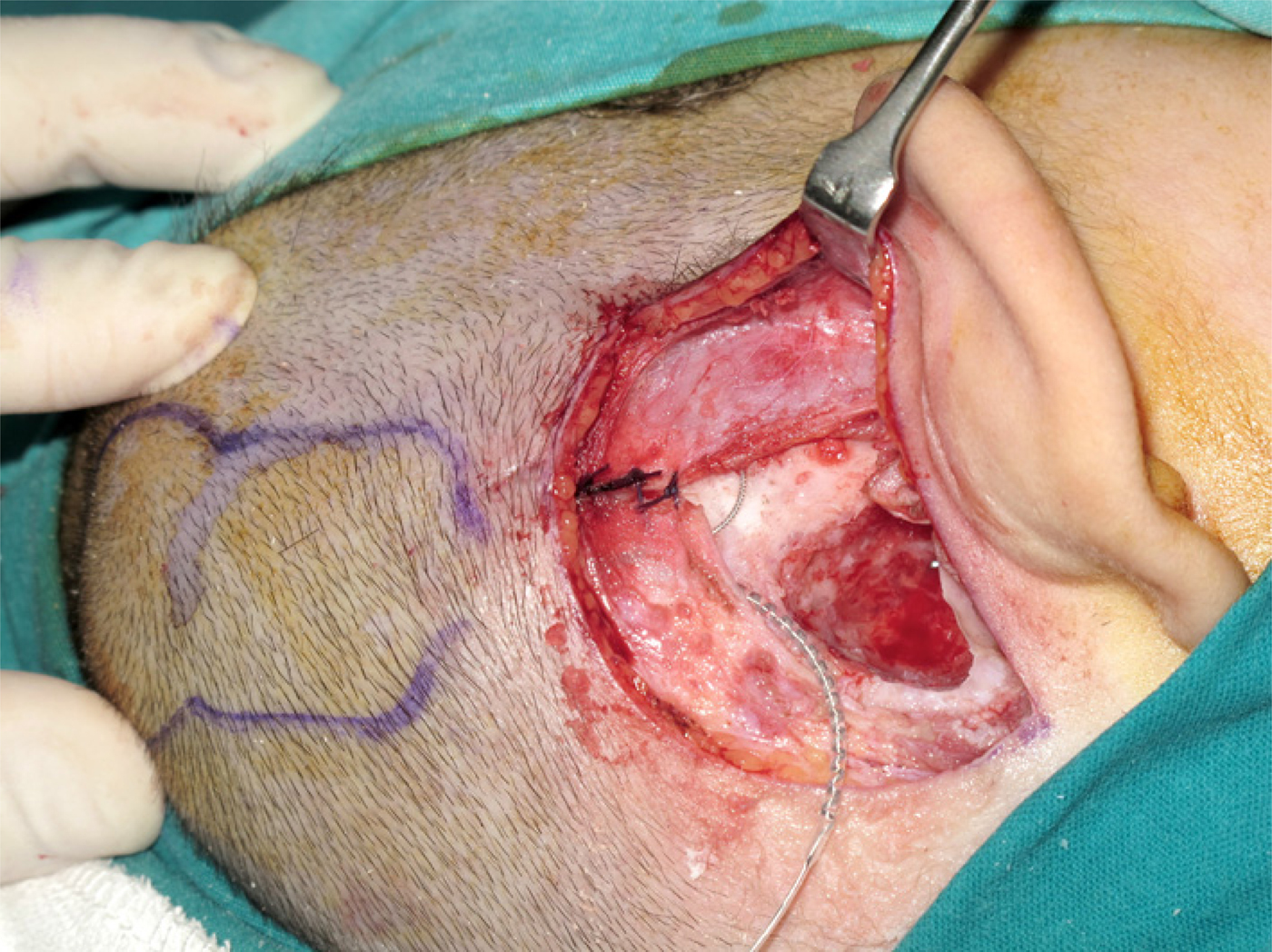
The first 3-0 polyglycolic acid suture was placed at the posterosuperior corner of the Palva flap, and the second suture was placed just posterior to the first suture (Figure 3). These sutures enhance the stabilisation of the receiver-stimulator by aiding entrapment of the silicon-coated connection point of the electrodes and the receiver-stimulator.
Posterior incision was sutured after placing receiver-stimulator.
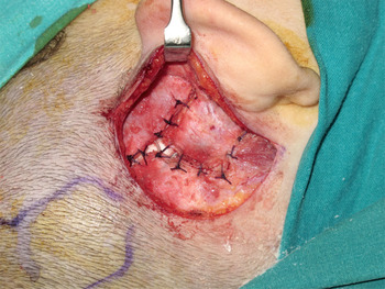
The electrodes were placed through the posterior tympanotomy and cochleostomy site. After electrophysiological tests, the remaining periosteal incisions were closed with 3-0 polyglycolic acid sutures (Figure 4).
Periosteal incisions were closed with 3-0 polyglycolic acid sutures.

Results
The patients were followed up for between 11 and 74 months (mean ± SD = 34 ± 19 months). The brands of implants used were Advanced Bionics (Valencia, California, USA) (59 patients), Medel (Innsbruck, Austria) (63 patients), Digisonic (Neurelec, Vallauris, France) (16 patients) and Nucleus (Cochlear, Lane Cove, Australia) (142 patients).
No intracranial complications were seen during or after the operations. In addition, we found no macroscopic migration of the implant.
We performed revision surgery in eight patients (3 per cent). The reasons for revision surgery were: ‘soft device failure’ (two patients), facial stimulation and paralysis (one patient), local flap failure (one patient), broken electrode (one patient), reference electrode failure (one patient), ‘hard device failure’ (one patient) and migration of the electrode towards the vestibule (one patient). In only one of the revision patients (0.4 per cent) was there a local flap failure and infection of the implant. This patient's implant was explanted and re-implantation was performed on the contralateral side.
Discussion
The subperiosteal pocket technique has become popular over the last few years. In 2009, Balkany et al. described the temporalis pocket technique. They reported the anatomical limitations of the temporalis pocket, which is bound ‘anteriorly by dense condensations of pericranium anteriorly at the temporal-parietal suture, posteroinferiorly at the lamboid suture, and anteroinferiorly by the bony ridge of the squamous suture’.Reference Balkany, Whitley, Shapira, Angeli, Brown and Eter2 We started to perform the subperiosteal pocket technique in 2008, before Balkany and colleagues' study. Our comparative clinical study showed that the mean operation time for the subperiosteal technique was 30 per cent shorter than that for the standard technique.Reference Güldiken, Orhan, Yiğit, Başaran, Polat and Güneş3
A large skin incision may cause flap necrosis because of the disruption of blood supply to the flap. Ray et al. reported that there were wound and flap problems in 2.3 per cent of cases with large skin incisions and in 1.1 per cent of cases with small skin incisions. The incidence of wound and flap problems was reduced with small skin incisions and minimal scalp mobilisation.Reference Ray, Gibson and Sanli4 We also used a small postauricular skin incision. As a result, we observed wound and flap problems in only one patient (0.4 per cent).
Ulug and Teker described a three-layer flap based on using the superficial musculoaponeurotic system as an additional, strong flap layer. They reported that this flap provided complete coverage and secure fixation for the implant, without any additional procedures and with reduced operation time.Reference Ulug and Teker5 The modified periosteal incision that we have described had a similar purpose of permitting secure fixation of the receiver-stimulator.
Balkany et al. reported that the anatomical limitations, including the lamboid suture, the bony ridge of the squamous suture and the pericranium, can enable fixation of the receiver-stimulator and prevent implant migration.Reference Balkany, Whitley, Shapira, Angeli, Brown and Eter2 Although there is little information about implant migration in the literature,Reference Balkany, Whitley, Shapira, Angeli, Brown and Eter2, Reference Bayazit, Goksu and Ozbilen6–Reference Orhan, Guldiken, Basaran, Ulusan, Polat and Çelik9 we observed early implant migration after implantation in two patients who had been operated on using the subperiosteal pocket technique but without the tailed Palva flap. We have not observed migration of the implant since we modified the periosteal incision (n = 280 at the time of writing). We believe that the first suture placed at the posterosuperior corner of the periosteal flap secures the receiver-stimulator and prevents migration in the early post-operative period. There is a minimal risk of implant migration after spontaneous bone bed formation.
In conclusion, the tailed Palva flap is a safe and reliable technique that provides secure fixation of the receiver-stimulator and prevents anterior migration in the early post-operative period.
Acknowledgement
The authors would like to acknowledge the efforts of Mehmet Çelik (Şerafeddin Sabuncuoğlu Education and Training Hospital, Amasya, Turkey).