Introduction
Concussions are often diagnosed based on their transient neurological, cognitive, and clinical symptoms due to the lack of evidence of an injury on traditional magnetic resonance imaging (MRI) or computerized tomography scans (Bazarian et al., Reference Bazarian, Zhong, Blyth, Zhu, Kavcic and Peterson2007; Hughes et al., Reference Hughes, Jackson, Mason, Berry, Hollis and Yates2004; Iverson, Lovell, Smith, & Franzen, Reference Iverson, Lovell, Smith and Franzen2000). Diffusion tensor imaging (DTI) is an advanced MRI technique that shows promise in elucidating a biological basis for this injury as it may be able to identify microstructural white matter (WM) changes caused by the shearing of axons due to the acceleration, deceleration, and rotational forces of a brain injury (Bigler, Reference Bigler2013). DTI quantifies the restriction of water flow in brain tissue, particularly WM fiber structure into indices like mean diffusivity (MD) and fractional anisotropy (FA). MD is a general diffusivity measure that likely reflects changes in any cellular structures that restrict the motion of water including cell membranes. Higher MD values may be associated with acquired deficits compared with intact WM (Neil, Miller, Mukherjee, & Huppi, Reference Neil, Miller, Mukherjee and Huppi2002). FA represents the overall directionality of flow within an area and is used as an index of overall WM health (Beaulieu et al., 1996). Lower FA may reflect damage to the myelin or axon membrane, reduced axonal packing density, and/or reduced axonal coherence. Because MD and FA can be greatly affected in regions where there are crossing fibers (Alexander, Lee, Lazar, & Field, Reference Alexander, Lee, Lazar and Field2007; Vos, Jones, Jeurissen, Viergever, & Leemans, Reference Vos, Jones, Jeurissen, Viergever and Leemans2012), other indices have been used in addition to determine more specific WM microstructural abnormalities. Axial diffusivity (AD) describes the principal direction of water flow and is thought to be related to axonal microstructure or changes in extra-axonal/extracellular space (Beaulieu & Allen, Reference Beaulieu and Allen1994; Glenn et al., Reference Glenn, Henry, Berman, Chang, Miller, Vigneron and Barkovich2003). In contrast, radial diffusivity (RD) represents the average diffusion perpendicular to an axon and is thought to be associated with changes in myelination or glial cell morphology (Song et al., Reference Song, Sun, Ramsbottom, Chang, Russell and Cross2002; Reference Song, Sun, Ju, Lin, Cross and Neufeld2003, Reference Song, Yoshino, Le, Lin, Sun, Cross and Armstrong2005).
Given that DTI is sensitive to axonal injuries characteristic in traumatic brain injury (TBI), it has been used to identify subtle brain abnormalities following concussions acutely after an injury. Studies have shown that lower FA and higher MD occur acutely after a concussion in a number of regions including the corpus callosum, internal capsule (Arfanakis et al., Reference Arfanakis, Haughton, Carew, Rogers, Dempsey and Meyerand2002), and centrum semiovale (Inglese et al., Reference Inglese, Makani, Johnson, Cohen, Silver, Gonen and Grossman2005). Lower FA and higher MD also have been associated with worse cognition, especially executive functioning skills (Lipton et al., Reference Lipton, Gulko, Zimmerman, Friedman, Kim, Gellella, Gold, Shifteh, Ardekani and Branch2009; Miles et al., Reference Miles, Grossman, Johnson, Babb, Diller and Inglese2008). Kumar et al. (Reference Kumar, Gupta, Husain, Chaudhry, Srivastava, Saksena and Rathore2009) showed lower FA as well as higher RD in the corpus callosum in people with mild and moderate TBIs within 2 weeks of their injury. Similar differences have been found at the semi-acute phase of recovery. Cubon and Putukian (Reference Cubon and Putukian2011) showed higher MD in the left inferior and superior longitudinal fasciculi, fronto-occipital fasciculus, internal capsule, and thalamic radiations at 1month post injury. Longitudinal analysis of collegiate athletes with a concussion indicated that higher RD and lower FA were evident at both 2 days and 2 months post injury in right hemisphere areas including the superior/inferior longitudinal fasciculi and internal capsule (Dettwiler et al., Reference Dettwiler, Murugavel, Cubon, Putukian, Echemendia, Cabrera and Osherson2014).
However, not all studies of this nature have shown diffusivity changes after injury. Zhang et al. (Reference Zhang, Johnson, Pennell, Ray, Sebastianelli and Slobounov2010) did not find DTI differences in the corpus callosum in student-athletes at 30 days post injury. Similarly, Lange, Iverson, Brubacher, Madler, & Heran (Reference Lange, Iverson, Brubacher, Madler and Heran2012) found no FA or MD differences in patients 6–8 weeks post concussion who initially sought emergency department treatment compared to orthopedic injury controls. Further, several studies have cited increases in FA post injury (Bazarian et al., Reference Bazarian, Zhong, Blyth, Zhu, Kavcic and Peterson2007; Lipton et al., Reference Lipton, Kim, Park, Hulkower, Gardin, Shifteh and Branch2012), which may be indicative of cytotoxic edema due to axonal swelling and more restricted water flow and may lead to poor clinical outcome (Kou et al., Reference Kou, Wu, Tong, Holshouser, Benson, Hu and Haacke2010). Alternatively, lower FA may indicate vasogenic edema, which will likely resolve over time. These discrepancies are likely due to a host of differences in the characteristics of the injury (e.g., location of impact, loss of consciousness versus not, negative MRI findings versus not, symptomatic versus not, different scanner strengths and sequences, and analysis strategies (e.g., whole-brain vs. region of interest based) across different regions of the brain with different dependent variables (e.g., FA, MD) (Shenton et al., Reference Shenton, Hamoda, Schneiderman, Bouix, Pasternak, Rathi, Vu, Purohit, Helmer, Koerte, Lin, Westin, Kikinis, Kubicki, Stern and Zafonte2012).
Several investigators have examined the chronic changes associated with WM metrics following a concussion, and its relationship with cognition. Lower FA and higher MD were shown in the corpus callosum, internal capsule, and subcortical areas at 8 months to 3 years post injury (Lipton et al., Reference Lipton, Gellella, Lo, Gold, Ardekani, Shifteh, Bello and Branch2008), while lower FA and higher AD were shown in the corticospinal tract, superior longitudinal fasciculus, and sagittal striatum 9 years post injury. Meta-analytic evidence of lower FA and higher MD in the corpus callosum has been supported with no evidence of publication bias and with minimal heterogeneity across empirical studies using a random effects model (Aoki, Inokuchi, Gunshin, Yahagi, & Suwa, Reference Aoki, Inokuchi, Gunshin, Yahagi and Suwa2012). Globally lower FA has been associated with slower reaction time (Niogi, Mukherjee, Ghajar, Johnson, Kolster, Sarkar et al., Reference Niogi, Mukherjee, Ghajar, Johnson, Kolster, Sarkar, Lee, Meeker, Zimmerman, Manley and McCandliss2008), while lower FA in the uncinate fasciculus has been associated with worse memory performance and lower FA in the left anterior corona radiata was correlated with worse attentional capacity (Niogi, Mukherjee, Ghajar Johnson, Kolster, Lee et al., Reference Niogi, Mukherjee, Ghajar, Johnson, Kolster, Lee, Suh, Zimmerman, Manley and McCandliss2008). Lower FA has also been associated with lower scores on a brief cognitive screening and IQ scores in those with chronic post-concussive symptoms in multiple brain regions (Wada, Asano, & Shinoda, Reference Wada, Asano and Shinoda2012).
WM changes are also evident in contact sport athletes in the absence of a concussion. Collegiate football players showed significant pre-season to post-season changes in FA and MD compared to non-athlete controls that persisted to a third MRI scanning session that took place 6 months after no-contact rest (Bazarian et al., Reference Bazarian, Zhu, Zhong, Janigro, Rozen, Roberts, Javien, Merchant-Borna, Abar and Blackman2014). The amount of FA change has been associated with the number of subconcussive impacts as detected by helmet-mounted accelerometers as well as memory ability (Bazarian et al., Reference Bazarian, Zhu, Zhong, Janigro, Rozen, Roberts, Javien, Merchant-Borna, Abar and Blackman2014; Davenport et al., Reference Davenport, Whitlow, Urban, Espeland, Jung, Rosenbaum, Gioia, Powers, Stitzel and Maldjian2014). Non-concussed collegiate contact sport athletes have also differed on MD compared to non-contact sport athletes in a number of brain regions, including the corpus callosum and hippocampus (McAllister et al., Reference McAllister, Ford, Flashman, Maerlender, Greenwald, Beckwith, Bolander, Tosteson, Turco, Raman and Jain2014). Further, changes in MD were associated with worse memory functioning.
Using DTI metrics, we examined microstructural characteristics of WM in a group of middle-aged former athletes who received at least two concussions in the context of their high school football experiences compared to control participants who also played high school football but never received a concussion. While acknowledging the somewhat ambivalent findings of longitudinal AD changes in concussion, based on the preponderance of previous literature and the cognitive impairments associated with diseases like Alzheimer’s and Chronic Traumatic Encephalopathy (CTE), we expected FA and AD to be lower and MD and RD to be higher in the concussion group compared to the control group on a whole-brain analysis and in a priori WM regions-of-interest (ROIs): corpus callosum, uncinate fasciculus, and anterior limb of the internal capsule (ALIC). We also expected correlations between these a priori WM tracts and cognitive performance due to the impact of WMdegeneration on neuropsychological functioning. Specifically, we expected that diffusion indices FA and AD would positively correlate with cognitive scores and MD and RD would negatively correlate with cognitive scores. Finally, in order to determine whether the relationship between DTI metrics and cognition differed between the concussion and control groups, exploratory moderation analyses were conducted.
Method
Participants
These data were a part of a larger dataset, for which other imaging modalities have already been published (Terry, Adams, Ferrara, & Miller, Reference Terry, Adams, Ferrara and Miller2015). Participants were recruited through newspaper and online advertisements, online news articles discussing the study, and football alumni listservs maintained through local coaches and athletic trainers. Participants were enrolled if they were right-handed, male, between the ages of 40 and 65, and did not endorse or exhibit any of the following exclusionary criteria: incompatibility with the MRI environment, history of substance abuse/dependence in the past 5 years, illiteracy, learning English as a second language, history of neurological disease, history of developmental learning disorder, history of schizophrenia or bipolar disorder, currently taking a psychotropic medication, and playing football beyond the high school level. Of the 145 individuals screened, 45 met all criteria and enrolled in the study by signing a consent form approved by the Institutional Review Board and in accordance with the Helsinki Declaration. Five subjects did not undergo scanning due to claustrophobia or late-identified MRI incompatibilities. Participants were given a small honorarium ($50) for their efforts.
Concussions were assessed using a two-step process. First, participants completed a self-report questionnaire designed for them to recall instances across their lifetime during which they received a head injury that met one or more of the following criteria set forth by the American Congress of Rehabilitation Medicine: (1) any period of loss of consciousness; (2) any loss of memory of events immediately before or after the accident; (3) any alteration in the mental state at the time of the accident (e.g., feeling dazed, disoriented or confused); and (4) focal neurological deficit(s) that may or may not be transient (Cassidy et al., Reference Cassidy, Carroll, Peloso, Borg, von Holst, Holm, Kraus and Coronado2004; Medicine, 1993). If two discrete head injuries were reported in the context of high school football, participants were administered the Acute Concussion Evaluation (ACE; Gioia & Collins, 2006). This systematic, evidence-based interview assesses the specific characteristics surrounding the injury, 22 symptoms and 5 signs associated with a concussion, and risk factors that may predict a prolonged recovery. This semi-structured clinical interview has an internal consistency of α = .82 and adequate content, predictive, and convergent validity (Gioia, Collins, & Isquith, 2008). Participants were excluded if they reported exactly one concussion or enduring a concussion outside of high school football. Based on this process, participants were divided into one of the two groups: those with a history of two or more concussions in the context of high school football, without any other lifetime concussive events (total N = 25; imaging N = 20), and those without any concussive events (total N = 20; imaging N = 20). This study was completed 22–47 years following football retirement.
Measures
Wechsler Test of Adult Reading (WTAR). This 50-item word reading test that estimates pre-morbid intellectual ability by incorporating participants’ performance with demographic variables (Green et al., Reference Green, Melo, Christensen, Ngo, Monette and Bradbury2008) was used to control for the potential confound of premorbid intellectual functioning differences between the concussed and control group. This measure has high test–retest reliability (Psychological Corporation, 2001). In a sample of older adults (mean age = 69.5, SD = 0.8), this test was highly correlated with objectively measured childhood intelligence assessed at an average age of 11, and was deemed to be an appropriate estimator of prior cognitive abilities (Dykiert & Deary, Reference Dykiert and Deary2013).
Symptom Assessment Scale (SAS). The SAS is a 22-item symptom list where the participant reflects on the previous 24 hr and identifies: (1) the presence or absence of each concussion-like symptom, (2) the duration of the symptom on a scale from 0 (briefly) to 6 (always) and, (3) the severity of the symptom on a scale from 0 (not severe at all) to 6 (as severe as possible). This scale has been used to assess concussion-like symptom reporting in athletes in prior studies (Broglio, Macciocchi, & Ferrara, Reference Broglio, Macciocchi and Ferrara2007).
Repeatable Battery for the Assessment of Neuropsychological Status (RBANS). The RBANS was used to assess overall cognitive functioning with twelve subtests, which yielded five Index Scores (i.e., Visuospatial/Constructional, Attention, Language, Immediate Memory, and Delayed Memory) and one Overall Scaled Score (Randolph, Tierney, Mohr, & Chase, Reference Randolph, Tierney, Mohr and Chase1998). Adequate reliability and convergent/divergent construct validity have been well established using patient groups (Randolph et al., Reference Randolph, Tierney, Mohr and Chase1998).
Green’s Medical Symptom Validity Test (MSVT) (Green, Reference Green2004). The MSVT is a short, computerized verbal memory test used to assess for adequate task engagement. Participants must have achieved a score considered to show effortful participation (85%) to continue with the study, as this criterion has effectively discriminated people suspected of malingering from cognitively normal people without financial incentives and people with true cognitive problems (Howe et al., Reference Howe, Anderson, Kaufman, Sachs and Loring2007). All participants (N = 45; 100%) met or exceeded this threshold.
Neuroimaging
Images were acquired using a General Electric (GE; Waukesha, WI) Signa HDx 3T MRI scanner, which is equipped with 16 RF receiver channels with TQ Engine gradients [amplitude, 45 mT/m (z-axis), 40 mT/m (x, y-axes); slew rate, 200 T/m/s] and an 8-channel phased-array head coil. 3D structural scans were acquired using a fast spoiled gradient recalled echo (FSPGR) protocol; TE <= 2 ms, TR = 7.5 ms, flip angle = 20°, 154 axial slices, slice thickness = 1 mm, and FOV = 256 mm × 256 mm. These images cover from the top of the head to the brainstem. Acquisition time for this sequence was 6 min and 20 s. Diffusion-weighted imaging (DWIs) scans were acquired axially using a single-shot diffusion-weighted SE-EPI sequence along the anterior commissure-posterior commissure line delineated from the FSPGR. Scan parameters included: TE ≤ 2 ms, TR = 15000 ms, 90° flip angle, 60 interleaved slices, slice gap = 0 mm, 2 mm isotropic voxels, acquisition matrix = 128 × 128, FOV = 256 mm × 256 mm, parallel acceleration factor = 2, b-value: 1000, and 30 optimized gradient directions with three b0 images. Total scan time for the DTI acquisition was 9 min and 38 s. To unwarp the DW images, we acquired a pair of magnitude and phase images: TE1 = 5.0 ms and TE2 = 7.2 ms, TR = 750 ms, 60 slices, slice gap = 0 mm, 2 mm isotropic voxels, acquisition matrix = 128 × 128, and FOV = 256 × 256 mm. Acquisition for the pair of images took approximately 2 min and 20 s. Additionally, several other scan sequences were collected on the participants but were not included in the current analysis. Total scanning time was approximately 55 min.
DTI analysis
DWI images were preprocessed using the Oxford Centre’s FMRIB Diffusion Toolbox (FDT) (Behrens et al., Reference Behrens, Woolrich, Jenkinson, Johansen-Berg, Nunes, Clare, Matthews, Brady and Smith2003), which corrects for eddy currents and motion during the scan, extracts the brain, and corrects distortion using the calculated fieldmaps. Given the potential impact of head motion to influence results (Weinberger & Radulescu, Reference Weinberger and Radulescu2015), head motion was extracted for each participant and compared across groups using M. Jenkinson’s script from the FMRIB Software Library (FSL) (https://www.jiscmail.ac.uk). In our sample, head motion did not statistically differ between the concussion and control groups [t(38) = 1.67, p = .10] and thus was not used as a covariate in subsequent analyses. Diffusion tensors were estimated for each voxel using DTIFIT. Tract-Based Spatial Statistics (TBSS; Smith et al., Reference Smith, Jenkinson, Johansen-Berg, Rueckert, Nichols, Mackay, Watkins, Ciccarelli, Cader, Matthews and Behrens2006), part of FSL (Smith et al., Reference Smith, Jenkinson, Woolrich, Beckmann, Behrens, Johansen-Berg, Bannister, De Luca, Drobnjak, Flitney, Niazy, Saunders, Vickers, Zhang, De Stefano, Brady and Matthews2004), was utilized to examine differences in DTI metrics between the concussion and control groups. First, TBSS determined which participant’s data was most representative of the entire sample and then affine-aligned it into standard space. Then, all other participants’ images were transformed into standard space by combining the nonlinear transform to the target FA image (FNIRT, using a b-spline representation of the registration warp field) (Rueckert et al., Reference Rueckert, Sonoda, Hayes, Hill, Leach and Hawkes1999) with the affine transform from that target to standard space (Andersson, et al., Reference Andersson, Jenkinson and Smith2007a, Reference Andersson, Jenkinson and Smith2007b). Next, the mean FA image was created and thinned to make a mean FA skeleton, which represented the centers of all tracts common to the group. Each subject’s aligned FA data was then projected onto this skeleton resulting in skeletonized FA images for each participant. Similarly, RD, MD, and AD data were projected onto the skeleton, creating skeletonized images for each diffusion parameter which were then used in voxelwise cross-subject statistical analyses.
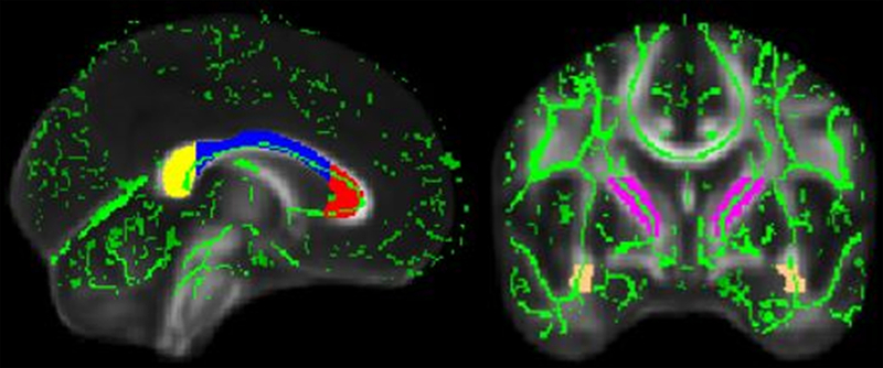
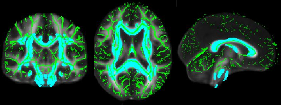
To analyze data for the a priori ROIs, the Johns Hopkins University (JHU) ICBM-DTI 91 White Matter Atlas (Mori et al., Reference Mori, Oishi, Jiang, Jiang, Li, Akhter, Hua, Faria, Mahmood, Woods, Toga, Pike, Neto, Evans, Zhang, Huang, Miller, van Zijl and Mazziotta2008) was used to create binary masks for the corpus callosum (divided into the genu, body, and splenium sections), ALIC, and uncinate fasciculus (see Figure 1). Studies comparing TBSS ROI analyses to voxel-based morphometry preprocessed ROIs and hand-drawn ROIs show that the inter-subject variability in TBSS is comparable to, if not better than, the other methods (Smith et al., Reference Smith, Jenkinson, Johansen-Berg, Rueckert, Nichols, Mackay, Watkins, Ciccarelli, Cader, Matthews and Behrens2006). Additionally, a binary mask was created for all WM tracts represented in the atlas and used in whole-brain global analyses (see Figure 2). To determine differences in diffusion parameters based on history of concussion, the concussion and control groups were compared using FSL’s RANDOMIZE function, which analyzed the skeletonized data in a nonparametric, permutation-based way, given that the null distribution for the data is unknown (Winkler et al., Reference Winkler, Ridgway, Webster, Smith and Nichols2014). All analyses were conducted under masked conditions using the global and ROI binary masks. Additionally, age was used as a covariate in additional analyses to ensure that significant results represented the effect of concussion history on WMmicrostructure, above and beyond normal aging changes. Output was viewed from a threshold-free cluster-enhancement (TFCE) framework for whole-brain and ROI analyses, which enhances detection of diffusivity differences by using neighborhood information to determine the statistical significance of a cluster of voxels instead of setting an a priori cluster threshold which may be unnecessarily conservative and lead to a Type II error. This method may be especially useful for ROI analyses because it is sensitive to finding true signal among noise (i.e., a small, focal area of strong signal in a large ROI) and provide more spatial detail than an analysis that examines the entire ROI (Smith & Nichols, Reference Smith and Nichols2009). For each instance of RANDOMIZE, 5000 null permutations were generated to test against the data. The two groups were compared using t-statistics at a TFCE-based family-wise error (FWE) corrected p-value of .05 for FA, RD, MD, and AD indices. A minimum cluster size of 8 voxels was used to increase the probability that findings are neuroanatomically meaningful and not spurious.
White Matter Regions-of-Interest (ROIs). The figure displays the binary masks used for ROI analyses for the corpus callosum (left) and the anterior limb of the internal capsule (ALIC) and uncinate fasciculus (right). Masks are overlaid on a single subject diffusion-weighted template in MNI space provided in FMRIB’s Software Library (FSL). The mean skeleton for the sample is superimposed on the figure in bright green.

Whole Brain. The figure displays the binary mask used for whole-brain analyses in the coronal (left), axial (middle) and sagittal views (right). The mask is overlaid on a single subject diffusion-weighted template in MNI space provided in FMRIB’s Software Library (FSL). The mean skeleton for the sample is superimposed on the figure in bright green.

Behavioral data analysis
To assess the relation between diffusion metrics and cognitive performance, binary masks were applied to the skeletonized FA, MD, RD, and AD images for each participant, and average diffusivity values were extracted for global WM and each ROI (i.e., corpus callosum, ALIC, and uncinate fasciculus). Previous research has shown global white matter FA to be sensitive to mild traumatic brain injury (mTBI) and associated with injury characteristics (Benson et al., Reference Benson, Meda, Vasudevan, Kou, Govindarajan, Hanks, Millis, Makki, Latif, Coplin, Meythaler and Haacke2007). When appropriate, right and left hemisphere values were averaged to create a single mean value for each ROI. Extracted data were then analyzed using the Statistical Package for Social Sciences (SPSS 24.0). Hypothesized relationships between ROIs and individual cognitive scores (i.e., RBANS Total Score and RBANS Index scores for Attention, Language, Visual-Spatial, Immediate Memory, and Delayed Memory) were assessed using bivariate Pearson correlations with a one-tailed test, given directional hypotheses. Additionally, moderation analyses were conducted using the PROCESS Macro (Hayes, Reference Hayes2013) to determine whether the relationship between diffusivity scores and RBANS performance differed between the concussion and control groups. Threshold for statistical significance was set at p < .01 to adjust for multiple comparisons.
Results
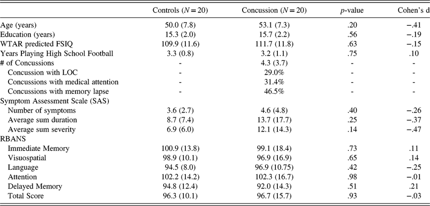
There were no statistically significant differences between the concussion and control groups with respect to age (t = 1.21, p = .20, Cohen’s d = −.41), years of formal education (t = .59, p = .56, d = −.19), pre-morbid IQ level as measured by the WTAR (t = .49, p = .63, d = −.15), and years playing high school football (t = .33, p = .75, d = .10) based on independent samples t-test analyses (Table 1). At the time of scanning, the two groups reported similar levels of concussion-like symptoms (see Table 1). Further, there were no statistically significant between-group differences on neuropsychological variables as measured by the RBANS (Table 1).
Group demographics

Note: Values are mean (SD). The median number of concussions for the group was 3. When two extreme values in the number of reported concussions were reduced to two standard deviations above the mean, the average number of concussions was 3.9. WTAR = Wechsler Test of Adult Reading; FSIQ = Full Scale Intelligence Quotient; LOC = Loss of consciousness; RBANS = Repeatable Battery for the Assessment of Neuropsychological Status.
DTI results
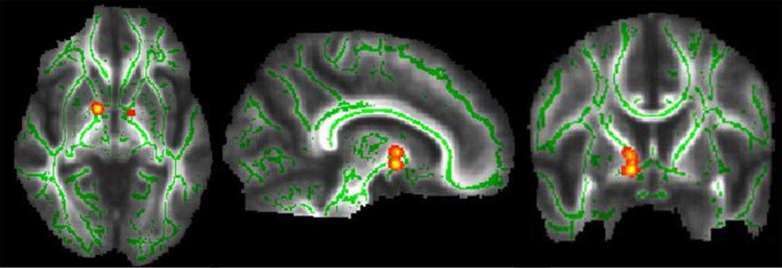
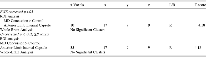
There were no significant group differences on whole-brain analysis that survived FWE-correction at the whole-brain level (Table 2). Using ROI analyses, the concussion group had higher MD in the right ALIC compared to controls (10 voxels, t = 4.18, p = .033 FWE-corrected). No other clusters survived the FWE-correction in the ROI analyses. Under a more liberal exploratory threshold (p < .001, uncorrected, ≥8 contiguous voxels), the concussion group again had a higher MD in the ALIC (35 voxels; See Table 2 and Figure 3). Using age as a covariate, there were no clusters that survived FWE-correction at the whole-brain or ROI level. There were also no clusters that were significant at the more liberal threshold (p < .001, uncorrected, ≥8 voxels) at either the whole-brain or the ROI level.
Tract-Based Spatial Statistics (TBSS) Results. The figure depicts the significant group difference between the concussion and control groups: lower FA in the anterior limb of the internal capsule (ALIC) depicted in the axial (left), sagittal (middle), and coronal (right) views. Results are displayed on the mean FA image for the sample with the mean skeleton superimposed in bright green.

White matter differences between concussion and control groups

Note: FA = factional anisotropy. RD = radial diffusivity. MD = mean diffusivity. L/R = left or right. x, y, and z coordinates are in MNI space (mm).
Levene’s test of equality of variance was used to examine the individual variability of the DTI metrics across the brain and the ROIs used in these analyses. There were no statistically significant differences in the variability of these metrics between the control group and the concussion group (ps > .05).
Behavioral results
Moderation analysis did not show any differences in the relationship between diffusion indices (i.e., FA, RD, MD, and AD) and RBANS scores between the concussion and control groups. Additionally, no significant correlations were found after correction for multiple comparisons. However, using a liberal threshold (p < .05, uncorrected), several significant correlations emerged between diffusion indices and RBANS scores which may be of future interest. In the concussion group, global FA was positively correlated with immediate memory (r = .501, p = .013, uncorrected), language (r = .483, p = .036, uncorrected), and global cognition as measured by the RBANS Total score (r = .514, p = .012, uncorrected). In the control group, FA in the genu of the corpus callosum was positively correlated with delayed memory (r = .464, p = .023, uncorrected), AD for the whole corpus callosum was positively correlated with visual-spatial performance (r = .486, p = .015, uncorrected), and AD in the ALIC was positively correlated with visual-spatial performance (r = .510, p = .011, uncorrected). No significant correlations were found for RD and MD indices.
Discussion
To our knowledge, this study is the first to evaluate the relationship between a history of multiple concussions sustained during high school football and significantly later-life WMmicrostructure in a cohort of middle-aged, former high school football players. Using a priori ROIs, we hypothesized that the concussion group would have lower FA/AD and higher MD/RD compared to control participants. Our findings suggest that former high school football players who endorsed two or more concussions in the context of high school football did not differ from control participants using a statistical threshold that corrected for multiple comparisons at the whole-brain level, nor did the two groups differ when examining the whole brain at a liberal threshold (p < .001, uncorrected, ≥8 voxels). While we found modest significant a priori effects in ROIs, specifically greater (worse) MD in the ALIC, little else survived following a conservative FWE correction or the liberal threshold (p < .001, uncorrected, ≥8 voxels). When covarying the analyses by age, there were no significant differences at either statistical threshold. While there were modest correlations between WM indices and cognitive performance within each group in our exploratory analyses, concussive history did not appear to influence the relationship between cognition and WM. There were no group differences in cognitive variables. Together, these modest results were as expected but attenuated. They suggest that multiple concussions may produce some small negative effects related to the microstructural characteristics of WM, particularly in the ALIC. Nevertheless, very few significant differences were found, even when being somewhat liberal statistically. Thus, they do not support large or widespread effects of multiple concussions on measures WMmicrostructure in this group of participants. These results suggest the likelihood of only minimal neuroanatomical changes in the brains of these past athletes potentially related to altered myelin micropathology.
DTI research examining a history of a remote concussion has been mixed. Some studies examining adult mTBI in civilians at least 1 year post injury have yielded positive findings, such as lower FA in the genu of the corpus callosum and posterior limb of the internal capsule in persistent cognitively impaired patients (Lo et al., Reference Lo, Shifteh, Gold, Bello and Lipton2009), and low FA in the anterior corona radiata and ALIC in those with chronic post-concussive symptoms compared to control participants (Dean et al., Reference Dean, Sato, Vieira, McNamara and Sterr2015). Other studies found no differences in diffusion metrics when comparing civilians who had a remote mTBI with chronic symptomatology to controls (Astafiev et al., Reference Astafiev, Shulman, Metcalf, Rengachary, MacDonald, Harrington, Maruta, Shimony, Ghajar, Diwakar, Huang, Lee and Corbetta2015; Maruta et al., Reference Maruta, Palacios, Zimmerman, Ghajar and Mukherjee2016). The findings related to remote concussions sustained in sports are also mixed. For instance, former collegiate football/hockey players aged 51–75 with a history of 1–5 concussions (the most recent of which was in early adulthood) had lower FA/higher MD in the corpus callosum, ALIC, and frontal–parietal regions compared to non-concussed former football/hockey players (Tremblay et al., Reference Tremblay, Henry, Bedetti, Larson-Dupuis, Gagnon, Evans, Théoret, Lassonde and De Beaumont2014). In non-depressed retired National Football League (NFL) players aged 41–79 with a history of 0–10 concussions, there were no significant FA differences compared to non-athlete controls (Strain et al., Reference Strain, Didehbani, Cullum, Mansinghani, Conover, Kraut, Hart and Womack2013). However, in the five athletes with current major depression, higher depression severity was associated with lower FA in multiple brain regions (Strain, et al., Reference Strain, Didehbani, Cullum, Mansinghani, Conover, Kraut, Hart and Womack2013). In a similar sample from the same research group, retired NFL players with cognitive impairment or depression had lower FA in frontal and parietal regions bilaterally, but those without cognitive impairment/depression did not differ from non-athlete controls on FA (Hart, et al., Reference Hart, Kraut, Womack, Strain, Didehbani, Bartz, Conover, Mansinghani, Lu and Cullum2013). Together, these results suggest that differences on DTI metrics are inconsistent and likely sample dependent, with seemingly more consistent positive results in those having a significant history of multiple concussions and current cognitive/mood difficulties. While no prior studies to our knowledge have examined former high school football players, it is important to consider that the overall head injury exposure in this sample is likely less than in participants in prior studies examining collegiate/professional collision sports. Further, the current sample was recruited from the general population (as opposed to a chronically symptomatic sample), reported relatively few concussion-like symptoms, and did not have lower cognitive test scores compared to controls.
Our results extend previous work suggesting the possibility of very subtle neuroanatomical changes in the brains of former high school football players, with the novel approach of following these former athletes several decades post concussion. Terry and colleagues previously found that these same former high school football players had left-hemisphere hypoactivation during a verbal learning and memory task using functional MRI, despite a lack of behavioral differences on the learning paradigm or learning/memory differences between the two groups on traditional neurocognitive tests (Terry et al., Reference Terry, Adams, Ferrara and Miller2015). List and colleagues showed cortical thinning in the right temporal lobe and left insula in a cohort of athletes that received at least two concussions at a minimum of 6 months post injury in the absence of differences between groups on neuropsychology test scores (List et al., Reference List, Ott, Bukowski, Lindenberg and Flöel2015). Our data suggest only very modest anatomical brain differences in former athletes with multiple concussions, several decades post concussion. These results, combined with a lack of neuropsychological test differences, suggest modest structural differences that may be related to multiple head injuries may be sustained many years prior in the absence of significant neuropsychological performance deficits.
Limitations, strengths, and future directions
There are several caveats and strengths in this study to take into account. We only examined high school football players who received their concussions in the context of this sport. These results may not be generalizable to other sports (i.e., soccer, hockey) or to civilian or military injuries due to different mechanisms of injury and rotational factors associated with each. This study did not assess/account for exposure to other contact sports. Further, this study may not be generalizable to female athletes given the notable differences between the genders in the recovery from a concussion (Zuckerman, Apple, Odom, Solomon, & Sills, Reference Zuckerman, Apple, Odom, Lee, Solomon and Sills2014). Also, the cross-sectional nature of this study limits our ability to associate early concussions with brain changes in a causal manner, though the groups did not statistically differ on many variables known to be associated with WM changes (i.e., age, sex, education, current concussion-like symptomatology). In this study, concussions were measured via self-report in a detailed but retrospective manner given most concussions were never reported to medical staff, and even if they were, those records likely are not available at present. Although an examiner confirmed group membership using a validated concussion interview (i.e., the ACE), which is more rigorous than many other studies, participants’ memory ability to report the details of the injuries that occurred before the age of 18, as well as nonspecific factors (e.g., personality style, knowledge about concussions) could have influenced the accuracy of their reporting. Notably, this study also employed an index of effort/task engagement (i.e., the MSVT) in case of exaggerated cognitive complaints. All participants performed adequately on this task, and thus, the cognitive results are thought to represent their true neurocognitive ability. While the tool we used to assess cognitive functioning (i.e., the RBANS) has been validated in brain injury samples (McKay, Wertheimer, Fichtenberg, & Casey, Reference McKay, Wertheimer, Fichtenberg and Casey2008), it was initially designed to be a screening measure and may lack the sensitivity to detect more subtle cognitive difficulties in this specific population.
Future studies should employ prospective methodologies to track adolescents who sustain concussions in a longitudinal fashion to help determine causal relationships and have a matched-control group. Future studies also will need to account for the impact of subconcussive blows. In the current study, we were only interested in the added impact of frank concussions, but both groups in this study were likely subject to repetitive head trauma related to their participation in tackle football. A non-contact sport control group should be recruited for studies that want to examine the effect of repetitive brain trauma. It may also be interesting to use DTI fiber tractography to examine the entire WM tract given that TBSS only examines the central skeleton of the parametric maps.
ACKNOWLEDGMENTS
This research was made possible by the charitable contributions of the John & Mary Franklin Foundation and the BioImaging Research Center at the University of Georgia.
CONFLICTS OF INTEREST
There are no other funding agencies or conflicts of interest to declare.