Implications
Reduced oestrus expression is one of the factors contributing to the current suboptimal reproductive efficiency in dairy cows. A recent series of publications that correlated the level of oestrous behaviour expression to gene expression levels in the cow brain and anterior pituitary are reviewed here and their findings placed in perspective of what is known about sexual behaviour from studies on other species. The improved understanding of the central genomic regulation of oestrous behaviour expression may in due time contribute to improved (genomic) selection strategies for appropriate oestrus expression in high-producing dairy cows.
Introduction
Maintaining good fertility and thereby optimum reproductive performance in dairy cows is of great economic importance for the dairy industry. Artificial insemination of cows is widely practiced in modern dairy farming. To ensure timely insemination and thereby maximize conception rates in the herd and profitability of the farm, it is important that the farmer does not miss the relatively short period in the oestrous cycle of the cow, called oestrus, when it is fertile and sexually receptive. In the absence of bulls in the farm, the farmer himself detects cows in oestrus by depending on observations of a number of physical and behavioural signs, collectively known as oestrous behaviour, that a cow exhibits only during oestrus or at a higher frequency during oestrus compared with the rest of its oestrous cycle. The physical signs include mucous discharges from the vulva, swelling of the vulva and reddening of the vaginal mucosa. The behavioural signs, studied in detail by Hurnik et al. (Reference Hurnik, King and Robertson1975) and Esslemont et al. (Reference Esslemont, Glencross, Bryant and Pope1980), include restlessness and mounting activities as well as increased levels of nudging, chin-resting, sniffing and licking of the genital area of other cows, and flehmen. Mounting activities include both mounting of other cows and being mounted by other cows. Only a cow in oestrus, when mounted, will show an immobilization reflex referred to as standing oestrus or standing heat. This is the only behavioural sign for true oestrus and is considered the primary sign of oestrus. All other behavioural signs of oestrus as well as the physical signs are useful to identify cows that are suspected to be in oestrus but are not conclusive. Hence, they are collectively referred to as secondary signs of oestrus.
Over the past 50 years, the percentage of Holstein Friesian dairy cows exhibiting standing oestrus (true oestrus) has declined from 80% to 50% and the average duration of true oestrus has progressively decreased from 15 to 18 h to <8 h (Dobson et al., Reference Dobson, Walker, Morris, Routly and Smith2008; Reames et al., Reference Reames, Hatler, Hayes, Ray and Silvia2011). In addition, the intensity with which secondary behavioural signs of oestrus are displayed have also steadily declined, even to the extent that some cows show no oestrus signs at all (‘silent’ oestrus). The reduced duration and intensity, or even the absence, of oestrous behaviour makes it difficult for dairy farmers to detect cows in oestrus, that is, oestrus may go undetected, or weak oestrous signs in luteal cows may be misinterpreted as ‘oestrus’. As timely and accurate oestrus detection is essential for successful artificial insemination, the decreased oestrus detection rates contribute significantly to the multi-factorial problem of suboptimal reproductive efficiency in modern dairy farming (Walsh et al., Reference Walsh, Williams and Evans2011). Using cow information, reproductive dynamics and economics representative for the dairy situation in the Netherlands, Inchaisri et al. (Reference Inchaisri, Jorritsma, Vos, Van der Weijden and Hogeveen2010) predicted that an improvement in the oestrus detection rate from 0.30 to 0.50 and from 0.50 to 0.70 will reduce the net economic loss by €53.29 and €11.20 per cow per year, respectively. Various oestrus detection methods aimed at improving detection rate have been developed, evaluated and compared (Diskin and Sreenan, Reference Diskin and Sreenan2000; Firk et al., Reference Firk, Stamer, Junge and Krieter2002; Roelofs et al., Reference Roelofs, López-Gatius, Hunter, Van Eerdenburg and Hanzen2010; Holman et al., Reference Holman, Thompson, Routly, Cameron, Jones, Grove-White, Smith and Dobson2011). Despite this, modern dairy farming would substantially benefit from an improved oestrus detectability via an increased expression of oestrous behaviour in cows, that is, if cows would generally show the various signs of oestrus during a longer period of time and at a higher intensity and if more cows would show standing oestrus.
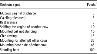
A number of studies have reported considerable variations in the durations of true oestrus among cows observed in herds (see review by Dobson et al., Reference Dobson, Walker, Morris, Routly and Smith2008). These studies do not directly discriminate between within-cow and between-cow variation in oestrus duration, as cows were generally observed for only one or two consecutive cycles. However, the fact that duration of oestrus is often seen to be associated with traits such as parity (Roelofs et al., Reference Roelofs, Van Eerdenburg, Soede and Kemp2005) or milk yield (Lopez et al., Reference Lopez, Satter and Wiltbank2004) suggests that there is a cow component in the variation in oestrous duration. Lopez et al. (Reference Lopez, Satter and Wiltbank2004) reported a negative correlation between the duration of true oestrus (recorded by a radio telemetry system) and the average milk production during the 10 days preceding the day of oestrus (r=−0.51; P<0.0001). At the same time, within a given range of milk production levels, there were still considerable variations in the durations of true oestrus among cows. For cows with an average milk yield of 25 to 30 kg/day, the duration of true oestrus varied from 6 to 27 h, whereas for cows with an average milk yield of 45 to 50 kg/day the duration of true oestrus varied from 0 to 18 h. In addition to the duration of true oestrus, the intensity with which typical behavioural oestrous signs (including standing-to-be-mounted) are displayed is also highly variable between cows (Dransfield et al., Reference Dransfield, Nebel, Pearson and Warnick1998; Yoshida and Nakao, Reference Yoshida and Nakao2005). The intensity of oestrus is often expressed as a cumulative score for weighted behavioural signs. The scoring method proposed by Van Eerdenburg et al. (Reference Van Eerdenburg, Loeffler and Van Vliet1996), shown in Table 1, has been frequently used. The intensity of oestrous behaviour expression is not only important for successful artificial insemination but also for high conception and calving rates once inseminated. Several studies have shown that cows with a more intense oestrous behaviour (including the standing response) have higher conception and calving rates (Dransfield et al., Reference Dransfield, Nebel, Pearson and Warnick1998; Garcia et al., Reference Garcia, Hultgren, Fällman, Geust, Algers, Stilwell, Gunnarsson and Rodriguez-Martinez2011; Gilmore et al., Reference Gilmore, Young, Patterson, Wylie, Law, Kilpatrick, Elliott and Mayne2011). Oestrous behaviour is thus a key fertility trait in cows.
The scoring method of Van Eerdenburg et al. (1996) for the quantification of the intensity of expression of oestrous behaviour in dairy cows

1 Each time a sign is observed during an observation period of 30 minutes, the assigned number of points is recorded. The sum of points per observation period is a measure for the intensity of expression of oestrous signs during the observation period. If the sum of the points exceeds 50 during two consecutive observation periods, a cow is considered to be in oestrus.
Understanding the background of individual differences in duration, and especially intensity of oestrous behaviour, may lead to opportunities to improve oestrus expression and reduce the fraction of cows in which intensity and duration of oestrus behaviour are too low for adequate and efficient heat detection. Here we focus on selected aspects of the physiological, neurophysiological and central genomic background of between- and within-cow variation in the expression of oestrous behaviour. We review recent information from literature concerning the interaction between milk production level, blood concentrations of ovarian steroids (oestradiol and progesterone) and the expression of oestrous behaviour. We also review the current knowledge on ovarian steroid-driven mechanisms in the central nervous system that relate to the expression of female sexual behaviour. Although much of this knowledge is based on research on species other than cattle, this review includes a series of recent studies in cattle and discusses how the results of these studies fit in, and connect with, the existing knowledge from studies on other species, especially rodents. Throughout this review, emphasis will be on endocrine, neuroendocrine and functional genomic aspects of female sexual (oestrous) behaviour. Thus, the aim of the current review is to integrate knowledge on endocrine regulation of oestrous behaviour of modern high-yielding dairy cows with knowledge on genomic regulation of oestrous behaviour in various animal species, including cattle. With this review, we hope to stimulate integrated physiological and molecular genetic research on the regulation of oestrous behaviour expression in dairy cows. Increased knowledge in this field may contribute to the development of tools for detecting oestrous behaviour or to improvement of oestrus expression through changes in animal management, or even to the development of breeding strategies for genetic improvement in the trait.
Ovarian steroid hormones and oestrous behaviour expression in dairy cows
Sexual arousal is dependent on neural (sensory and cognitive) factors, hormonal factors and genetic factors. It activates cognitive and physiological processes that can eventually lead to sexual behaviour (Schober and Pfaff, Reference Schober and Pfaff2007). Regarding hormonal factors, Schober and Pfaff (Reference Schober and Pfaff2007) stated that sexual arousal in female animals ‘is slavishly dependent on oestrogen and progesterone’. During the oestrous cycle, the sequential actions of the ovarian steroid hormones progesterone and oestradiol in the central nervous system cause cyclical fluctuations in the degree of sexual arousal and facilitate expression of oestrous behaviour just before ovulation. The actions of ovarian steroids on the chain of events constituting female sexual behaviour have been well studied in rodents (reviewed by Blaustein, Reference Blaustein2008 and Pfaff et al., Reference Pfaff, Waters, Khan, Zhang and Numan2011).
It has been suggested that the effect of oestradiol on oestrous behaviour in dairy cows is ‘all or none’: oestrous behaviour is induced as soon as a threshold of oestradiol concentration is reached and additional oestradiol above this threshold will not further enhance the duration and intensity of behavioural oestrus expression (Allrich, Reference Allrich1994). Although this might have been the case before intense selection for milk yield commenced, the situation seems different now. Several studies with modern high producing dairy cows indicate a relatively strong positive relationship between the oestradiol blood concentration during the late follicular phase and the duration and intensity of behavioural oestrus. For instance, Lyimo et al. (Reference Lyimo, Nielen, Ouweltjes, Kruip and Van Eerdenburg2000) (Table 2) found a significant correlation (r=0.56; P=0.023) between the maximum oestradiol concentration during the oestrous phase of the cycle and the total oestrus score as measured using the scoring method of Van Eerdenburg et al. (Reference Van Eerdenburg, Loeffler and Van Vliet1996). In the same study, the correlation of maximum oestradiol level with the length of the oestrous period was not significant (r=0.45; P=0.081). However, after correction for the variation between cows in progesterone concentration during dioestrus, both correlations were significant (r=0.66, P=0.006 for oestrous score and r=0.60, P=0.014 for duration of oestrus). In agreement with this, Lopez et al. (Reference Lopez, Satter and Wiltbank2004) (Table 2) reported a correlation coefficient of 0.57 (P<0.0001) between the oestradiol concentration on the day of oestrus and the duration of oestrus as recorded by a radio telemetry system. In addition, these authors found that the oestradiol concentration was negatively correlated with the average milk production of the 10 days before the day of oestrus (r=−0.57; P<0.0001). In low-producing cows, the oestradiol concentrations 1 day before oestrus and on the day of oestrus were 9.6±0.5 and 8.6±0.5 pg/ml, respectively, whereas in high producers these were lower at 7.7±0.7 and 6.8±0.5 pg/ml, respectively (low- and high-producing cows were defined based on whether the average milk production was below or above 39.1 kg/day in the 10 days before the day of oestrus).
Blood oestradiol concentration (mean and range) and correlations of oestradiol concentration with oestrous score, oestrus length and milk yield, respectively (r-values, with P-values between brackets)

1 Maximum level in oestrous phase in Lyimo; level on day of oestrus in Lopez.
2 Using the scoring method of Van Eendenburg et al. (1996).
3 Average milk production for the 10 days before the day of oestrus.
4 After correction for the variation between cows in progesterone concentration during dioestrus.
Regarding the role of progesterone in the expression of behavioural oestrus in ruminants, some conflicting results have been reported, especially when considering the experiments on ovariectomized ruminants treated with progesterone and/or oestrogens. Nevertheless, there is much evidence from studies with intact ruminants that suggests an important facilitatory role of progesterone priming. This role includes an increase in the oestradiol responsiveness of specific areas of the central nervous system (especially the hypothalamus) involved in the expression of behavioural oestrus. Several experiments on ovariectomized ewes treated with progesterone and/or oestradiol showed that progesterone priming is needed to induce complete oestrous behaviour after oestradiol injections (reviewed by Fabre-Nys and Gelez, Reference Fabre-Nys and Gelez2007). Similar results have been found in ovariectomized goats, although the effectiveness of progesterone priming was affected by seasonal breeding status and the time interval between the sequential progesterone and oestradiol treatments (reviewed by Katz, Reference Katz2007). As briefly reviewed by Davidge et al. (Reference Davidge, Wiebold, Senger and Hillers1987), experiments on ovariectomized heifers and cows have shown conflicting results about the influence of progesterone priming on oestradiol-induced oestrous behaviour. Although several studies cited by Davidge et al. (Reference Davidge, Wiebold, Senger and Hillers1987) have shown that oestrogen alone is sufficient to induce oestrus in ovariectomized heifers and cows, these studies did not attempt to determine the effect of progesterone on modifying the behavioural response. In their own study on ovariectomized cows, designed to test the hypothesis that progesterone before oestradiol-induced oestrus would have a regulating effect on the occurrence and intensity of oestrous behaviour, Davidge et al. (Reference Davidge, Wiebold, Senger and Hillers1987) found an inhibitory effect of progesterone. According to these authors, this unexpected result indicates that progesterone may not have the same effect on ovariectomized and intact cows.
In intact dairy cows, the importance of progesterone priming for the full display of oestrous behaviour is supported by the complete absence or short duration and low intensity of oestrous behaviour just before the first postpartum ovulation, an ovulation that is not preceded by a period of elevated progesterone blood levels (for references see Rhodes et al., Reference Rhodes, McDougall, Burke, Verkerk and MacMillan2003). Failure to display behavioural oestrus just before the first postpartum ovulation may be explained by the induction of a refractory state of critical brain areas by the high oestradiol concentrations towards the end of pregnancy. It has been proposed that progesterone can undo this refractory state (Kyle et al., Reference Kyle, Callahan and Allrich1992). It may be hypothesized that in cycling cows the elevated oestradiol concentrations during the follicular phase induces a similar refractory state that must be reset by the subsequent elevated progesterone concentrations during the luteal phase. Support for a significant influence of the level of circulating progesterone on the intensity of oestrous behaviour expression comes from a study on the oestrous behaviour of Mithun (Bos frontalis) cows (Dhali et al., Reference Dhali, Mishra, Karunakaran, Mech and Rajkhowa2006). In these cows, bulls are absolutely needed to detect oestrus. The only visible signs of oestrus are reddening and swelling of the vulva, restlessness and standing to be mounted by a bull. Several other typical behavioural signs of oestrus expressed in Bos taurus cows, including cow–cow interactions, are not shown by Mithun cows. These cows have preovulatory oestradiol levels that are actually slightly higher than those found in B. taurus cows. However, their peak progesterone concentrations during the oestrous cycle are on average <3 ng/ml, whereas in Holstein dairy cows the average concentrations reported in a number of recent studies were in the range of 6 to 8 ng/ml (Meier et al., Reference Meier, Roche, Kolver, Verkerk and Boston2009; Herzog et al., Reference Herzog, Brockhan-Lüdemann, Kaske, Beindorff, Paul, Niemann and Bollwein2010; Starbuck and Mann, Reference Starbuck and Mann2010). As in B. taurus cows, the lowest progesterone concentrations in Mithun cows (<0.5 ng/ml) were found on the day of oestrus (standing to be mounted by a bull). According to Dhali et al. (Reference Dhali, Mishra, Karunakaran, Mech and Rajkhowa2006), the relatively weak expression of behavioural oestrus in Mithun cows is most probably because of the overall low progesterone concentrations throughout the oestrous cycle. Considering this, the Mithun cow seems to be a suitable animal model to study the role of progesterone in the expression of oestrous behaviour. It could be of interest to study the effect of progesterone supplementation of Mithun cows during the luteal phase of the cycle on subsequent expression of the various oestrous signs normally observed in B. taurus cows.
It has been suggested that the decline of the blood progesterone concentration following luteal regression may affect the expression of oestrous behaviour in domestic ruminants in ways other than through its effect on follicle growth and therefore on oestradiol synthesis (Fabre-Nys and Gelez, Reference Fabre-Nys and Gelez2007). Experiments on ovariectomized French-Alpine goats that were sequentially treated with progesterone and oestrogen have shown that the decline in circulating progesterone concentrations before the rise of oestradiol may be more important than just the presence or absence of progesterone. In these goats, optimal expression of oestrous behaviour resulted when progesterone concentrations were maintained at luteal phase levels and then permitted to decline in a similar pattern as the decline which normally occurs during luteolysis (Katz, Reference Katz2007). Irrespective of the kind of facilitating action, progesterone also inhibits the expression of oestrous behaviour in most species if its level is elevated directly before or during oestrus. According to Fabre-Nys and Gelez (Reference Fabre-Nys and Gelez2007), the elevated progesterone concentrations in pregnant female ruminants explain why they rarely display oestrous behaviour. In cattle, it is observed that some pregnant cows do show oestrous behaviour with such intensity that they would be considered in oestrus (Dijkhuizen and Van Eerdenburg, Reference Dijkhuizen and Van Eerdenburg1997). However, this does not preclude an inhibitory effect of progesterone on oestrous behaviour in cyclic cows, as the progesterone responsiveness and the behaviour-related neurophysiological mechanisms in the brain might be different in pregnant cows from that in cyclic cows.
It is assumed that the decreasing duration and intensity of behavioural oestrus over the past 50 years has been accompanied by a concomitant decrease in the blood concentrations of the ovarian steroids oestradiol and progesterone (Wiltbank et al., Reference Wiltbank, Lopez, Sartori, Sangsritavong and Gümen2006). An increased metabolic clearance of ovarian steroids has been suggested as a cause of lower blood concentrations of these steroids in high-producing cows. On the basis of the results of experiments on lactating and non-lactating cows on different feeding levels, Sangsritavang et al. (Reference Sangsritavong, Combs, Sartori, Armentano and Wiltbank2002) concluded that the blood flow in the liver of high-producing dairy cows, which are at a continuous high plane of nutrition, may be chronically elevated, resulting in an increased metabolic clearance rate of oestradiol and progesterone. Further evidence in support of this conclusion comes from studies on Holstein cows (Rabiee et al., Reference Rabiee, Macmillan and Schwarzenberger2001), Zebu cows (Martins et al., Reference Martins, Mollo, Bastos, Guardieiro and Sartori2008) and ewes (Parr et al., Reference Parr, Davis, Miles and Squires1993a, and Reference Parr, Davis, Miles and Squires1993b). In comparison with a group of Holstein cows on a restricted pasture diet, Rabiee et al. (Reference Rabiee, Macmillan and Schwarzenberger2001) found in a comparable group of cows fed the same pasture diet ad libitum an increased excretion rate of progesterone to the faeces in association with a decreased concentration of peripheral progesterone. In a study involving Zebu cows with a low or a high feed intake, Martins et al. (Reference Martins, Mollo, Bastos, Guardieiro and Sartori2008) found no difference in the maximum diameter of the ovulatory follicle between the two groups, despite a lower preovulatory oestradiol surge in the cows with a high feed intake. This indicates a higher metabolism of oestradiol in the cows with a high feed intake. Furthermore, those authors reported no difference between the groups in progesterone concentration on day 7 after ovulation. Parr et al. (Reference Parr, Davis, Miles and Squires1993a and Reference Parr, Davis, Miles and Squires1993b) studied the metabolic clearance rate of progesterone in ovariectomized ewes fed different levels of the same ration. They showed a positive relationship between feeding level and the blood flow through the intestines and to the liver, as well as an increased metabolic clearance of progesterone with increasing intestinal and hepatic blood flow. Parr et al. (Reference Parr, Davis, Miles and Squires1993b) concluded that the association between the level of feed intake and the chronic rate of blood flow to the liver, coupled with the high efficiency at which the liver metabolizes progesterone, favours the hypothesis that the inverse relationship between the level of feed intake and concentration of progesterone in peripheral plasma is controlled by the rate of splanchnic blood flow. Further suggestive evidence for a higher rate of metabolism of ovarian steroids because of high milk yield and the associated increased feed intake in cows comes from the study by Sartori et al. (Reference Sartori, Haughian, Shaver, Rosa and Wiltbank2004) in which ovarian function and circulating ovarian steroids during oestrous cycles were compared between Holstein heifers and lactating dairy cows. In that study, the maximal serum oestradiol concentration preceding ovulation was found to be lower in cows than in heifers, despite the larger ovulatory follicles in cows. The maximal serum progesterone concentration too was lower for cows, whereas the maximal volume of the luteal tissue was lower for heifers. Other explanations for the relatively low blood concentrations of ovarian steroids in high-producing cows may be a lower steroidogenic activity of their ovarian follicles and corpora lutea (Wiltbank et al., Reference Wiltbank, Lopez, Sartori, Sangsritavong and Gümen2006) or the higher proportion of circulating ovarian steroids passing from blood into milk. The latter possibiliity has received little attention in literature. According to Erb et al. (Reference Erb, Chew and Keller1977), the amount of oestrogen secreted in milk is relatively small in comparison with that metabolized and excreted in urine and faeces. However, in high milk-producing cows, the amount of ovarian steroids secreted in milk may be biologically significant. For example, Lopez et al. (Reference Lopez, Bunch and Shipka2002) reported the average oestradiol levels in plasma and milk to be ~15 and 5 pg/ml, respectively in high-producing primiparous Holstein cows at oestrus in early lactation. On the basis of the latter value, cows producing 40 to 50 kg milk/day would secrete about 200 to 250 ng oestradiol per day in their milk just before oestrus.
Oestrogen signalling and gene expression in the brain in relation to female sexual behaviour
Oestradiol acts in the brain by a variety of genomic, non-genomic and growth factor-dependent mechanisms (Cardona-Gómez et al., Reference Cardona-Gómez, Mendez, DonCarlos, Azcoitia and Garcia-Segura2003; Vasudevan et al., Reference Vasudevan, Kow and Pfaff2005; Kelly and Qiu, Reference Kelly and Qiu2010). As far as the genomic mechanisms are concerned, oestradiol regulates transcription in the neurons and glia by the activation of classic nuclear oestrogen receptors (ERs). The two ERs, ER α and ER β , are products of different genes. They bind oestradiol with equal affinity but have different tissue distributions (Kuiper et al., Reference Kuiper, Carlsson, Grandien, Enmark, Häggblad, Nilsson and Gustafsson1997). The classic mode of genomic action of oestradiol through activation of ERs is complemented by alternative non-genomic actions, whereby oestradiol acts at the membrane or in the cytoplasm of neurons. These non-genomic actions of oestradiol are linked to the activation of different kinases and second messenger systems such as cyclic adenosine monophosphate (cAMP) and intracellular calcium (Ca2+), and activation of other transcriptional regulators such as cAMP response element-binding protein (Cardona-Gómez et al., Reference Cardona-Gómez, Mendez, DonCarlos, Azcoitia and Garcia-Segura2003; Vasudevan et al., Reference Vasudevan, Kow and Pfaff2005 and reviewed by Kelly and Qiu, Reference Kelly and Qiu2010). The effects of oestradiol in the brain may also be mediated by the activation of growth factor signalling (Cardona-Gómez et al., Reference Cardona-Gómez, Mendez, DonCarlos, Azcoitia and Garcia-Segura2003). The molecular and functional interactions of oestradiol and IGF-I in the brain have been reviewed by Cardona-Gómez et al. (Reference Cardona-Gómez, Mendez, DonCarlos, Azcoitia and Garcia-Segura2003). Molecular interactions include reciprocal regulation of ERs and IGF-I receptors (IGF-IRs), adrenergic α-1 receptor expression and activation, as well as MAPK and PI3K/Akt signalling pathway activation. Functional interactions between IGF and ER pathways affect axonal and dendritic growth, oestrous cycle-associated synaptic plasticity, gonadotropin release/ovulation, and reproductive behaviour/sexual receptivity. Reproductive success of the dairy cow is linked to its body energy reserves (Roche, Reference Roche2006), which may be compromised, especially during early and peak lactation periods. The metabolic responses in the cow to low-energy reserves involve signalling molecules and hormones (such as IGF-I and growth hormones) that are central not only to the somatotrophic axis that regulates the partitioning of energy/nutrients but also to the hypothalamic–pituitary–ovarian axis that regulates reproductive function (Chagas et al., Reference Chagas, Bass, Blache, Burke, Kay, Lindsay, Lucy, Martin, Meier, Rhodes, Roche, Thatcher and Webb2007; Garnsworthy et al., Reference Garnsworthy, Sinclair and Webb2008). Some examples of genes expressed in the cow brain that plays roles in feeding behaviour, metabolic rate and feed intake, and also found associated with oestrous behaviour, are described towards the end of the next section.
Cell nuclear actions of oestradiol in the ventromedial hypothalamic (VMH) neurons, the transcriptional consequences of oestradiol binding in those neurons and how those neurons then regulate neural circuits that lead to lordosis behaviour (i.e., sexual receptivity) has been partly elucidated in rodents (Pfaff et al., Reference Pfaff, Waters, Khan, Zhang and Numan2011). Primarily on the basis of the results of research by Pfaff and co-workers on lordosis behaviour in rodents, it has been proposed that oestradiol-activated genes in the VMH are organized in five modules referred to as GAPPS modules (Mong and Pfaff, Reference Mong and Pfaff2004; Pfaff, Reference Pfaff2005). Here, GAPPS stands for: Growth of hypothalamic neurons; Amplification of the oestrogen effects by progesterone; Preparative behaviours; Permissive actions on the sex behaviour circuitry; and Synchronization of mating behaviour with ovulation. The oestradiol-dependent growth effects on the neurons (first module) occur before lordosis behaviour is displayed. It permits oestradiol-facilitated, behaviour-directing hypothalamic neurons a greater range of input/output connections and thus physiological power. This module depends on ER α -activated gene transcription. The amplification of the oestradiol effect by progesterone (second module) depends on both ER α - and ER β -activated expression of the progesterone receptor (PGR) gene. The third module, preparations for mating by preceding behaviours includes ER α -dependent expression of the enkephalin and opioid receptor genes to cause partial analgesia, as well as ER α - and ER β -dependent transcription of the oxytocin and oxytocin receptor genes to reduce anxiety and aggression, and to improve social recognition. Analgesia and reduced anxiety allow the female animal to engage in courtship and mating behaviour, despite the risk of pain it may cause. The fourth module, permissive actions by hypothalamic neurons for the rest of the lordosis circuit to operate, includes the ER α -dependent increased transcription of the genes encoding the noradrenergic α-1b receptor and the muscarinic acetylcholine receptors. These receptors are important for noradrenergic and acetylcholinergic signalling, respectively. The increased expression of these receptors permits the neural circuit to activate lordosis behaviour. Finally, ER β -dependent transcription of the GnRH and GnRH receptor genes (fifth module) serves to synchronize mating behaviour with ovulation.
When considering the GAPPS modules in relation to the neuroendocrine regulation of oestrous behaviour in species other than rodents, it should be kept in mind that the Amplification module may be the most variable module among species. For example, a difference between rodents and ungulates is the occurrence of a progesterone peak during pro-oestrus in rodents but not in ungulates. The progesterone peak in rodents occurs after the oestradiol peak and coincides more or less with the preovulatory LH peak (Pawluski et al., Reference Pawluski, Brummelte, Barha, Crozier and Galea2009). The actions of this preovulatory progesterone peak are essential for the proceptive components of female sexual behaviour in rodents and for amplifying the stimulatory effects of oestradiol on receptivity (reviewed by Levine et al., Reference Levine, Chapell, Schneider, Sleiter and Szabo2001 and Blaustein, Reference Blaustein2008). Another important difference between ungulates and several groups within the Cricetidae and Muridae rodent families is that in these rodent species, development of a functional corpus luteum does not occur spontaneously after ovulation, as it does in ungulates, but requires stimuli associated with copulation. Thus, in these rodents, there is no luteal phase with elevated progesterone concentrations between subsequent oestrus periods and the inter-oestrus intervals are only 4 to 7 days long (Conaway, Reference Conaway1971). From this, it should not be concluded that the preovulatory progesterone peak in rodents is only important for potentiating receptivity (i.e., lordosis behaviour) in species without a true luteal phase. For example, female guinea pigs, which present 16-day-long oestrous cycles with a true luteal phase, have a preovulatory progesterone peak with a potentiating effect on lordosis behaviour (Delville and Blaustein, Reference Delville and Blaustein1991).
Oestrogen signalling and gene expression in the brain in relation to the expression of oestrous behaviour in dairy cows
To the best of our knowledge, the expression and distribution of ER β in the brain of cattle has not yet been studied, and only limited information is available about the expression and distribution of ER α . Tanaka et al. (Reference Tanaka, Shiina, Hayashi, Okamura, Kamomae and Kaneda2003) studied the distribution and morphology of immunoreactive ER α -containing cells in the brain of Holstein Friesian cattle (a 44-month-old dry cow, a 27-month-old cow that had calved 1 week before killing and a 3-month-old male calf). ER α signals were detected in the preoptic area (POA) of the rostral forebrain and the medial basal hypothalamic area (MBH). In the POA, they were located from the rostral part of the medial POA at the level of the vascular organ of the lamina terminalis to the region adjacent to the bed nucleus of the stria terminalis. In the MBH, the ER α signals were also detected in the arcuate nucleus and the periventricular and VMH nuclei. Tanaka et al. (Reference Tanaka, Shiina, Hayashi, Okamura, Kamomae and Kaneda2003) concluded that the distribution of ER α in the bovine hypothalamus closely parallels that of oestrogen-sensitive neurons in other mammalian species such as rats, guinea pigs and sheep (for references see Tanaka et al., Reference Tanaka, Shiina, Hayashi, Okamura, Kamomae and Kaneda2003). Changes in the distribution of immunoreactive ER α in the hypothalamic area during the oestrous cycle of lactating dairy cows have been studied by Van Eerdenburg et al. (Reference Van Eerdenburg, Daemen, Van der Beek and Van Leeuwen2000). During the luteal phase, ER α immunoreactivity was found in most of the nuclei of cells in the bed nucleus of the stria terminalis, medial POA, ventromedial hypothalamus and arcuate nucleus. During oestrus and metoestrus, ER α immunoreactivity could only be detected in a few cells in the caudal arcuate nucleus. According to Van Eerdenburg et al. (Reference Van Eerdenburg, Daemen, Van der Beek and Van Leeuwen2000), this decline in ER number at oestrus–metoestrus might have developed in the herds of wild cattle with numerous cows and only a few bulls to downregulate receptive behaviour once ovulation has occurred and thus to avoid non-fertile matings.
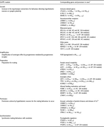
Relatively little is known about the genomic regulation of oestrous behaviour in dairy cows. However, in a recent series of studies, gene expression in the anterior pituitary and four brain areas (amygdala, hippocampus, dorsal hypothalamus and ventral hypothalamus) in oestrous and luteal phase cows, respectively, has been measured, and the relation with oestrous behaviour of these cows was analysed (Kommadath et al., Reference Kommadath, Mulder, De Wit, Woelders, Smits, Beerda, Veerkamp, Frijters and Te Pas2010, Reference Kommadath, Woelders, Beerda, Mulder, De Wit, Veerkamp, Te Pas and Smits2011 and Reference Kommadath, Te Pas and Smits2013; Kommadath, Reference Kommadath2012). Oestrous behaviour was recorded in 28 healthy primiparous Holstein Friesian cows in several consecutive cycles between 30 days in milk (DIM) until they were killed. The time of killing varied between 77 and 139 DIM. The oestrous behaviour recorded in these cows was quantified as heat scores according to the method of Van Eerdenburg et al. (Reference Van Eerdenburg, Loeffler and Van Vliet1996), and the scores from multiple consecutive cycles were averaged to obtain the average heat score per cow. Principal component analysis on individual cow cycle records indicated that there is a significant cow component in the heat score, that is, oestrous behaviour (heat score) to some extent was a consistent individual cow characteristic. Of the 28 cows, 14 were killed at the start of their oestrous cycle (day 0) and 14 around mid-cycle (day 12). Day 0 of the cycle was determined on the basis of ultrasonographical examinations of luteal regression and dominant follicle development, which were performed daily in the days before the expected day of oestrus (expected oestrus was based on the previous oestrus and previous cycle lengths), while using observations of vaginal mucous and oestrous behaviour as additional information. In these studies (Kommadath et al., Reference Kommadath, Mulder, De Wit, Woelders, Smits, Beerda, Veerkamp, Frijters and Te Pas2010, Reference Kommadath, Woelders, Beerda, Mulder, De Wit, Veerkamp, Te Pas and Smits2011 and Reference Kommadath, Te Pas and Smits2013; Kommadath, Reference Kommadath2012), a number of genes or gene clusters were found to have expression levels associated with the heat score. In the rest of this section, we describe how the results of these studies can be integrated within the framework of the GAPPS modules in rodents described in the previous section (see Table 3). The names of the gene symbols used here are given in Supplementary Table S1.
Oestrous behaviour-associated genes and processes in dairy cows grouped in the GAPPS modules described for female sexual behaviour in rodents (Mong and Pfaff, Reference Mong and Pfaff2004)

1
The relation of a gene with oestrus is described in brackets next
to the gene name. Symbols ‘+’,
‘–’, ‘
 ${\cup}$
’ and ‘
 ${\cap}$
’ denote that a positive linear, a
negative linear, a positive quadratic or a negative quadratic
association, respectively, was found between the gene expression
level in a brain area and the heat score of the cows (see
details in Kommadath et al., Reference Kommadath, Woelders, Beerda, Mulder, De Wit, Veerkamp, Te Pas and Smits2011). The brain areas
are amygdala (AM), hippocampus (HC), dorsal hypothalamus (DH),
ventral hypothalamus (VH) and anterior pituitary (AP).
Subscripts ((0), or (12) or
(0+12)) denote that the association
was found in cows killed in the oestrous phase (day 0), or in
the luteal phase (day 12) or when considering all cows. If a
gene is the member of a ‘consensus module’
(co-expressed genes in a pair of brain areas) whose
representative expression level had a positive (+) or
negative (−) association with oestrous behaviour,
this is indicated as e.g. ‘(+in AP : DH
module)’ (see details in Kommadath et
al., Reference Kommadath, Te Pas and Smits2013).
Data for PGR are from Kommadath, Reference Kommadath2012.
Several of the genes found associated with the heat score in cattle are related to synaptic plasticity (Kommadath et al., Reference Kommadath, Mulder, De Wit, Woelders, Smits, Beerda, Veerkamp, Frijters and Te Pas2010, Reference Kommadath, Woelders, Beerda, Mulder, De Wit, Veerkamp, Te Pas and Smits2011). These genes can therefore be considered to be part of the Growth module, which is characterized by oestradiol-dependent outgrowth of hypothalamic neurons. This ‘growth’ may also apply to other brain areas (amygdala and hippocampus) and the anterior pituitary, as genes related to synaptic plasticity were not limited to the hypothalamic area. Several ‘immune-related’ genes found in the association studies, such as CTLA4, IL1RL1, MARCO, FCRLA, IL33, CCL26 and CXCL10, may facilitate remodelling of synaptic networks. It has been shown that immunoglobulin superfamily proteins play important roles in brain developmental processes and the functioning of neuronal networks in adulthood because they provide the ideal structure for protein–protein interactions and thus cell–cell interactions (Rougon and Hobert, Reference Rougon and Hobert2003). In addition, other genes can be grouped in the Growth module. For instance, analysis of gene co-expression networks between the brain areas and anterior pituitary (Kommadath et al., Reference Kommadath, Te Pas and Smits2013) revealed gene clusters that correlated with oestrous behaviour, of which a number of hub genes have been reported to have functions related to neuronal growth or plasticity: NEFL (Terry-Lorenzo et al., Reference Terry-Lorenzo, Inoue, Connor, Haystead, Armbruster, Gupta, Oliver and Shenolikar2000), NDRG2 (Takahashi et al., Reference Takahashi, Yamada, Ohata, Honda and Yamada2005), THY1 (Rege and Hagood, Reference Rege and Hagood2006) and GAP43 (Gispen et al., Reference Gispen, Nielander, De Graan, Oestreicher, Schrama and Schotman1991). The gene NEFL, for example, regulates the dephosphorylation of phosphoproteins implicated in synaptic plasticity. Furthermore, several ribosomal genes associated with oestrous behaviour in the association studies (RPL24; Kommadath et al., Reference Kommadath, Woelders, Beerda, Mulder, De Wit, Veerkamp, Te Pas and Smits2011) or the co-expression studies (RPL14, RPL18, RPS11, RPS18; Kommadath et al., Reference Kommadath, Te Pas and Smits2013) across several brain areas. The increased synthesis of ribosomal RNA in the ventromedial hypothalamus has been reported to be one of the early effects of oestrogen administered subcutaneously in ovariectomized rats and represents a primary event in the activation of neuronal cells and neuronal pathways involved in female reproductive behaviour (Jones et al., Reference Jones, Harrington, Chikaraishi and Pfaff1990). Oestrogen-driven expansion of dendrites and synapses follows from the stimulation of synthesis of ribosomal RNA (Mong and Pfaff, Reference Mong and Pfaff2004; Pfaff, Reference Pfaff2005).
The second module in GAPPS is the Amplification of the oestrogen effect by progesterone, mediated by the nuclear PGR. In cattle, the expression of PGR in the anterior pituitary was found to be upregulated on day 0 (oestrous phase) compared with day 12 (luteal phase) of the oestrous cycle (Kommadath, Reference Kommadath2012). The expression of PGR was not found to be associated with the heat score of the cows. Furthermore, in cattle, progesterone levels in cows remain low during oestrus. Nevertheless, an increased expression of PGR on day 0 could suggest a relation of the PGR with oestrous behaviour, perhaps via ligand-independent pathways (reviewed by Mani and Blaustein, Reference Mani and Blaustein2012).
Several genes can also be identified that may be grouped within the Preparation module. For instance, the expression of oxytocin and arginine vasopressin genes in several brain areas of cows were associated with oestrous behaviour (Kommadath et al., Reference Kommadath, Mulder, De Wit, Woelders, Smits, Beerda, Veerkamp, Frijters and Te Pas2010 and Reference Kommadath, Woelders, Beerda, Mulder, De Wit, Veerkamp, Te Pas and Smits2011). Oxytocin, produced by the supraoptic and paraventricular nuclei of the hypothalamus, is released within the brain where it acts on specific oxytocin receptors to elicit effects such as female sexual receptivity, grooming behaviour and partner bonding (Leng et al., Reference Leng, Meddle and Douglas2008). In the presence of oestrogen, oxytocin exerts an anxiolytic effect, mediated by increases in oxytocin-binding density in the lateral septum, thereby favouring courtship and mating (McCarthy et al., Reference McCarthy, McDonald, Brooks and Goldman1997; Mong and Pfaff, Reference Mong and Pfaff2004). Similar to oxytocin, vasopressin is associated with sexual behaviour and bonding and its expression is under the control of oestrogen and progesterone (Patisaul et al., Reference Patisaul, Scordalakes, Young and Rissman2003; Kalamatianos et al., Reference Kalamatianos, Kalló, Goubillon and Coen2004; Curley and Keverne, Reference Curley and Keverne2005; Donaldson and Young, Reference Donaldson and Young2008). Genes that may also be grouped in the Preparation module are POMC, MCHR1, CCK, DRD2, HTR2A and GABRA6, whose expression levels in at least one of the brain areas are associated with oestrous behaviour score (Kommadath et al., Reference Kommadath, Woelders, Beerda, Mulder, De Wit, Veerkamp, Te Pas and Smits2011). These genes are known to modulate emotional states such as anxiety and satiety (Rex et al., Reference Rex, Marsden and Fink1997; Marsh et al., Reference Marsh, Weingarth, Novi, Chen, Trumbauer, Chen, Guan, Jiang, Feng, Camacho, Shen, Frazier, Yu, Metzger, Kuca, Shearman, Gopal-Truter, MacNeil, Strack, MacIntyre, Van der Ploeg and Qian2002; Uhart et al., Reference Uhart, McCaul, Oswald, Choi and Wand2004; Millington, Reference Millington2007). The link between fertility and appetite is evident from the finding of the POMC and MCHR1 genes, both of which play roles in feeding behaviour, metabolic rate and feed intake (Marsh et al., Reference Marsh, Weingarth, Novi, Chen, Trumbauer, Chen, Guan, Jiang, Feng, Camacho, Shen, Frazier, Yu, Metzger, Kuca, Shearman, Gopal-Truter, MacNeil, Strack, MacIntyre, Van der Ploeg and Qian2002; Millington, Reference Millington2007). It is known that interactions between monoamines (dopamine, serotonin, noradrenaline) and steroid hormones play a major role in the integration of reproductive behaviour and gonadal function (Fabre-Nys, Reference Fabre-Nys1998). In ewes, dopamine-mediated D2 receptor (DRD2) signalling in the mediobasal hypothalamus is known to affect female sexual motivation and receptivity (Fabre-Nys et al., Reference Fabre-Nys, Chesneau, De La Riva, Hinton, Locatelli, Ohkura and Kendrick2003). Furthermore, the perception and awareness of male-related cues differ with the stage of oestrous cycle, with releases of monoamines (linked to serotonin (HTR2A) and DRD2) and γ-aminobutyric acid (GABA) (linked to GABRA6) in the mediobasal hypothalamus being triggered by such cues only when ewes are in oestrus (Fabre-Nys et al., Reference Fabre-Nys, Ohkura and Kendrick1997). Studies on female rats and hamsters have shown the inhibitory and facilitatory effects of serotonin receptor agonists and antagonists on the hypothalamic regulation of sexual receptivity (Uphouse, Reference Uphouse2000; Caldwell and Albers, Reference Caldwell and Albers2002). This regulation is also mediated by GABAergic neurons interacting with serotonin-containing neurons. Other genes that may be grouped in the Preparation module are those known from studies on other species to play a role in emotional responses and that were found to be associated with the heat score of cows (Kommadath et al., Reference Kommadath, Woelders, Beerda, Mulder, De Wit, Veerkamp, Te Pas and Smits2011), for example, TTR, MOBP, LTA4H and KCNN2. TTR has been linked to anxiety (Sousa et al., Reference Sousa, Grandela, Fernández-Ruiz, De Miguel, De Sousa, Magalhães, Saraiva, Sousa and Palha2004), MOBP to mood disorders (Sokolov, Reference Sokolov2007), LTA4H to depression (Zhao et al., Reference Zhao, Quyyumi, Patel, Zafari, Veledar, Onufrak, Shallenberger, Jones and Vaccarino2009), and KCNN2 to anxiety and stress responses (Mitra et al., Reference Mitra, Ferguson and Sapolsky2009).
A number of oestrous behaviour score-associated genes (Kommadath et al., Reference Kommadath, Mulder, De Wit, Woelders, Smits, Beerda, Veerkamp, Frijters and Te Pas2010 and Reference Kommadath, Woelders, Beerda, Mulder, De Wit, Veerkamp, Te Pas and Smits2011) may be grouped in the fourth GAPPS module – Permission. For instance, the association between oestrous behaviour scores and the expression of acetylcholinesterase (ACHE) and several cholinergic receptors (CHRM1, CHRM3 and CHRNA5) can be explained by the effect of the neurotransmitter acetylcholine on arousal, plasticity and reward. The products of the muscarinic cholinergic receptor genes, CHRM1 and CHRM3, are Gq-protein-coupled receptors whose activation releases intracellular Ca2+ via the phospholipase C – inositol 1,4,5-trisphosphate signalling pathway (Billups et al., Reference Billups, Billups, Challiss and Nahorski2006). The genes for phospholipase C and inositol triphosphate kinase (PLCB2, ITPKA) and several protein kinases were also found associated with oestrous behaviour scores. These findings can be explained based on the hypothesis put forward by Kow and Pfaff (Reference Kow and Pfaff2004) that the membrane actions of oestrogen can modulate the genomic actions of oestrogen and that this transcriptional potentiation is mediated via signalling pathways requiring the activation of certain protein kinases and increased intracellular Ca2+.
Finally, the heat score-associated genes PTGDS, PTGIS and PTGFR (Kommadath et al., Reference Kommadath, Woelders, Beerda, Mulder, De Wit, Veerkamp, Te Pas and Smits2011 and Reference Kommadath, Te Pas and Smits2013) may be grouped in the last GAPPS module (Synchronization). These genes regulate prostaglandin functioning. Prostaglandins are known to be under the influence of oestrogen (Amateau and McCarthy, Reference Amateau and McCarthy2002) and are capable of directly affecting the neurons that synthesize and secrete gonadotropin-releasing hormone (Jasoni et al., Reference Jasoni, Todman, Han and Herbison2005; Clasadonte et al., Reference Clasadonte, Poulain, Hanchate, Corfas, Ojeda and Prevot2011).
Conclusion
Current research findings indicate that the decreased expression of oestrous behaviour seen in high-producing dairy cows is related to decreased blood concentrations of ovarian steroids, which in turn may partly be explained by an increased rate of hepatic metabolization of ovarian steroids in high-producing dairy cows. A better understanding of the between-cow variation in the ovarian steroid-driven central genomic regulation of oestrous behaviour in dairy cows may contribute to the development of tools and management strategies to improve oestrous behaviour detection and therewith reproductive efficiency. In addition, this understanding may facilitate the development of a (genomic) selection strategy for improving the trait of oestrous behaviour expression, thereby improving reproductive efficiency in high-producing dairy cows. A firm base of understanding of the ovarian steroid-driven central genomic regulation of female sexual behaviour exists in rodents. The recent studies on cattle reviewed in this paper have now contributed first steps towards such understanding in cattle and point to a functional clustering of oestrous behaviour-associated genes in cattle within the GAPPS modules described earlier for female sexual behaviour in rodents.
Acknowledgements
This work was co-financed by the European Commission funded projects SABRE (www.sabre-eu.eu/) (contract no. FOOD-CT-2006-016250) and EADGENE (www.eadgene.info/) (contract no. FOOD-CT-2004-506416), the Dutch Ministry of Agriculture, Nature and Food Quality (www.minlnv.nl) (KB-04-004-012) and the International cattle breeding co-operative – CRV BV (www.crv4all.com/). The text represents the authors’ views and does not necessarily represent a position of the European Commission who will not be liable for the use made of such information.