INTRODUCTION
Malaria is one of the most devastating diseases of the world, particularly in tropical countries. Annually, around 300–500 million of people are affected by this disease worldwide and of them approximately 1–3 million die (Shiff, Reference Shiff2002; Sachs and Malaney, Reference Sachs and Malaney2002; Miller et al. Reference Miller, Baruch, Marsh and Doumbo2002). Severe acute malaria (SAM) that can lead to death, may clinically present either as a neurological syndrome (cerebral malaria; CM), as multi-organ failure or severe anaemia leading to hypoxia and acidosis (Miller et al. Reference Miller, Baruch, Marsh and Doumbo2002; Helbok et al. Reference Helbok, Dent, Nacher, Lackner, Treeprasertsuk, Krudsood, Wilairatana, Silachamroon, Looareesuwan and Schmutzhard2005; Maitland and Newton, Reference Maitland and Newton2005). P. berghei ANKA in selected strains of mice leads to a form of CM and has been used as a model of P. falciparum-induced CM, representing 20–25% of all SAM cases (Greenwood and Mutabingwa, Reference Greenwood and Mutabingwa2002; Snow et al. Reference Snow, Guerra, Noor, Myint and Hay2005), for extensively analysing the molecular and cellular mechanisms involved. In contrast, the 3-fold more frequent non-neurological SAM has received much less attention.
Plasmodium induces stage-specific pathological changes, which are asymptomatic during liver stage and symptomatic during the erythrocyte stage. Infection of inbred mouse strains with Plasmodium chabaudi chabaudi (AS) results in non-lethal or lethal infections, depending on the genetic background of the host. With DBA/2 mice, which are susceptible to P. c. chabaudi (AS) infection, it is possible to dissect disease mechanisms and putative therapeutic measures can be tested.
We have previously reported that experimental infection with P. c. chabaudi, while readily controlled in BALB/c mice, is fatal for most DBA/2 animals (Seixas and Ostler, Reference Seixas and Ostler2005). Interestingly, DBA/2 mice die by day 9–11 of infection, with controlled parasitaemia and no neurological symptoms. Prior to death, circulating TNF-α and IL-12 reach high levels (Seixas and Ostler, Reference Seixas and Ostler2005). TNF-α is a potent pro-inflammatory molecule and has been implicated as a crucial mediator of toxic shock following systemic infections by gram-negative bacteria (Vassalli, Reference Vassalli1992; Spooner et al. Reference Spooner, Markowitz and Saravolatz1992). We have thus hypothesized that SAM in this model could have a similar mechanism. To address this we analysed, by histology, different organs of DBA/2 mice after infection to analyse their function and correlate with the severity of the disease in this model. We observed marked liver injury in the susceptible compared to the resistant strain of mice. Some studies have shown that during the erythrocyte stage liver injury could develop, although merozoites are not capable of infecting liver parenchymal cells (Tsutsui et al. Reference Tsutsui, Kayagaki, Kuida, Nakano, Hayashi, Takeda, Matsui, Kashiwamura, Hada, Akira, Yagita, Okamura and Nakanishi1999, Reference Tsutsui, Matsui, Okamura and Nakanishi2000; Naka et al. Reference Naka, Tsutsui, Fujimoto, Kawazoe, Kohzaki, Morita, Nakagawa, Narazaki, Adachi, Yoshimoto, Nakanishi and Kishimoto2001; Adachi et al. Reference Adachi, Tsutsui, Kashiwamura, Seki, Nakano, Takeuchi, Takeda, Okumura, Van Kaer, Okamura, Akira and Nakanishi2001, Reference Adachi, Tsutsui, Seki, Nakano, Takeda, Okumura, Van Kaer and Nakanishi2004). However, large numbers of infected erythrocytes pass through spleen and liver and the injury induced by the parasite is probably caused by the local production of cytokines.
We have now investigated some of the molecular and cellular mechanisms leading to death induced by P. chabaudi in DBA/2 mice, aiming at providing an experimental model for human non-neurological SAM. The analysis has revealed that susceptible mice die of hepatic failure, associated with liver necrosis. Furthermore, if infected mice (DBA/2) are treated with neutralizing monoclonal anti-TNF-α antibodies the majority survive infection, show much reduced liver pathological changes, while continuing to clear parasites from the blood as untreated animals. These findings support the view that TNF-α-related multifocal liver lesions are a major cause of death in this model and may open new therapeutic possibilities in fatal human malaria.
MATERIALS AND METHODS
Experimental animals and parasites
All mouse strains used were bred and maintained under specific pathogen-free conditions in our animal facilities. BALB/c and DBA/2 (both H-2d) female mice, 8–12 weeks of age were infected intraperitoneally (i.p.) with 106 blood stages of Plasmodium chabaudi chabaudi (AS). Courses of infection were monitored by examination of Giemsa-stained blood smears from tail blood throughout the experimental period.
Detection of TNF-α by ELISA
Plasma samples of infected mice were tested for TNF-α by performing ELISA. Antibody pairs (capture and detection) and cytokine standards were purchased from R&D Systems and the assay followed the manufacturer's instructions.
In vivo antibody treatment
The anti-TNF-α mAb used for in vivo depletion experiments was purified in house from the XT3.11 Hybridoma. Purified Rat IgG (Sigma) was used as an isotype control antibody. Anti-TNF-α antibody and Rat IgG (0·2 mg/mouse) were injected i.p. into DBA/2 mice (10 mice/group) every day beginning on day 5 until day 9 of infection. The efficacy of depletion during the experiment was assessed by ELISA using plasma samples.
Histopathological observations
Liver samples were obtained from DBA/2 and BALB/c mice infected with P. c. chabaudi or treated with anti-TNF-α antibody. These samples were fixed in 10% formaldehyde and then embedded in paraffin. For further histological examination, sections were stained with haematoxylin-eosin (H&E). Fragments of the liver of these animals were processed by routine techniques for electron microscopical observation.
The first set of observations was performed in fragments of liver, kidney, lung and brain of DBA/2 mice, 8–10 days after infection. Samples were fixed with formalin, embedded in paraffin and stained by conventional H&E.
The subsequent histopathological observations were made in liver, the only organ presenting significant changes, of both mice strains (DBA/2 and BALB/c). Mice were euthanized (only DBA/2 mice), 2, 4, 6, 8, 10 and 12 days after infection. Livers were fixed in formalin, embedded in paraffin and stained with H&E. Small fragments of the livers from most of the mice were fixed in 2·5% glutaraldehyde in sodium cacodylate buffer, 0·1 m, pH 7·2, and post-fixed in 1% osmium tetroxide in the same buffer and in 1% aqueous uranyl acetate. The fragments were embedded in Epoxy resin and the thin sections stained in uranyl acetate and lead citrate, and observed in a CM10 Philips electron microscope.
In the third set of morphological observations the livers from DBA/2 infected mice treated with anti-TNF-α or control antibody inoculated, were euthanized 8 days after infection, fixed in formalin and processed for optical microscopical observations as indicated above. Histological sections of liver from control antibody and anti-TNF-α treated DBA/2 mice, euthanized on day 8 post-infection, were observed using a square lattice grid with 100 points (corresponding to an area of 0·01 mm2) in the ocular eyepiece of the optical microscope with a magnification of ×100 (objective ×10).
Liver damage assessment and haemoglobin concentration
To assess hepatic function and cellular injury following infection with P. c. chabaudi, liver-specific enzymes were measured. Plasma samples from DBA/2 and BALB/c mice at days 6, 8 and 10 after infection were analysed for aspartate aminotransferase (AST) and alanine aminotransferase (ALT).
Haemoglobin (Hb) of 2 μl of tail-vein blood suspended in 500 μl Drabkin's solution (Sigma) was measured by absorbance at 540 nm. Values were converted to mg per ml by means of a standard curve of human Hb (Sigma) dissolved in Drabkin's solution.
RESULTS
Liver injury in DBA/2 and BALB/c mice
DBA/2 and BALB/c mice were infected with P. c. chabaudi and at different time-points after infection, liver specimens and plasma were sampled for histological and biochemical analysis.
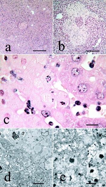
Brain, kidney, and lung of DBA/2-infected mice had no pathological alterations (data not shown), but liver sections showed extensive histopathological changes. Starting on day 6 of infection, the livers of all susceptible mice showed areas of coagulative bridging necrosis. The lesions were mostly located in the acinar zones 2 and 3. Apoptotic cells were scarce and isolated. The cytoplasm of the hepatocytes showed micro-vesicular transformation. This unusual pattern of liver necrosis, present in almost every section, increased in number and size from day 6 to day 10 of infection (Fig. 1a and b). In contrast, liver sections of BALB/c mice showed much less severe lesions on day 6 and 8 of infection, being absent by day 10 (data not shown). There were no signs of inflammation or cellular infiltrates in the liver samples of either BALB/c or DBA/2 mice, at any time-point. This pattern, characterized by the absence of inflammatory reaction, associated with the morphology of affected cells, necrosis in acinar zones 2 and 3, is strongly suggestive of an ischaemic pathogenesis (Burt et al. Reference Burt, Portmann and MacSween2002).
Histological analysis of mice infected with Plasmodium chabaudi chabaudi. Livers were removed and processed for optical (a–c) and electron microscopical observation (d and e). (a) Haematoxylin-eosin (H&E)-stained section of a liver from a BALB/c mouse at day 6 after infection, showing 2 small areas of necrosis. (b) H&E-stained liver section from a DBA/2 mouse on day 10 after infection, showing extensive and confluent areas of necrosis. (c) Viable hepatocytes, showing cytoplasmic micro-vacuolization, are present at the right. At the left, necrotic hepatocytes are present, with nuclei shadows and homogenization of the cytoplasm. Two apoptotic hepatocytes are present at the centre of the image. (d) Ultrastructural view of the clear-cut transition between viable and necrotic areas of the liver. (e) Ultrastructural detail of the cytoplasm of the necrotic hepatocytes. Images are representative profiles (sections) from 6 animals per group.

High-power optical microscopical and electron microscopical observations showed that the altered hepatocytes were sharply demarcated from adjacent viable cells, which present variable numbers of small neutral lipid inclusions (Fig.1c and d). Hepatocytes showed lysis of the different cellular compartments. Ultrastructural features of apoptosis, suggested by the presence of condensed and fragmented nuclei and homogenized cytoplasm were detected in some of the hepatocytes present in the periphery of the areas of necrosis.
Neutralization of TNF-α in vivo alters DBA/2 susceptibility to P. c. chabaudi infection
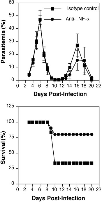
Since levels of TNF-α were found previously to be higher in the plasma of susceptible mice compared to resistant animals, we determined whether neutralization of TNF-α in vivo could prevent DBA/2 mice from death. Treatment of mice with either anti-TNF-α or control antibody had no effect on the parasitaemia course after P. c. chabaudi infection (Fig. 2). However, while DBA/2 mice treated with control antibody showed a typical survival curve (Fig. 2) with 70% mortality, 80% of mice treated with anti-TNF-α antibody were able to survive the infection.
TNF-α neutralization protects DBA/2 mice from a lethal infection. Course of infection and survival rate of DBA/2 mice infected with Plasmodium chabaudi chabaudi and treated with 0·2 mg of anti-TNF-α antibody or rat IgG as control every day from day 5 to day 9 after infection. Parasitaemias are presented as the mean of 10 animals per group±the standard error of the mean (s.e.m.). DBA/2 mice treated with anti-TNF-α antibody survived significantly better than mice treated with control antibody, P<0·05 (log-rank test).

Anti-TNF-α treatment reduces liver injury in DBA/2 mice
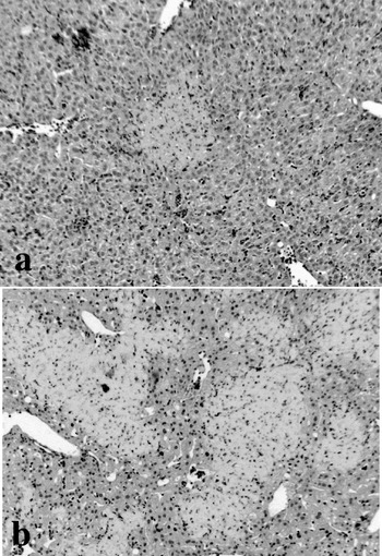
As we observed that anti-TNF-α treatment rescued DBA/2 mice from death, it was important to analyse whether TNF-α was involved in the liver injury that is observed in P. c. chabaudi-infected mice. For that, mice were infected and treated with anti-TNF-α antibody or rat IgG as control, and at days 6 and 8 after infection liver samples were taken for H&E staining. On day 8 after infection, DBA/2 mice presented extensive areas of cells undergoing apoptosis (Fig. 3b). However, in the group of DBA/2 mice treated with the antibody (Fig. 3a), although a few cells on those conditions are observed, there was a significant reduction and the mice are able to survive the infection. We also quantified the influence of treatment with anti-TNF-α antibody on liver necrosis in DBA/2 mice. The percentage of the intersections of necrotic liver over the total liver area, measured in 20–30 random areas, was of 13·7% (s.d., 4·4) in the control animals and of 5·3% (s.d., 5·0) in the anti-TNF-α treated animals. The difference between the means was statistically significant (P<0·05).
Histological analysis of DBA/2 mice infected with Plasmodium chabaudi chabaudi and treated with either control or anti-TNF-α antibody. Livers were removed from both groups of mice and stained with H&E. Liver section from a DBA/2 mouse infected and treated with anti-TNF-α antibody showing small area of necrosis (a). Liver section from a DBA/2 mouse infected and treated with control antibody showing extensive areas of necrosis (b). Images are representative profiles (sections) from 6 animals per group.

Biochemical liver assessment and haemoglobin concentration
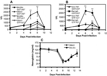
Liver injury was further quantified by measuring serum levels of the liver-specific enzymes aspartate aminotransferase (AST) and alanine aminotransferase (ALT). Plasma samples of infected DBA/2 and BALB/c mice (Fig. 4A) and DBA/2 mice after anti-TNF-α treatment (Fig. 4B) were processed for biochemical markers of hepatic cell destruction.
Biochemical parameters of the liver of BALB/c, DBA/2 (A) and DBA/2 mice treated with anti-TNF-α antibody or rat IgG control (B) and haemoglobin levels of BALB/c and DBA/2 mice (C) at different days after infection with Plasmodium chabaudi chabaudi. Data are from 7 animals per group±the standard error of the mean (s.e.m.). * P<0·05, ** P<0·01, Student's t-test, when comparing BALB/c and DBA/2 mice or DBA/2 mice with and without treatment with anti-TNF-α.

As can be seen in Fig. 4A, susceptible DBA/2 mice showed highly increased levels of aspartate aminotransferase (AST) and alanine aminotransferase (ALT), conventional biochemical markers of liver damage. The same was observed in DBA/2 mice treated with control antibody (Fig. 4B) while anti-TNF-α treatment induces a marked reduction in the levels of AST and ALT. Since severe anaemia is also a major cause of mortality in malaria and it is thought that TNF depresses bone-marrow and hence red blood cell replacement (Clark and Chaudhri, Reference Clark and Chaudhri1988; Miller et al. Reference Miller, Silverman, Kullgren and Mahlmann1989; McDevitt et al. Reference McDevitt, Xie, Gordeuk and Bucala2004), we analysed the haemoglobin concentration in BALB/c and DBA/2 mice after infection (Fig. 4C). Both strains of mice showed a decrease in haemoglobin around day 8 after infection and recovered after that (Fig. 4C). There was no significant difference between the two strains of mice during the course of infection.
DISCUSSION
In the present study we have provided evidence that death in DBA/2 mice infected with P. c. chabaudi is associated to TNF-α-related liver lesions.
We analysed, by histology, different organs of DBA/2 mice after infection to analyse their histopathological alterations and correlate them with the severity of the disease in this model. We observed marked liver injury in the susceptible compared to the resistant BALB/c strain of mice. Some studies have shown that during the erythrocyte stage liver injury could develop, although merozoites do not infect liver cells (Adachi et al. Reference Adachi, Tsutsui, Kashiwamura, Seki, Nakano, Takeuchi, Takeda, Okumura, Van Kaer, Okamura, Akira and Nakanishi2001, Reference Adachi, Tsutsui, Seki, Nakano, Takeda, Okumura, Van Kaer and Nakanishi2004; Guha et al. Reference Guha, Kumar, Choubey, Maity and Bandyopadhyay2006). However, large numbers of infected erythrocytes pass through spleen and liver and the injury induced by the parasite is probably caused by the local production of cytokines. TNF-α is a potent pro-inflammatory molecule and has been implicated as a mediator of toxic shock (Vassalli, Reference Vassalli1992; Spooner et al. Reference Spooner, Markowitz and Saravolatz1992). Lipopolysaccharide, a major constituent of the outer membrane of Gram-negative bacteria, provokes liver injury in the host via local activation of pro-inflammatory cytokines, although it does not possess liver tropism (Laskin and Pendino, Reference Laskin and Pendino1995; Tsutsui et al. Reference Tsutsui, Kayagaki, Kuida, Nakano, Hayashi, Takeda, Matsui, Kashiwamura, Hada, Akira, Yagita, Okamura and Nakanishi1999, Reference Tsutsui, Matsui, Okamura and Nakanishi2000; Naka et al. Reference Naka, Tsutsui, Fujimoto, Kawazoe, Kohzaki, Morita, Nakagawa, Narazaki, Adachi, Yoshimoto, Nakanishi and Kishimoto2001). If liver damage was indeed the cause of death, anti-TNF-α treatment was expected to reduce hepatocyte cell death. This experiment could also provide insight into the molecular mechanism mediating the liver damage. In this experimental model we show that treatment with anti-TNF antibody reduces liver injury and increases the survival of the susceptible strain. We did not observe major infiltration of cells into the liver and it has been shown before that liver damage caused by TNF occurred in mice even when leukocyte infiltration was inhibited (Schlayer et al. Reference Schlayer, Laaf, Peters, Schaefer, Decker, Wisse, Knook and Decker1989; Leist et al. Reference Leist, Gantner, Bohlinger, Germann, Tiegs and Wendel1994). High in vivo concentrations of TNF-α correlate with hyperparasitaemia, hypoglycaemia, fever, anaemia, renal impairment and pulmonary oedema in human malaria (Shaffer et al. Reference Shaffer, Grau, Hedberg, Davachi, Lyamba, Hightower, Breman and Phuc1991; Krishna et al. Reference Krishna, Waller, Ter Kuile, Kwiatkowski, Crawley, Craddock, Nosten, Chapman, Brewster, Holloway and White1994; Odeh, Reference Odeh2001). Our results show an effect of TNF-α on susceptibility of DBA/2 mice to infection with P. c. chabaudi. Using anti-TNF-α treatment we were able to increase survival in DBA/2 mice and reduce the liver damage presented by these mice after infection. Using other mouse strains, it was shown that treatment of IL-10 knockout mice with antibodies to neutralize TNF-α in vivo allowed mice to survive (Li et al. Reference Li, Sanni, Omer, Riley and Langhorne2003). Our results suggest that the anti-TNF-α treatment is acting on the liver damage to increase survival but we cannot exclude that TNF might also be acting in other ways.
Since severe anaemia is also a major cause of mortality in malaria and it is thought that TNF depresses bone marrow and hence red blood cell replacement (Odeh, Reference Odeh2001; McDevitt et al. Reference McDevitt, Xie, Gordeuk and Bucala2004), we analysed the haemoglobin concentration in BALB/c and DBA/2 mice after infection. Although TNF-α has been shown to induce bone-marrow suppression and erythrophagocytic activity, in our experimental model of susceptibility to malaria in DBA/2 mice there is no difference in anaemia with the resistant BALB/c strain. Although cytokines like TNF-α can suppress erythropoiesis, other factors may contribute to ineffective erythropoiesis. Several studies using in vivo neutralizing antibodies have indicated that IFN-γ and TNF-α are not the key cytokines involved in inhibition of erythropoiesis (Yap and Stevenson, Reference Yap and Stevenson1991, Reference Yap and Stevenson1994). Studies have shown that TNF-α knockout mice suffered more severe anaemia than WT mice (Hernandez-Valladares et al. Reference Hernandez-Valladares, Naessens, Musoke, Sekikawa, Rihet, Ole-Moiyoi, Busher and Iraqi2006) suggesting that TNF deficiency does not prevent mice from developing anaemia. The severity of the morphological features of dyserythropoiesis suggests that factors other than pro-inflammatory cytokines may contribute to inadequate erythropoiesis during malaria infection. Haemozoin present in the circulation or in the bone marrow, is capable of directly inhibiting erythropoiesis (Casals-Pascual et al. Reference Casals-Pascual, Kai, Cheung, Williams, Lowe, Nyanoti, Williams, Maitland, Molyneux, Newton, Peshu, Watt and Roberts2006). Haemozoin and TNF-α might have additive effects on the development of erythroid cells. In the mouse, both lethal and non-lethal malaria infections induce ineffective erythropoiesis with alterations in erythropoietic progenitor and precursor populations as well as in the sites of erythropoiesis. Decrease in mouse bone-marrow cellularity is seen with reductions in cellularity proportional to the severity of the infection (Lamikanra et al. Reference Lamikanra, Brown, Potocnik, Casals-Pascual, Langhorne and Roberts2007). We previously observed a marked decrease in bone-marrow cellularity in DBA/2 mice as compared to BALB/c mice. The decrease in bone-marrow cellularity observed in both strains is compensated by increased erythropoietic activity in spleen, however, the increase in splenic erythroblasts is lower in the DBA/2 mice as compared to BALB/c (Seixas and Ostler, Reference Seixas and Ostler2005). Marked splenic erythropoietic response is probably required for survival of the mice. TNF-α might be produced in the liver and induce the production of acute-phase proteins. TNF-α might be exerting its action in different ways, since TNF mediates inflammation such as an acute-phase response and generation of ROS (Bouharoun-Tayoun et al. Reference Bouharoun-Tayoun, Oeuvray, Lunel and Druilhe1995). Although our results indicate that the anti-TNF-α antibody treatment is acting by preventing liver damage, we cannot exclude the possibility that this treatment is also preventing anaemia.
TNF-α is also linked to IL-12 and IFN-γ and all these cytokines are increased in primary infection with P. chabaudi. During the acute phase of both human and mouse infections there is a strong pro-inflammatory response, which results in increases in TNF-α and IFN-γ (Miller et al. Reference Miller, Silverman, Kullgren and Mahlmann1989; Kwiatkowski et al. Reference Kwiatkowski, Hill, Sambou, Twumasi, Castracane, Manogue, Cerami, Brewster and Greenwood1990; Yap and Stevenson, Reference Yap and Stevenson1994). Secretion of IL-12 from macrophages and dendritic cells induces production of IFN-γ from NK cells, and B and T cells (Trinchieri and Gerosa, Reference Trinchieri and Gerosa1996). Studies using models of sepsis with mice lacking IFN-γ have shown that IFN-γ has an important role in the up-regulation of TNF-α production in vivo (Kamijo et al. Reference Kamijo, Le, Shapiro, Havell, Huang, Aguet, Bosland and Vilcek1993). Parasite antigen-stimulated cells produce high levels of IFN-γ, which may up-regulate the production of TNF-α in the susceptible mouse strain. We previously observed increased TNF-α levels in the serum of DBA/2 mice (Seixas and Ostler, Reference Seixas and Ostler2005) but the levels might also be increased in the liver and have a deleterious effect on the outcome of the disease. The observed differences in liver injury between BALB/c and DBA/2 mice might be due to immunoregulatory cytokines and not only to the direct TNF-α-inducing effect.
Hepatic dysfunction has been described in human SAM, and is believed to be involved in the morbidity of the disease (Deller et al. Reference Deller, Cifarelli, Berque and Buchanan1967; Srivastava et al. Reference Srivastava, Khanduri, Lakhtakia, Pandey and Choudhuri1996; Kochar et al. Reference Kochar, Agarwal, Kochar, Jain, Rawat, Pokharna, Kachhawa and Srivastava2003). In human malarias as well as in experimental models different degrees of hepatocyte necrosis, focal and zonal have been described (Deller et al. Reference Deller, Cifarelli, Berque and Buchanan1967; Rodriguez-Acosta et al. Reference Rodriguez-Acosta, Finol, Pulido-Mendez, Marquez, Andrade, Gonzalez, Aguilar, Giron and Pinto1998; Joshi et al. Reference Joshi, Tandon, Acharya, Babu and Tandon1986). The present observations establish a novel model for the genetic and molecular analyses of this situation, also opening the possibility to test putative therapeutic interventions. In the experimental model of fatal infection of C57BL/6 mice with P. berghei, the liver injury is caused by a cytotoxic T cell-mediated perforin-dependent mechanism (Adachi et al. Reference Adachi, Tsutsui, Seki, Nakano, Takeda, Okumura, Van Kaer and Nakanishi2004). ‘Malaria hepatitis’ is a term used to describe hepatocytic dysfunction in severe and complicated malaria; however, histologically severe hepatic inflammation has never been documented (Anand and Puri, Reference Anand and Puri2005). In our model, the cellular inflammatory reaction is inconspicuous, hepatocytic necrosis and apoptosis being the leading finding, a process that is mediated by circulating TNF-α. Some recent work relates liver cell apoptosis in malaria experimental infection with oxidative stress (Guha et al. Reference Guha, Kumar, Choubey, Maity and Bandyopadhyay2006), that can be induced by increasing levels of TNF-α (Shen and Pervaiz, Reference Shen and Pervaiz2006). The basis of the differential response of DBA/2 and BALB/c mice could be due either to variable levels of TNF-α production, or else, to differential susceptibility of liver cells to TNF-α. The first alternative is more likely, given the divergent serum concentrations of this cytokine upon infection of both strains resulting in the observed different degrees of hepatic lesion.
Several studies have shown that in patients with ‘malaria hepatitis’, although their clinical presentation is not different from other patients with malaria, the incidence of complications such as CM, renal failure, pulmonary oedema and shock is higher and mortality is also higher (Wilairatana et al. Reference Wilairatana, Looareesuwan and Charoenlarp1994; Murthy et al. Reference Murthy, Sahay, Sreenivas, Sundaram and Shantaram1998). The fact that hepatocytic dysfunction and raised hepatic enzymes are detected in patients with P. falciparum malaria indicates a more severe illness with a higher incidence of complications and poor diagnosis (Bhalla et al. Reference Bhalla, Suri and Singh2006). Due to this fact, this situation should be promptly recognized in order to define methods of action. Using the model we describe here it will be possible to dissect further the causes of the hepatocytic dysfunction and define therapeutic measures to act efficiently to prevent complications and poor diagnosis.
We thank Rosa Santos for production of the anti-TNF-α antibody and Dolores Bonaparte for mouse production. We greatly acknowledge the assistance of Pedro Branco for electron microscopy work. This work was supported by a project grant on malaria from the ‘Fundação Calouste Gulbenkian’, Portugal. E. S. was awarded a fellowship from ‘Fundação para a Ciência e a Tecnologia’, Portugal.