




No CrossRef data available.
Article contents
Mode of action of a potentially important excretory–secretory product from Giardia lamblia in mice enterocytes
Published online by Cambridge University Press: 03 March 2005
Abstract
Giardia, a common enteric protozoan parasite is a well-recognized cause of diarrhoeal illness. The detailed mechanism of diarrhoea due to this infection is not well understood. A 58 kDa enterotoxin (ESP) was purified from the excretory–secretory product of the parasite. The present study was designed to investigate the mode of action of this enterotoxin of G. lamblia in mice enterocytes. An increase in cyclic adenosine monophosphate level, as well as intracellular Ca2+ concentration, was observed in the ESP-triggered enterocytes. The levels of phospholipase Cγ1 and inositol triphosphate were found to be upregulated. The activity of protein kinase C (PKC) in the enterocytes was also enhanced following stimulation with the ESP. An increase in the level of reactive oxygen species in ESP-stimulated cells correlated well with the decline in the activity of antioxidant enzymes (superoxide dismutase and catalase). The significantly high levels of nitrite and citrulline indicated the generation of reactive nitrogen intermediates in the ESP-triggered enterocytes. Thus, ESP could induce cross-talk among the different signal transduction pathways in the enterocytes, which could together bring about a common secretory response.
Keywords
Information
- Type
- Research Article
- Information
- Copyright
- © 2005 Cambridge University Press
INTRODUCTION
Infection with the protozoan parasite Giardia lamblia (syn. Giardia duodenalis) is emerging as the leading non-viral cause of infectious diarrhoea in children (Thompson et al. 2000). The symptoms of Giardia infection are highly variable and some individuals may shed infectious cysts in their faeces without showing any clinical symptoms (Farthing, 1997). This variation in symptoms is not fully understood, although host factors and strain variation of the parasite are both likely to be involved (Thompson et al. 2000). It has been estimated that there are closer to 1000 million cases of giardiasis at one time, contributing to 2·5 million deaths annually from diarrhoeal disease (WHO press release, 1998).
The intracellular messengers proposed to regulate directly the small intestinal electrolyte transport include cyclic adenosine monophosphate (cAMP), cyclic guanosine monophosphate (cGMP) and Ca2+(Hardcastle et al. 1984; Donowitz et al. 1989), the levels of which were altered by the extracellular messengers like microbial toxins. The intracellular calcium [Ca2+]i has been established as an important regulator of small intestinal active Na+ and Cl− transport (Donowitz & Welsh, 1987). The release of [Ca2+]i from intracellular stores can be mediated through cAMP or inositol triphosphate (IP3) (Hardcastle et al. 1984). Direct evidence of synergism between cAMP and the Ca2+ mediated secretory mechanism in the intestine has been reported (Cartwright et al. 1985). Another Ca2+ sensitive pathway implicated in this signalling network is known to be mediated by protein kinase C (PKC) (Donowitz et al. 1989) which has been reported to play a central role in the electrolyte imbalance in intestinal infection. PKC, an intracellular Ca2+ and phospholipid-dependent enzyme, is known to be activated by diacylglycerol (DAG) which is transiently produced from inositol bisphosphate by phospholipase Cγ1 (PLCγ1) in response to extracellular signals. Several authors have reported that under appropriate conditions protein kinase C activation and Ca2+ mobilization could act synergistically to elicit a physiological response (Kawamoto & Hidaka, 1984).
Reactive oxygen species (ROS) have been implicated in the diseases of the gastrointestinal tract (Naya et al. 1997). ROS are responsible for causing tissue damage and the extent of oxidative damage depends on the balance between the pro-oxidants and the anti-oxidants. A significant decrease in the level of antioxidants in enterocytes during V. cholerae 0139 infection could lead to lipid peroxidation of the membrane components and thus might contribute to the changes in membrane permeability and ultimately fluid secretion (Gorowara, Sapru & Ganguly, 1998). NO has been known to act as both secretogogue as well as an absorbagogue in cholera toxin (CT) induced secretion (Turvill, Mourad & Farthing, 1999). Hence, radical species of nitrogen and oxygen may be important modulators of the response of intestinal epithelial cells to external signals.
The pathogenesis of giardiasis is not clearly understood, but villous atrophy and reduction of the absorptive area of the small intestine have been reported which result from brush border deficiency responsible for malabsorption (Jimenez et al. 2004). The mechanism of action of Giardia-induced diarrhoea has been studied in detail. The intracellular messengers like cAMP, Ca2+ and PKC have been implicated in electrolyte imbalance in giardiasis (Gorowara et al. 1992). However, in diarrhoeagenic conditions toxins are considered to play an important role in the induction of fluid secretion (Rout et al. 1974). Studies in vitro and in vivo have suggested that the parasite is able to produce toxin(s) but there is no direct evidence regarding the toxin-mediated electrolyte imbalance in the case of giardiasis. Thus, in the present investigation, an attempt has been made to evaluate the level of different intracellular messengers and free radicals in the ESP-triggered mice enterocytes. The outcome of such a study would be helpful for the better understanding of the mode of action of the ESP, in Giardia-induced diarrhoea as well as for the development of various strategies for prevention as well as treatment of the disease.
MATERIALS AND METHODS
Chemicals
In the present study chemicals used were of analytical and molecular biology grade. Radio-isotope labelled chemicals were obtained from Bhabha Atomic Research Centre, Trombay (Mumbai, India) and Radiochemical Centre, Amersham (UK).
Laboratory animals
Specific-pathogen free Inbred Balb/c mice (10–15 g) obtained from the Central Animal House of the Post-graduate Institute of Medical Education and Research (Chandigarh, India) were used for the study.
Axenic culture of G. lamblia
The trophozoites of G. lamblia, Portland-1 (P-1) strain, were maintained axenically in modified TYI-S-33 medium at 37 °C (Diamond, Harlow & Cunnick, 1978) and the mass cultivation of the parasite was done (Shant et al. 2002).
Purification of excretory–secretory product (ESP)
The ESP was purified by the method of Shant et al. (2002). Briefly, the crude protein was fractioned at 4 °C, with ammonium sulphate saturations from the culture supernatant in Minimum Essential Medium (MEM). Each fraction was dialysed extensively against 1 mM Tris-HCl (pH 7·2) at 4 °C and lyophilized. The fractions showing biological activity were pooled and further purified on Superdex 200 HR 10/30 gel filtration column in the FPLC system (Pharmacia, Sweden).
Isolation of IgG of anti-ESP sera
The antiserum against the purified ESP was raised in rabbits and the IgG fraction (IgGES) of the anti-ESP sera was purified using protein A-Sepharose CL 4B column (Shant et al. 2002). The ELISA titre of the IgGES was 1[ratio ]5000 with 1 μg of purified ESP.
Isolation of enterocytes
Enterocytes were isolated from mice small intestine as described by Pinkus (1981) with modifications as reported by Toyoda, Lee & Lebenthal (1985). The animals were sacrificed and the small intestine of each animal was quickly excised. The intestine was opened longitudinally, cut into small pieces and enterocytes were isolated by chelation-elution.
Study on the mode of action of the ESP in mice enterocytes
The viable enterocytes (106 cells/ml) were triggered with the purified ESP (2 μg) for different time-periods. Enterocytes without the ESP served as control in all the assays. The enterocytes triggered with commercial CT (2 μg) were taken as positive control. Most of the assays were done in the presence and absence of GM1 (1 μg) or IgGES (polyclonal antibody raised against ESP in rabbits) [diluted to 1[ratio ]2500 in 20 mM Tris-HCl, (pH 7·2) containing 150 mM NaCl (TBS)] to assess the effect of the ESP on different parameters of signal transduction. Further, specific inhibitors of different intracellular mediators were used in respective assays to reveal the authenticity of the results. All the assays were performed in triplicate.
Estimation of intracellular free calcium concentration [Ca2+]i
[Ca2+]i was estimated in enterocytes by the method of Pace & Galan (1994). For this, isolated enterocytes were washed 3 times with 20 mM HEPES buffer (pH 7·4) containing 145 mM NaCl, 5 mM KCl, 1 mM Na2HPO4, 1 mM CaCl2, 0·5 mM MgSO4 and 5 mM glucose. The pellet was resuspended in HEPES buffer, distributed in different test tubes and triggered with the purified ESP for different time-periods. The cells were centrifuged (1000 g, 10 min at 4 °C) to remove the excess toxin, resuspended in HEPES buffer and loaded with 2 μM Fura-2/AM (Sigma, Chemicals, dissolved in dimethyl sulfoxide) at 37 °C for 45 min. The unabsorbed dye was removed by washing the cells in HEPES buffer (thrice). Finally, the enterocytes were suspended in the same buffer and fluorescence measurements of the cell suspension were performed at an excitation wavelength of 340 nm. The emission spectrum was recorded at 510 nm. Fura-2 is a fluorescent dye, which specifically binds to Ca2+. After measuring the basal fluorescence (F), 2 mM EGTA (prepared in 1 M Tris buffer, pH 8·8) was added to the suspension to adjust the pH of the solution to 8·3. A rapid and sustained decrease in fluorescence after addition of EGTA was attributed to extra Fura-2/AM. Digitonin (10 nM) was added to permeablize the cells and release the trapped dye, which resulted in a maximum decrease in fluorescence (Fmin). Then 5 mM CaCl2 was added to saturate the Fura-2/AM and EGTA. The resultant fluorescence signal was designated as Fmax. The [Ca2+]i was calculated by the equation:

To study the role of intracellular calcium stores, in a separate set of experiments the cells were pre-incubated with (i) dantrolene, a drug known to trap Ca2+ in intracellular stores (20 μM, 15 min, 37 °C), (ii) thapsigargin, an endoplasmic reticulum calcium–ATPase inhibitor (40 μM, 15 min, 37 °C), (iii) verapamil, an L-type Ca2+ channel blocker (50 μM, 15 min, 37 °C), (iv) calciseptin, an endoplasmic reticulum (ER) L-type Ca2+ channel (Ca2+–ATPase) blocker (1 μM, 15 min, 37 °C) and treated with or without the ESP for 5 min. The [Ca2+]i was then measured in each set of experiments as described above.
Measurement of cyclic adenosine 3′,5′-monophosphate (cAMP) levels
The cAMP levels were measured in the enterocytes by use of an RIA kit (Amersham International, Radiochemical Centre, Amersham, UK, code TRK 432) according to the manufacturers' instructions. The assay is based on the competition between unlabelled cAMP and a fixed quantity of 3H-labelled cAMP for binding to a protein that has high specificity and affinity for cAMP. Measurement of the protein-bound radioactivity enables calculation of the amount of unlabelled cAMP in the sample. In this method, the enterocytes were suspended in TBS (pH 7·2) and triggered with the purified ESP. The cells were centrifuged and cold 0·1 M HCl (0·1 ml) was added to the cell pellet followed by incubation for 15 min at 37 °C. The cell debris was removed by centrifugation (500 g, 10 min) and the supernatant was collected. Subsequently the supernatant was titrated to pH 7·2 with 0·1 M NaOH and used for estimation. Results were expressed as pmol of cAMP/mg protein from a standard curve plotted with Co/Cx (where Co is the cpm bound in the absence of unlabelled cAMP and Cx is the cpm bound in presence of standard or unlabeled cAMP) versus pmol standard. A separate set of experiments was conducted with the enterocytes pre-incubated with 2′,5′-dideoxyadenosine (DDA), a specific inhibitor of adenylate cyclase and the level of cAMP was measured as described above.
Assessment of phospholipase C γ1 (PLCγ1) level
The level of phospholipase Cγ1 in the mice enterocytes was assessed by Western immunoblot. Briefly, the enterocytes were triggered with the ESP for different time-periods. They were then sonicated and debris was removed by centrifugation (1000 g, 10 min). The supernatant was analysed for the expression of PLC γ1 using affinity-purified rabbit polyclonal antibody raised against PLCγ1 [PLC γ1(1249):SC 81;Santa Cruz Biotechnology, Inc] as the primary antibody (diluted to 1[ratio ]1000 in TBS containing 1% BSA) and HRP-conjugated swine immunoglobulins to rabbit immunoglobulins (Dakopatts, Denmark) as the secondary antibody (diluted to 1[ratio ]1000 in TBS-BSA). For quantitative results, flow cytometric analysis of whole cells triggered with the ESP pre-incubated in the presence and absence of IgGES/GM1 was performed with the primary antibody (diluted to 1[ratio ]1000 in TBS). The FITC-conjugated swine immunoglobulins to rabbit immunoglobulins (Dakopatts, Denmark) was used as secondary antibody (diluted to 1[ratio ]1000) using the Cell Quest program on a FACScan (Becton and Dickinson, USA). Results were expressed as the percentage of labelled cells, which could be directly correlated to the level of PLCγ1 in the enterocytes.
Inositol triphosphate (IP3) turnover
The IP3 turnover in the enterocytes was measured using labelled myo[3H]-inositol according to the method of Oldham (1990). Briefly, enterocytes were taken in TBS and incubated with the purified ESP for different time-periods at 37 °C. The cells were then centrifuged (1000 g, 10 min at 4 °C) to remove the excess toxin. This was followed by treatment with lithium chloride (10 mM, 1 ml) for 20 min. Subsequently the cells were incubated with myo[3H]-inositol for 45 min at 37 °C. The cell suspension was centrifuged (250 g, 10 min) to wash off excess labelled inositol, and the cells were suspended in TBS (1 ml). The suspension was recentrifuged (250 g, 10 min). The cell pellet was suspended in TBS (7 ml), treated with perchloric acid (20%, 0·2 ml) and incubated on ice for 20 min. Proteins were removed by centrifugation (200 g, 20 min, 4 °C) and the supernatant was collected. Siliconized glassware was used in subsequent steps to minimize the loss of inositol phosphates. Supernatant was titrated to pH 7·5 with ice-cold KOH (10 M) and incubated on ice for 15 min. Precipitated KClO4 was removed by centrifugation (2000 g, 20 min, 4 °C). The supernatant was applied on Amprep™ mini columns (SAX, 100 mg, Amersham), pre-conditioned with 1 M KHCO3 (5 ml) and distilled water (15 ml). Elution was done in a stepwise manner by 5 ml of each eluent [distilled water, 50 mM KHCO3 (to elute IP1), 100 mM KHCO3 (to elute IP2), 170 mM KHCO3 (to elute IP3) and 250 mM KHCO3 (to elute IP4), respectively]. Each eluate (1 ml/5 ml) was transferred to a scintillation vial containing aqueous scintillation fluid (7 ml) and counted. Radioactivity incorporated was determined by liquid scintillation counter (Rackbeta, 1214).
Measurement of protein kinase C (PKC) level
The protein kinase C activity in the enterocytes was measured by using a PepTag® Non-radioactive protein kinase C Assay kit (Promega, USA, Cat. No. V5330), according to the manufacturers' instructions. The PepTag® Assay utilizes bright coloured fluorescent peptide substrate i.e. Pep Tag® C1 peptide, PLSRTLSVAAK that is highly specific for the kinase. The hot pink colour is imparted by the addition of a dye molecule to the PepTag® peptide substrate. Phosphorylation by PKC of this specific substrate alters the peptide's net charge from +1 to −1. This change in the net charge of the substrate allows the phosphorylated and the non-phosphorylated forms of the substrate to be rapidly separated on an agarose (1%) gel at neutral pH. The phosphorylated substrate was extracted from the gel, heated at 95 °C, solubilized, acidified with glacial acetic acid and finally evaluated by measuring the optical density at 550 nm. This system detects up to 2 ng of protein kinase C. For this assay, the enterocytes were isolated by the method of Pinkus (1981). The cells were suspended in 20 mM TBS (pH 7·2) and incubated with the ESP for different time-periods. Each tube containing the cells was centrifuged (100 g, 10 min at 4 °C) to remove the excess toxin. The cells were resuspended in TBS (pH 7·2) and sonicated. The debris was removed by centrifugation (500 g, 10 min) and the supernatant of each tube was used for the estimation of PKC. The activity of PKC was expressed as units/106 cells. In a separate set of experiments, the cells were pre-treated with H7, a serine-threonine kinase inhibitor at 6 μM concentration and incubated with the ESP. The activity of PKC was measured as above.
Estimation of total free oxygen radicals
Total free oxygen radicals released from the enterocytes was measured by estimating the luminal-dependent chemiluminescence response of the enterocytes (Cheung, Archibald & Robinson, 1984). This method is based on the principle that mammalian cells undergoing a respiratory burst whilst coming into contact with the stimulating agent, release various ROS which emit light when electrons come down from the excited state to the ground level. The biological reaction produces chemiluminescence (CL) that is less than 100 photons/second/cell. Thus luminol (5-amino-2,3-dihydro-1,4-phthalazinedine) is used as chemiluminogenic probe for the amplification of the luminescence to 103 to 104 times by its conversion to phthalate anions. Latex acts as a non-specific stimulant of CL. Light emitted during CL absorbs maximally at 425 nm. In this assay, the isolated enterocytes (106 cells/ml) suspended in HBSS were taken in the wells of chemiluminescence plate. The enterocytes were incubated with or without the ESP. Background counts (A) were recorded for 1 min in Berthold luminometer (BioLumat, LKB 9500C) set at integration mode at 37 °C. Luminol (20 μl) was added and the counts (B) were recorded for 1 min. Latex (20 μl, 0·81 μm diameter, Difco Labs, USA) was added to the suspension and mixed properly. Counts were taken after every 1 min until a peak was attained which was recorded as ‘C’. Care was taken to minimize the time lag for taking counts. Results were expressed as counts per minute (cpm)/106 cells and calculated as chemiluminescence index: (C−A)/(B−A).
Catalase assay
Catalase was assayed in the enterocytes by the method of Beers & Sizer (1984). The assay is based on the principle that catalase causes time-dependent decomposition of H2O2 which can be monitored spectrophotometrically at 240 nm. The change in absorbance was read at 240 nm for 60 sec at 10 sec intervals. The specific activity was calculated using a molar absorbance index (ε) 43·6 for H2O2 and expressed as nmole/mg protein/min. In this assay, the enterocytes were incubated with the ESP for different time-periods and washed in PBS. The cells were disrupted by sonication and debris was removed by centrifugation. Catalase was estimated in the supernatant fractions. The assay system was comprised of 3 ml of 150 mM phosphate buffer (pH 7·0) containing 0·16 ml H2O2 (30% v/v) and 20 μl of the supernatant was added to the reaction mixture. The rate of decomposition of H2O2 by catalase was measured by recording the time required for decrease of the absorbance by 0·05 at 240 nm.
Superoxide dismutase (SOD) assay
SOD activity was measured using the method of Kono (1978). This assay is based on the rate of nitrobluetetrazolium (NBT) dye reduction by superoxide anion radicals (generated by photooxidation of hydroxylamine hydrochloride) in the presence of the enzyme, SOD. The absorbance of the reduced NBT was recorded at 560 nm. In this assay, the enterocytes were incubated with the ESP for different time-periods and washed in PBS. The cells were disrupted by sonication and debris removed by centrifugation. SOD activity was estimated in the supernatant fractions. Small aliquots of the supernatant were added to the reaction mixture containing 50 mM Na2CO3/0·1 M EDTA (pH 10·0) containing 96 μM NBT, 0·6% Triton X-100 and 20 μM NH2OH/HCl. The decline in the rate of NBT reduction in the presence of SOD in the supernatant was assessed. One unit of SOD was taken as the inverse of the amount of protein (mg) required to inhibit the reduction of NBT by 50%. Finally, the activity was expressed as IU/106 cells.
Measurement of nitric oxide (NO)
NO released from the enterocytes incubated with the ESP was measured by the determination of nitrite and citrulline levels in culture supernatant of the cells.
Nitrite levels were measured by the method of Green et al. (1982). In this assay, nitrites in the sample react with a Griess reagent to form a purple azo dye. The colour of the product (dye) was developed by incubation in a 60 °C water bath followed by cooling in a 0 °C water bath. Finally, the absorbance was monitored at 546 nm. In this assay, the enterocytes were incubated in the presence or absence of the purified ESP for different time-periods and the excess toxin was removed by washing the cells. The cells in each tube were then incubated in RPMI containing L-arginine (1 mM) at 37 °C for 2 h. The cell suspensions were centrifuged (500 g, 10 min) and supernatant of each tube was taken for nitrite estimation. The supernatant (1 ml) was added to an equal volume of reagent C [prepared by mixing N-1-napthylethylene diamine dihydrochloride (0·1%) along with sulphanilamide (1%) and phosphoric acid (5%) at a ratio 1[ratio ]1 v/v)] followed by incubation for 10 min, at room temperature and the absorbance was recorded at 546 nm. Blank and standards (1–10 nmol) were also run in parallel. The results were expressed as μmole of nitrite formed/106 cells.
Citrulline levels were measured by the method of Boyde & Rahmattulah (1980). In this method, citrulline, a carbamido compound produced from L-arginine (obtained from the acid hydrolysis of proteins/peptides) forms a purple- coloured complex with diacetyl monoxime in acid solution which can be monitored spectrophotometrically at 530 nm. For the estimation of citrulline, enterocytes were incubated in the presence of the ESP for different time-periods and the excess toxin was removed by washing the cells. The cells in each tube were then incubated in RPMI containing L-arginine (1 mM) at 37 °C for 2 h. The cell suspensions were centrifuged and supernatant of each tube was taken for citrulline estimation. The supernatant of each tube (50 μl) was treated with 0·1 M HCl (450 μl) and 1·5 ml of reagent C [prepared by mixing 2 parts of reagent A (550 ml of DW, 250 ml of H2SO4 (95–98%), 200 ml of o-phosphoric acid (85%) and 250 mg of ferric chloride) and 1 part of reagent B (0·5% diacetyl monoxime and 0·01% thiosemicarbazide)]. Tubes containing the reaction mixture were immersed in a water bath at 100 °C for 5 min followed by cooling to room temperature. The absorbance was measured at 530 nm. Blank and standards (25–300 nmol) were run simultaneously. Results were expressed as μmole of citrulline formed/106 cells.
RESULTS
To understand the mechanism of action of the 58 kDa ESP in mice enterocytes, different parameters of signal transduction were studied.
The [Ca2+]i in mice enterocytes triggered with the ESP (2 μg) at different time-intervals (30 sec, 1 min, 5 min, 10 min, 20 min and 30 min) revealed a 6·6-fold increase at 1 min as compared to that in control enterocytes (Table 1). Fig. 1 depicts that in the presence of dantrolene (20 μM)/verapamil (50 μM)/calciseptine (1 μM)/thapsigargin (40 μM), the ESP-induced [Ca2+]i in the enterocytes (106 cells) was significantly reduced to 45·3±1·3 nM/222·8±10·5 nM/338·6±16·4 nM/117·1±19·0 nM respectively as compared to that in ESP triggered cells (389·8±18·8 nM) [control value in each set was subtracted from the respective test value]. Further, in enterocytes triggered with ESP pre-incubated with IgGES/GM1, the [Ca2+]i was found to be 168·3±10·1 nM/138·1±25·6 nM respectively [control values subtracted were cells+IgGES/cells+GM1 i.e. 81·2+2·5/89·4+3·8 respectively]. The CT (positive control) could induce a 4·8-fold increased [Ca2+]i (370·0±8·0 nM) in the enterocytes as compared to that of control cells (76·7±3·6 nM).

Fig. 1. Values are expressed as mean±S.D. The [Ca2+]i levels in enterocytes (106) isolated from mouse intestines and triggered with 2 μg of purified ESP (5 min) in the presence of different inhibitors/channel blockers/IgGES. The cells were washed and labelled with Fura2-AM as described in the Materials and Methods section. The [Ca2+]i was calculated by using KD value of Fura2-AM as 224. The optimum dose of the ESP was then selected. The results were expressed in mM/106 cells. The levels of significance are *P<0·05, **P<0·01, as compared to ESP.
Table 1. Intracellular free calcium concentrations in enterocytes stimulated with ESP (Results are expressed as [Ca2+]i levels in nM/1×106 enterocytes. Values are expressed as mean±S.D.)

The level of cAMP (pmol/mg protein) in enterocytes triggered with ESP or ESP pre-incubated with IgGES/GM1 is depicted in Table 2. A statistically significant (P<0·01) increase in cAMP level in enterocytes triggered with ESP for 5 min was noticed as compared to that in control enterocytes. However, the level was decreased in the presence of IgGES/GM1. In the presence of 250 μM DDA the cAMP level was reduced in the ESP triggered enterocytes. The CT-triggered enterocytes (positive control), revealed a significant (P<0·001) increase in the level of cAMP as compared to that of control cells.
Table 2. cAMP levels in enterocytes triggered with ESP in the presence and absence of inhibitors/IgGes (Values are expressed as mean±S.D.)

The time-profile study of PLCγ1 in the Western immunoblot (Fig. 2) revealed an increase in the level of this enzyme within 1 min of the ESP triggering of the enterocytes. However, the expression of PLCγ1 could be reduced by 60·03±5·0% and 39·12±2·1% respectively, when the cells were triggered with the ESP pre-incubated separately with IgGES and GM1 (the level of PLCγ1 in enterocytes stimulated with ESP taken as 100%) as assessed by flow cytometric analysis.

Fig. 2. Western immunoblot showing phospholipase C γ1 activity in mouse enterocyte membranes triggered with the ESP at different time-periods. The enterocytes were triggered with the ESP, sonicated and centrifuged. The supernatant was analysed for the expression of PLC γ1 using affinity-purified rabbit polyclonal antibody to PLC γ1 [PLC γ1(1249):SC 81; Santa Cruz Biotechnology, Inc] as the primary antibody (1[ratio ]1000). Lane a, control; lanes b, c, d and e, 15 s, 30 s, 1 min and 2 min respectively; lane f, cholera toxin.
The IP3 turnover (cpm/106 cells) was measured in control enterocytes as well as in cells triggered separately with the purified ESP and CT (positive control) (Fig. 3). A gradual increase in the IP3 level was observed on triggering the cells with the purified ESP for 0 min, 1 min and 5 min (743·6±56·2, 3366·0±168·5 and 6603·2±188·1 respectively). Then a gradual decline in the value of IP3 was noticed in the enterocytes stimulated with the purified ESP for 10 min, 20 min and 30 min (5375·2±382·9, 5050·2±258·1 and 3629·7±204.8 respectively). Maximum IP3 formed in ESP induced enterocyte at 5 min (6603·2±188·1) was significantly higher (P<0·001) as compared to that of control enterocytes (743·6±56·2). The IP3 level in CT (positive control) triggered cells was maximum (5129·9±126·1) at 10 min.

Fig. 3. Values are expressed as mean±S.D. The mouse enterocytes were incubated with purified ESP (2 μg) and CT (positive control) separately at different times and the IP3 turnover was estimated in cpm/106 cells. The level of significance is ***P<0·001 as compared to control.
The protein kinase C (PKC) activity (units/106 cells) was studied in ESP triggered mice enterocytes for different time-periods (Fig. 4). A statistically significant (P<0·001) increase in the PKC activity was observed at 1 min (0·93±0·05) as compared to that in the control cells (0·11±0·03). The PKC activity was reduced to a value below detectable range when the cells were triggered with the ESP pre-incubated separately in the presence of IgGES and GM1. Further, complete reduction in the PKC activity was observed in the ESP-triggered enterocytes, pre-incubated with 6 μMof H-7. CT, the positive control could increase the PKC activity (1·04±0·31) significantly (P<0·001) in the enterocytes as compared to that of control cells.

Fig. 4. Values are expressed as mean±S.D. The PKC activity in mouse enterocytes (106 cells) was measured by using a Pep Tag®non- radioactive protein kinase C assay kit (Promega, USA). For this assay, the enterocytes were incubated with the ESP for different time-periods and the PKC activity was estimated in the cell lysate as per manufacturers' instructions. The activity of PKC was expressed as units/106 cells. The level of significance is ***P<0·001 as compared to control.
Chemiluminescence response [Chemiluminescence index (CI)] was measured in the ESP triggered enterocytes at different time-periods (Fig. 5). Release of ROS was highest and statistically significant (P<0·05) at 1 min (3·03±0·23) as compared to that of control cells (1·47±0·02). The levels were reduced significantly when the enterocytes were triggered with the ESP pre-incubated separately with IgGES and GM1. The values were 1·85±0·07 and 1·40±0·06 respectively. The CT (positive control) could also release ROS (3·00±0·20) of statistically significant level (P<0·05).

Fig. 5. Values are expressed as mean±S.D. The ROS levels in ESP (2 μg)-treated mouse enterocytes were estimated by the luminal-dependent chemiluminescence response. Results were expressed as counts per minute (cpm)/106 cells and calculated as chemiluminescence index: (C−A)/(B−A). The level of significance is *P<0·05 as compared to control.
The catalase activity (nmole/mg protein/min) and the SOD activity (IU/106 cells) in the ESP-triggered mice enterocytes at different time-periods are shown in Table 3. A 2·5-fold decrease in the catalase activity and a statistically significant (P<0·05) reduction in the SOD activity was observed in the enterocytes at 30 sec as compared to that of the control cells. However, when enterocytes were triggered with ESP pre-treated with IgGES, the activity of both the enzymes was increased. The CT (positive control) could decrease the activity of catalase and SOD in the enterocytes significantly as compared to that in the control cells.
Table 3. Antioxidant enzyme activity in enterocytes triggered with ESP at different times (Values are expressed as mean±S.D.)

The levels of nitrite and citrulline (μmole/106 cells) in the supernatant of the ESP-triggered enterocytes at different time-periods are shown in Table 4. A significant increase in the levels of both nitrite and citrulline was found in the enterocytes at 5 min as compared to that of control cells. However, the levels were found to be partially decreased in the enterocytes stimulated with ESP pre-incubated separately with GM1 and IgGES. The CT (positive control) revealed a significant increase in the levels of both nitrite and citrulline in the enterocytes as compared to that in the control cells.
Table 4. Nitrite and citrulline levels in enterocytes triggered with ESP at different times (Values are expressed as mean±S.D.)

DISCUSSION
Giardiasis is a relatively common protozoal infection of the intestinal tract in man and animals. Despite the considerable morbidity caused by giardiasis, little is known about the parasite and the pathophysiology of diarrhoea associated with it. The parasite is not invasive, but it is capable of causing brush border alteration, diarrhoea and malabsorption (Jimenez et al. 2004).
Some Giardia surface antigens are known to be excreted/secreted during parasite growth in vitro (Nash, Gillin & Smith, 1983; Papanastasiou et al. 1997; Adam, 2001). However, the exact nature as well as the biological significance of these excretory secretory antigens in regulation of the infection was not elucidated. The role of putative Giardia toxins or metabolic ES products as toxins is speculative, although there is mounting evidence that Giardia trophozoites could produce toxins. The culture filtrates of Giardia have been reported to damage fibroblasts in culture and reduce salt and water absorption from the perfused loops of rats (Katelaris et al. 1988). One of the most observed changes in experimental giardiasis is inhibition of the activities of several digestive enzymes, including sucrase and maltase during acute-phase infection. A correlation exists between the infectious load and the extent of enzyme inhibition, which suggests that the effects might be caused by factors released by live parasites (Eckmann & Gillin, 2001). The parasite could cause diffuse shortening of enterocyte microvilli, thereby inhibiting microvillus enzyme activity and nutrient transport (Buret, Gall & Olson, 1990). Further, sonicated trophozoites and culture media have been shown to cause increased smooth muscle contractility in gerbils and were cytotoxic to Chinese hamster ovary cells (Olson, Morck & Ceri, 1996). Recently, in our laboratory, a glycoprotein (ESP) has been identified in the excretory-secretory component of G. lamblia, Portland-1 strain. The purified ESP could accumulate fluid in the intestine of sealed adult mice and also induce morphological changes in HEp-2 cells. The IgGES showed cross-reactivity with the binding subunit of cholera toxin. Also GM1, the known receptor for cholera toxin showed a good extent of binding to the ESP (Shant et al. 2002). All this evidence clearly indicated the toxic nature of the ESP. Thus, in the previous investigation (Shant et al. 2004) and also in the present study we made an attempt to understand the mechanism of action of the ESP, keeping the classical cholera toxin pathway in mind. In G. lamblia-infected gerbils, glucose-stimulated absorption of sodium and water was decreased as compared to that in uninfected controls. G. lamblia infection of gerbils also accelerated gastrointestinal transit and smooth muscle contractility, which could play a role in the pathogenesis of giardial diarrhoea (Buret et al. 1992). In contrast to gerbils, infection of mice with G. lamblia has been reported to cause net secretion of sodium and chloride in the basal state, whereas control animals showed net absorption of these ions under the same conditions (Gorowara et al. 1992). In the previous paper (Shant et al. 2004) it was reported that the ESP could bind to a 41 kDa glycoprotein (receptor?) on the enterocyte membrane and activate a G-protein-mediated signal transduction pathway. The reduced GTPase activity seemed to be responsible for the increase in the adenylate cyclase-dependent cAMP level and thereby PKA activity. This could be, in part, responsible for the alteration in the electrolyte transport observed (secretion of chloride and no absorption of sodium).
Calcium has been established as an intracellular regulator of small intestinal as well as colonic transmembrane electrolyte transport (Donowitz et al. 1989). In the present investigation an increase in [Ca2+]i was observed in the ESP triggered mice enterocytes. The increased intracellular Ca2+ may have its source either from increased Ca2+ entry from the extracellular milieu to the cell or from the release of Ca2+ from intracellular stores. In our study to understand the source of intracellular Ca2+, various Ca2+ channel blockers were used. Dantrolene is known to trap calcium in intracellular stores. In the present study, the intracellular calcium stores seem to have major involvement since maximum reduction in the concentration of cytosolic calcium was observed in enterocytes triggered with the ESP in presence of dantrolene. This result is in good agreement with the previous report (Gorowara et al. 1992) in which it was shown that 2·5 μM serosal dantrolene could result in a significant increase in the absorption of Na+, Cl− and Ca2+ in G. lamblia-induced infection. The involvement of intracellular Ca2+ stores was also observed in V. cholerae non-01 heat stable enterotoxin induced a response in rat enterocytes (Hoque et al. 2001). The stimulation of enterocytes with a Ca2+ mobilizing agent thapsigargin, followed by triggering with the ESP indicated major involvement of this channel in the ESP induced rise in [Ca2+]i. Further, with calciseptin, an appreciable level of intracellular Ca2+ was observed in ESP-triggered enterocytes, which might be due to the involvement of other Ca2+ channels. The [Ca2+]i was reduced significantly in the presence of verapamil as compared to that in the presence of calciseptin. These observations have clearly indicated that the release of Ca2+ from intracellular stores plays a very significant role in the ESP induced alteration of the intracellular calcium concentration. However, the probable involvement of the plasma membrane L-type Ca2+ channels also cannot be ignored.
Trimeric guanine nucleotide-binding proteins (G-proteins) function as the key regulatory elements in a number of transmembrane signalling cascades (Svoboda & Novotny, 2002). These G-proteins can activate adenylate cyclase or phospholipase C present on the inner face of membrane. Adenylate cyclase converts ATP into cAMP; whereas phospholipase C cleaves membrane lipid phosphatidylinositol 4,5-bisphosphate (PIP2) into DAG and IP3. The IP3 as well as cAMP can lead to an increase in intracellular calcium concentration and imbalance in electrolyte transport across the membrane.
It has been reported that in G. lamblia infection there was an increase in cAMP levels and cAMP could produce its effect by mobilizing calcium from intracellular stores, which in turn could regulate the electrolyte transport (Gorowara et al. 1992). In the present study, the cAMP level was found to be upregulated in ESP-triggered mice enterocytes. The authenticity of our finding has been further substantiated by a reduction in the level of cAMP in the presence of DDA. The cAMP level was shown to be elevated in Salmonella enterotoxin (S-LT) treated cells (Peterson et al. 1983). Further, a large and sustained increase in the cAMP concentration was observed in vascular smooth muscle cells after 2h of cholera toxin treatment (Sachinidis et al. 2000).
Ruschkowski, Rosenshine & Finlay (1992) have suggested that S. typhimurium could stimulate host phospholipase C (PLC) activity, resulting in the cleavage of PIP2 into IP3 and DAG. The IP3 produced as a result of phospholipid hydrolysis in turn could mobilize Ca2+ release from the intracellular stores, which might be responsible for several changes including disruption of the microvillus structure, loss of transepithelial electrical resistance as well as the localized rearrangement in actin filaments and related proteins like villin in the host cell. This pathway has also been implicated in the cholera toxin induced diarrhoea (Sears & Kaper, 1996; Gorowara et al. 1998). Bhatnagar et al. (1999) have shown that the cytotoxic activity of anthrax lethal toxin in J774A.1 cells could be inhibited in the presence of neomycin, a phospholipase C inhibitor. Our results have also indicated the involvement of PLCγ1 in the ESP-triggered mice enterocytes. Gorowara et al. (1992) have reported that G. lamblia trophozoites as well as their ES extracts could bring about a rise in IP3 levels in mouse enterocytes. This observation is in good agreement with our present findings, in which a significant increase in the IP3 level was observed after 5 min in enterocytes triggered with the ESP. However, the concentration of intracellular Ca2+ was found to be upregulated after 1–5 min of ESP stimulation. Thus, the early response of Ca2+ could be attributed to the involvement of other signalling pathways including adenylate cyclase dependent cAMP mediated route of signal transduction (since the level of cAMP was also maximum at 1 min) in ESP treated enterocytes.
Several workers have proposed the role of PKC in the stimulation of intestinal secretion (Donowitz et al. 1989). Khurana et al. (1991) have reported that the S-LT induced fluid secretion in rat enterocytes could be mediated through PKC. Further, it was shown by Kaur et al. (1997) that an appreciable level of PKC in the membrane fraction of enterocytes isolated from Shigella toxin treated rabbit ileum could be reduced in the presence of H7, an inhibitor of PKC. In the present investigation, it was found that H7 could decrease the activity of PKC in the ESP-triggered enterocytes. The two possibilities regarding PKC activation may be (a) mobilization of Ca2+ from intracellular stores by cAMP that in turn may activate PKC (b) phospholipid hydrolysis by PLCγ, which may induce PKC activation through DAG along with Ca2+ mobilization by IP3 (Peterson et al. 1983). A synergism between the PKC activation and alterations in the cAMP levels has also been well documented ( Sugden et al. 1985). Ganguly et al. (2001) have provided evidence regarding the role of Ca2+ in the translocation of PKC-α from the cytosol to the membrane of enterocytes treated with E. coli heat-stable enterotoxin. However, in ESP-treated mice enterocytes, it would be difficult to comment on whether cAMP could cause the release of Ca2+ from intracellular stores that then activated PKC, or phospholipid hydrolysis by PLCγ could induce PKC activation through DAG and Ca2+ mobilization by IP3, which ultimately acted synergistically to cause direct phosphorylation of the catalytic subunit of adenylate cyclase. Further, Gorowara et al. (1992) have also demonstrated the role of PKC in the stimulation of intestinal ion secretion in G. lamblia induced infection. It has been suggested that in G. lamblia infection, protein kinase C might be located at the cross-over point of various pathways involving Ca2+, cyclic nucleotides, inositol phospholipids and prostaglandins.
The gastrointestinal tract is a rich source of antioxidant enzymes which are known to detoxify the effect of ROS. However, when the rate of production of ROS exceeds the capacity of the antioxidant defences, substantial tissue damage occurs. Mehta, Singh & Ganguly (1999) have investigated the role of ROS in mediating the enterocyte damage during in vitro exposure to S-LT. McNeil, Knox & Proudfoot (2002) have observed that parasite rejection could be inhibited by the administration of antioxidants, which strongly suggested the involvement of oxygen-derived free radicals in intestinal parasite rejection. All the previous reports are in good agreement with our present findings, in which the level of ROS was found to be elevated accompanied by a significant decrease in the activity of SOD and catalase in enterocytes triggered with the ESP.
Nitrites and citrulline are known to be the end-products of the oxidative metabolism of the labile NO in vivo and their quantitation is regarded as an indicator of NO generation (Stuehr & Marletta, 1985). Excessive NO produced during inflammation may cause pathophysiological changes and tissue damage (Tabouret et al. 2001). Studies by Forsythe et al. (2002) on rat intestinal epithelial cells have shown that enterocytes could be the target as well as producer of NO. The involvement of NO in several intestinal inflammatory diseases due to cytoskeletal alteration has also been demonstrated (Vignoli et al. 2001). Our results have clearly revealed that in the ESP-triggered enterocytes there is an increased generation of both ROS and NO. Thus, the generation of peroxynitrite anion may possibly be due to the reaction between ROS and NO, which can induce tissue injury through lipid peroxidation and the oxidation of sulfhydryl groups in the membrane component (Beckman et al. 1990).
The alteration in the level of most of the intracellular mediators could be inhibited in the presence of GM1 as well as IgGES. In most of the cases, the maximum inhibition was observed in the presence of IgGES. This observation has clearly indicated that the antibody-binding epitope of the ESP might have a precise role in cell signalling as compared to the GM1-binding epitope. As per literature, the GM1-binding epitope might be involved only in the recognition of the cell surface receptor (Minke et al. 1999). However, complete reversal of the level of intracellular mediators was not observed in enterocytes stimulated with the ESP pre-incubated in the presence of either IgGES or GM1 which suggested that somehow both the IgGES and GM1 specific epitopes of the ESP are essential for transducing signals within the cells.
Thus, it can be suggested that the ESP could induce the activation of PLCγ1 in mice enterocytes that could lead to the generation of IP3 and DAG. The IP3 could then mobilize Ca2+ release from intracellular stores. The activation of PKC could be due to the presence of DAG, resulting in the activation of adenylate cyclase as well as generation of ROS. The adenylate cyclase could increase cAMP levels and hence [Ca2+]i causing electrolyte imbalance. ROS could induce electrolyte imbalance through lipid peroxidation. NO generated by the iNOS, activated in the presence of intracellular Ca2+ could also lead to lipid peroxidation and thus electrolyte imbalance. So it can be proposed that the ESP, an enterotoxin of G. lamblia, could induce cross-talk among diverse signal transduction pathways (Fig. 6) that might result in secretion and/or malabsorption.

Fig. 6. Cross-talk among diverse signal transduction pathways induced in mouse enterocytes by ESP from Giardia lamblia. Activation of PLCγ1 in the ESP- stimulated mice enterocytes could lead to the hydrolysis of PIP2 into IP3 and DAG. IP3 mobilized Ca2+ from intracellular stores whereas DAG activated PKC leading to electrolyte imbalance in the intestine by phosphorylation of transport carriers and conductance channels. PKC could also activate adenylate cyclase thereby increasing cAMP levels and hence [Ca2+]i. PKC could also cause generation of ROS leading to electrolyte imbalance through lipid peroxidation. NO generated by the iNOS, activated in the presence of intracellular Ca2+ could also lead to lipid peroxidation and thereby cause electrolyte imbalance.
The authors acknowledge the financial assistance provided by CSIR, New Delhi to J.S.
References
REFERENCES
ADAM, R. D. (
2001).
Biology of Giardia lamblia.
Clinical Microbiology Reviews
14,
447–475.CrossRefGoogle Scholar
BECKMAN, J. S.,
BECKMAN, T. W.,
CHEN, J.,
MARSHALL, P. A. &
FREEMAN, B. A. (
1990).
Apparent hydroxyl radical production by peroxynitrite: implications for endothelial injury from nitric oxide and superoxide.
Proceedings of the National Academy of Sciences, USA
87,
1620–1624.CrossRefGoogle Scholar
BEERS, R. F. &
SIZER, J. W. (
1984).
A spectophotometric method for measuring the breakdown of hydrogen peroxide by catalase.
Journal of Biological Chemistry
195,
133–140.Google Scholar
BHATNAGAR, R.,
AHUJA, N.,
GOILA, R.,
BATRA, S.,
WAHEED, S. M. &
GUPTA, P. (
1999).
Activation of phospholipase C and protein kinase C is required for expression of anthrax lethal toxin cytotoxicity in J774A.1 cells.
Cell Signal
11,
111–116.CrossRefGoogle Scholar
BOYDE, T. R. &
RAHMATULLAH, M. (
1980).
Optimization of conditions for the colorimetric determination of citrulline, using diacetyl monoxime.
Analytical Biochemistry
107,
424–431.CrossRefGoogle Scholar
BURET, A.,
GALL, D. G. &
OLSON, M. E. (
1990).
Effects of murine giardiasis on growth, intestinal morphology, and disaccharidase activity.
Journal of Parasitology
76,
403–409.CrossRefGoogle Scholar
BURET, A.,
HARDIN, J. A.,
OLSON, M. E. &
GALL, D. G. (
1992).
Pathophysiology of small intestinal malabsorption in gerbils infected with Giardia lamblia.
Gastroenterology
103,
506–513.CrossRefGoogle Scholar
CARTWRIGHT, C. A.,
McROBERTS, J. A.,
MANDEL, K. G. &
DHARMSATHAPHORN, K. (
1985).
Synergistic action of cyclic adenosine monophosphate- and calcium-mediated chloride secretion in a colonic epithelial cell line.
Journal of Clinical Investigation
76,
1837–1842.CrossRefGoogle Scholar
CHEUNG, K.,
ARCHIBALD, A. C. &
ROBINSON, M. F. (
1984).
Luminol-dependent chemiluminescence produced by neutrophils stimulated by immune complexes.
Australian Journal of Experimental Biology and Medical Science
62,
403–419.CrossRefGoogle Scholar
DIAMOND, L. S.,
HARLOW, D. R. &
CUNNICK, C. C. (
1978).
A new medium for the axenic cultivation of Entamoeba histolytica and other Entamoeba.
Transactions of the Royal Society of Tropical Medicine and Hygiene
72,
431–432.CrossRefGoogle Scholar
DONOWITZ, M.,
COHEN, M. E.,
GOULD, M. &
SHARP, G. W. (
1989).
Elevated intracellular Ca2+ acts through protein kinase C to regulate rabbit ileal NaCl absorption. Evidence for sequential control by Ca2+/calmodulin and protein kinase C.
Journal of Clinical Investigation
83,
1953–1962.Google Scholar
DONOWITZ, M. &
WELSH, M. J. (
1987).
Regulation of Mammalian Small Intestinal Electrolyte Secretion.
Raven Press,
New York.
ECKMANN, L. &
GILLIN, F. D. (
2001).
Microbes and microbial toxins: paradigms for microbial-mucosal interactions I. Pathophysiological aspects of enteric infections with the lumen-dwelling protozoan pathogen Giardia lamblia.
American Journal of Physiology, Gastrointestinal and Liver Physiology
280,
G1–G6.Google Scholar
FARTHING, M. J. (
1997).
The molecular pathogenesis of giardiasis.
Journal of Pediatric Gastroenterology and Nutrition
24,
79–88.CrossRefGoogle Scholar
FORSYTHE, R. M.,
XU, D. Z.,
LU, Q. &
DEITCH, E. A. (
2002).
Lipopolysaccharide-induced enterocyte-derived nitric oxide induces intestinal monolayer permeability in an autocrine fashion.
Shock
17,
180–184.CrossRefGoogle Scholar
GANGULY, U.,
CHAUDHURY, A. G.,
BASU, A. &
SEN, P. C. (
2001).
STa-induced translocation of protein kinase C from cytosol to membrane in rat enterocytes.
FEMS Microbiology Letters
204,
65–69.CrossRefGoogle Scholar
GOROWARA, S.,
SAPRU, S. &
GANGULY, N. K. (
1998).
Role of intracellular second messengers and reactive oxygen species in the pathophysiology of V. cholera O139 treated rabbit ileum.
Biochimica et Biophysica Acta
1407,
21–30.CrossRefGoogle Scholar
GOROWARA, S.,
GANGULY, N. K.,
MAHAJAN, R. C. &
WALIA, B. N. (
1992).
Study on the mechanism of Giardia lamblia induced diarrhoea in mice.
Biochimica et Biophysica Acta
1138,
122–126.CrossRefGoogle Scholar
GREEN, L. C.,
WAGNER, D. A.,
GLOGOWSKI, J.,
SKIPPER, P. L.,
WISHNOK, J. S. &
TANNENBAUM, S. R. (
1982).
Analysis of nitrate, nitrite, and [15N]nitrate in biological fluids.
Analytical Biochemistry
126,
131–138.CrossRefGoogle Scholar
HARDCASTLE, J.,
HARDCASTLE, P. T. &
NOBLE, J. M. (
1984).
The involvement of calcium in the intestinal response to secretagogues in the rat.
Journal of Physiology
355,
465–478.CrossRefGoogle Scholar
HOQUE, K. M.,
PAL, A.,
NAIR, G. B.,
CHATTOPADHYAY, S. &
CHAKRABARTI, M. K. (
2001).
Evidence of calcium influx across the plasma membrane depends upon the initial rise of cytosolic calcium with activation of IP(3) in rat enterocytes by heat-stable enterotoxin of Vibrio cholerae non-O1.
FEMS Microbiology Letters
196,
45–50.CrossRefGoogle Scholar
JIMENEZ, J. C.,
FONTAINE, J.,
GRZYCH, J. M.,
DEI-CAS, E. &
CAPRON, M. (
2004).
Systemic and mucosal responses to oral administration of excretory and secretory antigens from Giardia intestinalis.
Clinical and Diagnostic Laboratory Immunology
11,
152–160.CrossRefGoogle Scholar
KATELARIS, P. H.,
SIDHU, G. S.,
OAKENFULL, D. G. &
NGU, M. C. (
1988).
Pathogenesis of diarrhoea caused by Giardia lamblia: evidence for an exotoxin.
Journal of Gastroenterology and Hepatology
3 (Suppl. I), A4(abstract).Google Scholar
KAUR, T.,
SINGH, S.,
VERMA, M. &
GANGULY, N. K. (
1997).
Calcium and protein kinase C play a significant role in response to Shigella toxin in rabbit ileum both in vivo and in vitro.
Biochimica et Biophysica Acta
1361,
75–91.CrossRefGoogle Scholar
KAWAMOTO, S. &
HIDAKA, H. (
1984).
1-(5-Isoquinolinesulfonyl)-2-methylpiperazine (H-7) is a selective inhibitor of protein kinase C in rabbit platelets.
Biochemical and Biophysical Research Communications
125,
258–264.CrossRefGoogle Scholar
KHURANA, S.,
GANGULY, N. K.,
KHULLAR, M.,
PANIGRAHI, D. &
WALIA, B. N. (
1991).
Studies on the mechanism of Salmonella typhimurium enterotoxin-induced diarrhoea.
Biochimica et Biophysica Acta
1097,
171–176.CrossRefGoogle Scholar
KONO, Y. (
1978).
Generation of superoxide radical during autoxidation of hydroxylamine and an assay for superoxide dismutase.
Archives of Biochemistry and Biophysics
186,
189–195.CrossRefGoogle Scholar
McNEIL, K. S.,
KNOX, D. P. &
PROUDFOOT, L. (
2002).
Anti-inflammatory responses and oxidative stress in Nippostrongylus brasiliensis-induced pulmonary inflammation.
Parasite Immunology
24,
15–22.CrossRefGoogle Scholar
MEHTA, A.,
SINGH, S. &
GANGULY, N. K. (
1999).
Effect of Salmonella typhimurium enterotoxin (S-LT) on lipid peroxidation and cell viability levels of isolated rat enterocytes.
Molecular and Cellular Biochemistry
196,
175–181.CrossRefGoogle Scholar
MINKE, W. E.,
ROACH, C.,
HOL, W. G. &
VERLINDE, C. L. (
1999).
Structure-based exploration of the ganglioside GM1 binding sites of Escherichia coli heat-labile enterotoxin and cholera toxin for the discovery of receptor antagonists.
Biochemistry
38,
5684–5692.CrossRefGoogle Scholar
NASH, T. E.,
GILLIN, F. D. &
SMITH, P. D. (
1983).
Excretory-secretory products of Giardia lamblia.
Journal of Immunology
131,
2004–2010.Google Scholar
NAYA, M. J.,
PEREBOOM, D.,
ORTEGO, J.,
ALDA, J. O. &
LANAS, A. (
1997).
Superoxide anions produced by inflammatory cells play an important part in the pathogenesis of acid and pepsin induced oesophagitis in rabbits.
Gut
40,
175–181.CrossRefGoogle Scholar
OLDHAM, K. G. (
1990).
Polyphosphoinositide turnover. In
Receptor Effector Coupling: A Practical Approach
(ed. Hulme, E. C.), pp.
99–102.
Oxford University Press,
Oxford.
OLSON, M. E.,
MORCK, D. W. &
CERI, H. (
1996).
The efficacy of a Giardia lamblia vaccine in kittens.
Canadian Journal of Veterinary Research
60,
249–256.Google Scholar
PACE, J. L. &
GALAN, J. E. (
1994).
Measurement of free intracellular calcium levels in epithelial cells as consequence of bacterial invasion.
Methods in Enzymology
236,
482–490.CrossRefGoogle Scholar
PAPANASTASIOU, P.,
BRUDERER, T.,
LI, Y.,
BOMMELI, C. &
KOHLER, P. (
1997).
Primary structure and biochemical properties of a variant-specific surface protein of Giardia.
Molecular and Biochemical Parasitology
86,
13–27.CrossRefGoogle Scholar
PETERSON, J. W.,
MOLINA, N. C.,
HOUSTON, C. W. &
FADER, R. C. (
1983).
Elevated cAMP in intestinal epithelial cells during experimental cholera and salmonellosis.
Toxicon
21,
761–775.CrossRefGoogle Scholar
PINKUS, L. M. (
1981).
Separation and use of enterocytes.
Methods in Enzymology
77,
154–162.CrossRefGoogle Scholar
ROUT, W. R.,
FORMAL, S. B.,
DAMMIN, G. J. &
GIANNELLA, R. A. (
1974).
Pathophysiology of Salmonella diarrhea in the Rhesus monkey: intestinal transport, morphological and bacteriological studies.
Gastroenterology
67,
59–70.Google Scholar
RUSCHKOWSKI, S.,
ROSENSHINE, I. &
FINLAY, B. B. (
1992).
Salmonella typhimurium induces an inositol phosphate flux in infected epithelial cells.
FEMS Microbiology Letters
74,
121–126.CrossRefGoogle Scholar
SACHINIDIS, A.,
SEUL, C.,
GOUNI-BERTHOLD, I.,
SEEWALD, S.,
KO, Y.,
VETTER, H.,
FINGERLE, J. &
HOPPE, J. (
2000).
Cholera toxin treatment of vascular smooth muscle cells decreases smooth muscle alpha-actin content and abolishes the platelet-derived growth factor-BB-stimulated DNA synthesis.
British Journal of Pharmacology
130,
1561–1570.CrossRefGoogle Scholar
SEARS, C. L. &
KAPER, J. B. (
1996).
Enteric bacterial toxins: mechanisms of action and linkage to intestinal secretion.
Microbiological Reviews
60,
167–215.Google Scholar
SHANT, J.,
GHOSH, S.,
BHATTACHARYYA, S.,
GANGULY, N. K. &
MAJUMDAR, S. (
2004).
The alteration in signal transduction parameters induced by excretory-secretory product from G. lamblia.
Parasitology
129,
421–430.CrossRefGoogle Scholar
SHANT, J.,
BHATTACHARYYA, S.,
GHOSH, S.,
GANGULY, N. K. &
MAJUMDAR, S. (
2002).
A potentially important excretory-secretory product of Giardia lamblia.
Experimental Parasitology
102,
178–186.CrossRefGoogle Scholar
STUEHR, D. J. &
MARLETTA, M. A. (
1985).
Mammalian nitrate biosynthesis: mouse macrophages produce nitrite and nitrate in response to Escherichia coli lipopolysaccharide.
Proceedings of the National Academy of Sciences, USA
82,
7738–7742.CrossRefGoogle Scholar
SUGDEN, D.,
VANECEK, J.,
KLEIN, D. C.,
THOMAS, T. P. &
ANDERSON, W. B. (
1985).
Activation of protein kinase C potentiates isoprenaline-induced cyclic AMP accumulation in rat pinealocytes.
Nature, London
314,
359–361.CrossRefGoogle Scholar
SVOBODA, P. &
NOVOTNY, J. (
2002).
Hormone-induced subcellular redistribution of trimeric G proteins.
Cellular and Molecular Life Sciences
59,
501–512.CrossRefGoogle Scholar
TABOURET, G.,
VOULDOUKIS, I.,
DURANTON, C.,
PREVOT, F.,
BERGEAUD, J. P.,
DORCHIES, P.,
MAZIER, D. &
JACQUIET, P. (
2001).
Oestrus ovis (Diptera: Oestridae): effects of larval excretory/secretory products on nitric oxide production by murine RAW 264·7 macrophages.
Parasite Immunology
23,
111–119.CrossRefGoogle Scholar
THOMPSON, R. C.,
HOPKINS, R. M. &
HOMAN, W. L. (
2000).
Nomenclature and genetic groupings of Giardia infecting mammals.
Parasitology Today
16,
210–213.CrossRefGoogle Scholar
TOYODA, S.,
LEE, P. C. &
LEBENTHAL, E. (
1985).
Physiological factors controlling release of enterokinase from rat enterocytes.
Digestive Disease Science
30,
1174–1180.CrossRefGoogle Scholar
TURVILL, J. L.,
MOURAD, F. H. &
FARTHING, M. J. (
1999).
Proabsorptive and prosecretory roles for nitric oxide in cholera toxin induced secretion.
Gut
44,
33–39.CrossRefGoogle Scholar
VIGNOLI, A. L.,
SRIVASTAVA, R. C.,
STAMMATI, A.,
TURCO, L.,
TANORI, M. &
ZUCCO, F. (
2001).
Nitric oxide production in Caco-2 cells exposed to different inducers, inhibitors and natural toxins.
Toxicology In Vitro
15,
289–295.CrossRefGoogle Scholar
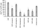
Fig. 1. Values are expressed as mean±S.D. The [Ca2+]i levels in enterocytes (106) isolated from mouse intestines and triggered with 2 μg of purified ESP (5 min) in the presence of different inhibitors/channel blockers/IgGES. The cells were washed and labelled with Fura2-AM as described in the Materials and Methods section. The [Ca2+]i was calculated by using KD value of Fura2-AM as 224. The optimum dose of the ESP was then selected. The results were expressed in mM/106 cells. The levels of significance are *P<0·05, **P<0·01, as compared to ESP.
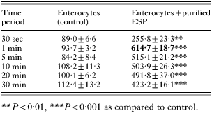
Table 1. Intracellular free calcium concentrations in enterocytes stimulated with ESP
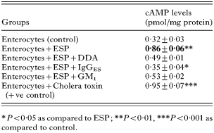
Table 2. cAMP levels in enterocytes triggered with ESP in the presence and absence of inhibitors/IgGes
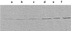
Fig. 2. Western immunoblot showing phospholipase C γ1 activity in mouse enterocyte membranes triggered with the ESP at different time-periods. The enterocytes were triggered with the ESP, sonicated and centrifuged. The supernatant was analysed for the expression of PLC γ1 using affinity-purified rabbit polyclonal antibody to PLC γ1 [PLC γ1(1249):SC 81; Santa Cruz Biotechnology, Inc] as the primary antibody (1[ratio ]1000). Lane a, control; lanes b, c, d and e, 15 s, 30 s, 1 min and 2 min respectively; lane f, cholera toxin.
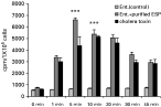
Fig. 3. Values are expressed as mean±S.D. The mouse enterocytes were incubated with purified ESP (2 μg) and CT (positive control) separately at different times and the IP3 turnover was estimated in cpm/106 cells. The level of significance is ***P<0·001 as compared to control.
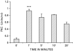
Fig. 4. Values are expressed as mean±S.D. The PKC activity in mouse enterocytes (106 cells) was measured by using a Pep Tag®non- radioactive protein kinase C assay kit (Promega, USA). For this assay, the enterocytes were incubated with the ESP for different time-periods and the PKC activity was estimated in the cell lysate as per manufacturers' instructions. The activity of PKC was expressed as units/106 cells. The level of significance is ***P<0·001 as compared to control.
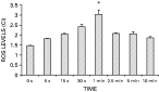
Fig. 5. Values are expressed as mean±S.D. The ROS levels in ESP (2 μg)-treated mouse enterocytes were estimated by the luminal-dependent chemiluminescence response. Results were expressed as counts per minute (cpm)/106 cells and calculated as chemiluminescence index: (C−A)/(B−A). The level of significance is *P<0·05 as compared to control.
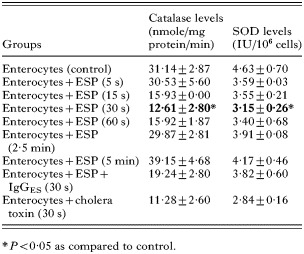
Table 3. Antioxidant enzyme activity in enterocytes triggered with ESP at different times
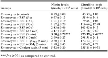
Table 4. Nitrite and citrulline levels in enterocytes triggered with ESP at different times
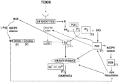
Fig. 6. Cross-talk among diverse signal transduction pathways induced in mouse enterocytes by ESP from Giardia lamblia. Activation of PLCγ1 in the ESP- stimulated mice enterocytes could lead to the hydrolysis of PIP2 into IP3 and DAG. IP3 mobilized Ca2+ from intracellular stores whereas DAG activated PKC leading to electrolyte imbalance in the intestine by phosphorylation of transport carriers and conductance channels. PKC could also activate adenylate cyclase thereby increasing cAMP levels and hence [Ca2+]i. PKC could also cause generation of ROS leading to electrolyte imbalance through lipid peroxidation. NO generated by the iNOS, activated in the presence of intracellular Ca2+ could also lead to lipid peroxidation and thereby cause electrolyte imbalance.
 You have
Access
- 9
- Cited by