INTRODUCTION
The conservation of deep-sea coral habitats has become a topic of increased concern as marine conservation agencies have begun to recognize the importance of these environments and their associated biota (e.g. Husebø et al., Reference Husebø, Nøttestad, Fosså, Furevik and Jørgensen2002; Reed et al., Reference Reed, Weaver and Pomponi2006). This coincides with the use of Remotely Operated Vehicles (ROVs) to provide extremely detailed, hi-resolution video and imagery of organisms in the deep-sea and more directed collections of deep-sea faunas from these areas (e.g. Mah et al., Reference Mah, Nizinski and Lundsten2010).
Asteroids are ecologically important (e.g. Birkeland, Reference Birkeland1974) and their feeding habits have long been a topic of interest to a wide range of marine researchers (e.g. Sloan, Reference Sloan1980; Jangoux, Reference Jangoux, Jangoux and Lawrence1982). However, most in situ observations have been limited to shallow-water taxa. Relatively few observations of deep-sea asteroids have been recorded. Recent work by Gale et al. (Reference Gale, Hamel and Mercier2013) has studied observations of relatively deep-sea species off the coast of the North Atlantic, including some lab-based experimental studies. Expeditionary work undertaken by the R/V Okeanos Explorer, operated by the National Oceanic and Atmospheric Administration (NOAA), has captured multiple instances of feeding by deep-sea asteroids displaying predatory behaviour on deep-sea corals. This complements the material present in museum collections and that recorded from specimens collected during various oceanographic expeditions.
The two genera of hippasterine asteroids, Hippasteria and Evoplosoma, are the most commonly encountered corallivorous asteroids in deep-sea coral settings. These two genera, as well as other members of the Hippasterinae (Goniasteridae) have been documented as predators on several different taxa of deep-sea corals (see Mah et al., Reference Mah, Nizinski and Lundsten2010, Reference Mah, Neill, Eleaume and Foltz2014). In situ observations remain relatively few and so I present further video data on this behaviour in order to demonstrate its occurrence and to seek further insight into the feeding mode in these poorly understood species.
An undescribed species of Evoplosoma discovered by the 2004 NOAA ‘Mountains in the Sea’ Expedition in addition to new museum data from New Zealand and Paris forms the basis for this paper. Additional material has prompted several changes to the classification of species within Evoplosoma and Hippasteria. In order to compliment the observed feeding behaviour of the undescribed Evoplosoma species described herein, I include a brief taxonomic overview of some Atlantic deep-sea goniasterid species that have shown corallivorous behaviour.
MATERIALS AND METHODS
The specimens detailed below were obtained from several institutions, including the California Academy of Sciences in San Francisco, CA (CASIZ), the New Zealand Institute of Water and Atmosphere in Wellington, New Zealand (NIWA) and the Muséum National d'Histoire Naturelle in Paris (MNHN). The specimen described below is housed at the National Museum of Natural History in Washington DC (NMNH).
In addition to the comparative overview of the corallivore Evoplosoma and the description of Evoplosoma watlingi n. sp., range extensions and feeding observations of North Atlantic coral feeding Goniasteridae have also been included. Although several of these species have been established in the Atlantic (Clark & Downey, Reference Clark and Downey1992), these records embellish occurrence data on seamounts, canyons and other features. All of the species listed below reflect either a range extension or feeding observation, save for Sthenaster emmae Mah et al., Reference Mah, Nizinski and Lundsten2010, which is included for the sake of completeness.
The locality data below includes the general place name, coordinates, depth and any other information which was available. Range extensions under species headings are boldfaced. The locality data for the images is expressed first, followed by the image descriptor for each observation. Note that the video observations outlined below refer to locality followed by different image descriptors, of which more than one descriptor often refers to the same observation. Two different image descriptors are used. This includes ‘expl XXX’, which is how the images are identified on the NOAA Photo Library website (http://www.photolib.noaa.gov/brs/nuind1.htm) and a second longer thread of numbers and letters, which refers to the exact image data from the NOAA image data server.
The figures used to represent taxonomic detail are included in the Systematics section, whereas those that describe feeding are listed in the Discussion section.
SYSTEMATICS
Family GONIASTERIDAE Forbes, 1841
Circeaster americanus (A.H. Clark, 1916)
Lydiaster americanus A.H. Clark, 1916, p. 141; 1954, p. 376.
Circeaster occidentalis H.L. Clark, 1941, p. 46, pl. 4, Figure 2. A.H. Clark, 1954, p. 376.
Circeaster americanus Halpern Reference Halpern1970a, p. 265, Figures 26–28. Halpern Reference Halpern1970b, p. 174. Downey 1973, p. 47, 55, pls. 21A, B. Walenkamp 1979, p. 52, Figures 14 & 18, pl. 12, Figures 1 & 2. Clark & Downey, Reference Clark and Downey1992, p. 237. Clark, Reference Clark1993, p. 250; Mah, Reference Mah2006, p. 943, Figure 7.
COMMENTS
The individual indicated below agrees in most respects with species descriptions in Mah (Reference Mah2006) and Clark and Downey (Reference Clark and Downey1992), save for the presence of well-defined pointed granules forming a serial edge on the marginal plates. This image was based on a specimen from the Gulf of Mexico, unlike most examined specimens that are known from the Caribbean. Consistent with this hypothesis is that at least one Caribbean specimen (e.g. USNM E38689) shows an angular edge with granules on the lateral sides of the marginal plates. Several specimens show eroded damage on the distal arm tips, suggesting that the absence of these granules in descriptions may also be the result of trawl collection.
Full taxonomic summaries of this species are found in Clark & Downey (Reference Clark and Downey1992) and Halpern (Reference Halpern1970a, Reference Halpernb) with further information on variation and synonymy in Mah (Reference Mah2006). Based on Mah's (Reference Mah2006) morphology-based phylogenetic analysis, Circeaster americanus is most closely related to the Pacific species, Circeaster pullus and Circeaster sandrae.
OCCURRENCE
Tropical Atlantic, Florida, Louisiana, Gulf of Mexico, North Carolina and the Caribbean, including Columbia, the Grenadines, Leeward Islands, Nicaragua, Netherlands Antilles, St. Vincent, Suriname and Venezuela. Manning Seamount, North Atlantic. Depth range of this species: 500–1450 m. Extended to 1675.4 m.
TAXONOMIC SUMMARY
R/r = 2.4–40. Disk swollen, large. Arms elongate. Interradial arcs linear to curved. Disk plates with granules, round, hemispherical scattered over disk surface. Arm plates two to three times larger than disk plates and bare relative to plates on disk. Arm plate size variable but most examined specimens show abrupt demarcation between large and small plates (e.g. USNM E19076) versus smaller specimens (e.g. E38689), which show a more gradual gradation. Clam-shaped pedicellariae on abactinal plate surface.
Marginal plates wide. Number of marginal plates: 52–58 (at R = 9.7). Marginal plate centre surface covered with coarse, round to pointed granules. Superomarginal plates abutted over midline. Arms elongate, tapering, with tips upturned.
Actinal plates composed of four to five chevrons, irregularly covered with coarse pointed to round granules, densely packed. Adambulacral furrow spines four to seven (mostly five or six), thick and angular in cross-section. Row of two to three enlarged subambulacral spines (about 2–3× as thick as the furrow spines) present adjacent to furrow spines. Bivalve pedicellariae present on marginal, actinal and adambulacral plates. Pedicellariae present on most actinal plates, especially those proximal to mouth.
VIDEO OBSERVATIONS
Images expl 2389 and 2410, Manning Seamount; coordinates: 38°13.0599′N 60°30.6657′W; water depth: 1333 m; 2005 North Atlantic Stepping Stones Science Party, IFE, URI-IAO; NOAA/OAR/OER. Image expl 7294, NOAA Okeanos Explorer, Gulf of Mexico 2012 Expedition (EX1202L2_IMG_20120323T184023Z_ROVHD_COR_SPO_ASR; coordinates: 27.9161°N -86.03553°W; water depth: 1675.2 m. EX1202L2_IMG_20120323T184058Z _ROVHD _COR_SPO_ASR; coordinates: 27.91617°N -86.03555°W; water depth: 1675.5 m. EX1202L2_IMG_ 20120323T184144Z_ROVHD_ COR_SPO_ASR; coordinates: 27.91615°N -86.0356°W; water depth: 1675.5 m. EX1202L2_IMG_ 20120323T184423Z_ROVHD_SPO_ASR; coordinates: 27.91161°N -86.03554°W; water depth: 1675.6 m). Images 7642–7643 (Figure 1), Gulf of Mexico, NOAA Okeanos Explorer, Gulf of Mexico 2012 Expedition (EX1202L2_IMG_ 20120323T185600Z_ ROVHD_ COR_ASR; coordinates: 27.91613°N -86.03569°W; water depth: 1673.6 m. EX1202L2_IMG_ 20120323T185641Z_ROVHD_ COR_ASR; coordinates: 27.91617°N -86.03563°W; water depth: 1673.5 m. EX1202L2_IMG_ 20120323T185722Z _ROVHD_COR_ASR; coordinates: 27.91615 -86.03561; water depth: 1673.5 m. EX1202L2_IMG_20120323T185746Z_ ROVHD_COR_ASR; coordinates: 27.91615°N -86.03561°W; water depth: 1673.5 m). Image expl. 8125, NOAA Okeanos Explorer, Gulf of Mexico 2012 Expedition; coordinates: 26.3033°N -93.4363°W; water depth: 1934.5 m.
Image Descriptor. EX1304L2_IMG_ 20130804T205244Z_ ROVHD_COR_ASR_EAT_COR.
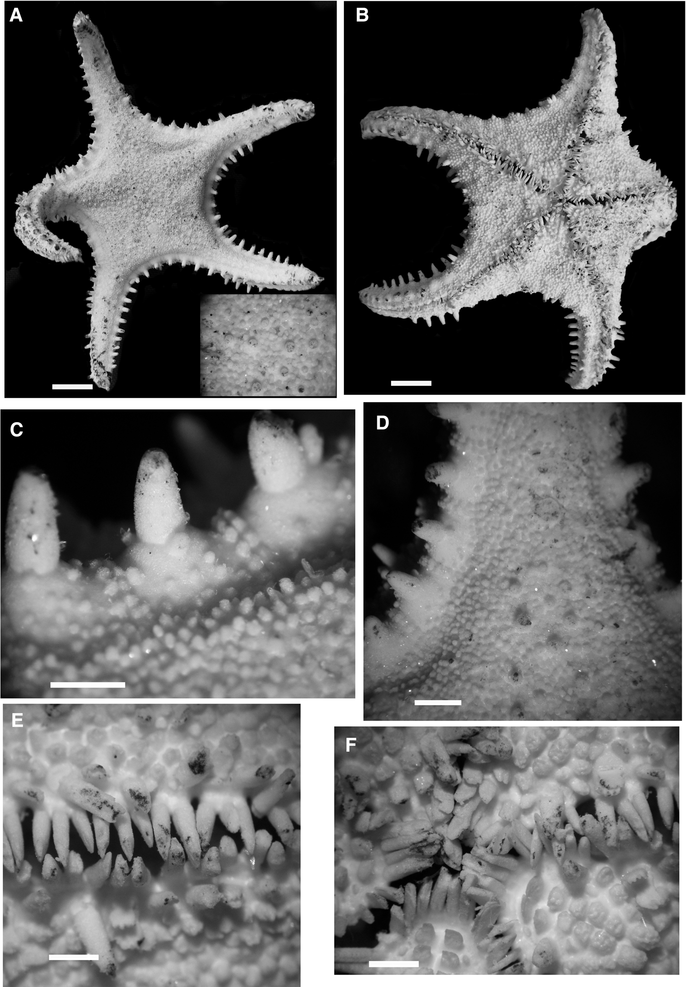
Evoplosoma tasmanica n. comb. NIWA 86966. R = 3.9 cm. (A, B) Scale bar = 1.0 cm, (C–F) scale bar = 3.0 mm. (A) Abactinal surface. Inset shows details of abactinal accessories (granules, spines, etc.). (B) Oral surface. (C) Marginal plate spines showing surface and adjacent abactinal accessory covering. (D) Abactinal surface showing surface and surface of superomarginal plates. (E) Adambulacral spination showing furrow spines and adjacent actinal plate surface accessories. (F) Oral region showing spination, etc. Photos by Kate Neill, NIWA.

Evoplosoma Fisher, Reference Fisher1906
Fisher, Reference Fisher1906, p. 1065; Koehler, Reference Koehler1909, p. 96; Spencer & Wright, Reference Spencer, Wright and Moore1966, p. U58; Clark & Downey, Reference Clark and Downey1992, p. 241; A.M. Clark, Reference Clark1993, p. 253; Mah et al., Reference Mah, Nizinski and Lundsten2010, p. 278.
OCCURRENCE
In addition to records outlined below and by Mah et al. (Reference Mah, Nizinski and Lundsten2010), further images that do not permit species-level identification suggest a wider occurrence for Evoplosoma in the North Atlantic than specimen collections would suggest.
OBSERVATIONS
September–October 2014 observations by the R/V Okeanos Explorer in the North Atlantic show one or several unidentified species of Evoplosoma (possibly Evoplosoma scorpio or Evoplosoma watlingi n. sp.) from Hendrickson Canyon, Kelvin (dive EX1404-L3, dive #9, 29 September 2014, at approximately 63.74°N 38.85°W and at a water depth of 2062.54 m), and Atlantis II seamount (dive EX1404-L3, dive #7, 27 September 2014, at 63.74°N 38.85°W, at water depths of 2062.54 m and 2640 m). Another observation of Evoplosoma sp. predation was recorded by the E/V Nautilus on Noroit Seamount in the Caribbean Sea (Mah, personal observation, 10 September 2014).
DIAGNOSIS
Body strongly stellate. Arm narrow, elongated. R/r ranges from 2.3 to 4.11 (most between 3.0 and 4.0). Interradial arcs straight to weakly curved.
Abactinal plates, flat and platform-like. Carinal series poorly distinguished. Abactinal plates tightly articulated. Body covered by tissue layer with pulpy texture that overlies plates and spines (seen more clearly in wet specimens). Prominent spines on abactinal, superomarginal, inferomarginal and actinal plates in most species. Spine morphology variable from blunt conical to pointed, to cylindrical or small and spinelet-like. Granules with spiny tips in most species, with some having rounded surfaces. Granules present but with variable abundance among species. Tong-like pedicellariae with serrated valves present or absent on abactinal, marginal or actinal surface.
Marginal plates generally quadrate in shape, some showing direct 1:1 superomarginal/inferomarginal correspondence but others being more offset. Marginals relatively numerous ranging in number from 30–70 per interradius. Some species with bare marginal plate surface, but most with even to dense granule covering. Granules varying from having rounded surface to pointed or prismatic edges. Large prominent spine or spines known in all but one species. Large single spines observed as a linear series in several species. Spinelets or multiple shorter spines observed on marginal plate surfaces of other species. Pedicellariae variably present on either supero or inferomarginal series.
Actinal intermediate regions relatively small with fewer than six rows present (three or four present in most species). Actinal plate boundaries obscured by pulpy tissue layer and/or granulation. Granules round or with spiny edges present on all species. Primary spines present on actinal plate surface in most species.
Furrow spines varying in number from two or three, to seven to 12. Spines generally compressed, quadrate to polygonal in cross section. Tips varying from smooth and blunt to jagged with furrowed tips. Subambulacral spination variable but a felipedal (clamp-like bivalve) pedicellariae present among the subambulacrals in most species. Subambulacrals varying but spination ranging from blunt spines, pointed spinelets to pointed or rounded granules.
Based on in situ observations herein and from prior accounts (e.g. Mah et al., Reference Mah, Nizinski and Lundsten2010), the colour of most species ranges from yellow to deep orange.
INCLUDED SPECIES
Evoplosoma augusti Koehler, Reference Koehler1909, Evoplosoma claguei Mah et al., Reference Mah, Nizinski and Lundsten2010, Evoplosoma forcipifera Fisher, Reference Fisher1906 (type species), Evoplosoma scorpio Downey, Reference Downey1981, Evoplosoma tasmanica nov. comb., Evoplosoma timorensis Aziz & Jangoux, Reference Aziz and Jangoux1985, Evoplosoma virgo Downey, Reference Downey1982, Evoplosoma voratus Mah et al., Reference Mah, Nizinski and Lundsten2010, Evoplosoma watlingi n. sp.
KEY FOR THE SPECIES OF EVOPLOSOMA
0. Numerous granules and/or spinelets covering abactinal, marginal, actinal plate surfaces. Prominent spines on marginal plates present or absent (irregularly distributed when present). Known occurrence: Indian, Pacific and Atlantic.
0′. Marginal plate surface bare except for a large prominent and blunt spine, present on both marginal series on most, if not all plates. Granules/spinelets absent. Known occurrence: North and Central Pacific. 4
1. Primary spines (i.e. spines rising above the plane of the surface) absent on abactinal, actinal surface and partially absent from all but distalmost marginal plates. Subambulacral spines absent, replaced by pedicellariae. Body surface covered by spiny granules. Body strongly stellate with elongate arms. R/r = 3.5. Known occurrence: Gulf of Mexico. Evoplosoma virgo Downey, Reference Downey1982
1′. Marginal plates with short primary spines, five on superomarginals, one to four on inferomarginals. Pedicellariae also present on marginal plates. Abactinal surface with blunt, pointed tubercular spines. Body more weakly pentagonal, with broad disk and triangular arms. R/r = 2.6. Known occurrence: Indian Ocean (no identified specimens known, holotype unavailable). Evoplosoma augusti Koehler, Reference Koehler1909
1″. Marginal plates with large primary spines present (large conical or blunt spines rising above plane of abactinal surface) in series. Body surface covered by granules (either round or spiny), sometimes densely. Large primary spines also abundant on abactinal and actinal surface. Body stellate with elongate arms. R/r > 3.0 Known occurrence: North Atlantic and South Pacific. 2
2. Dense round, smooth granules covering abactinal, marginal and actinal surfaces. Abactinal surface covered by evenly distributed conical spines. Furrow spines, four to five. Both furrow and subambulacral spines round in cross-section with conical tips. Known occurrence: North Atlantic-Bear Seamount. Evoplosoma watlingi nov. sp.
2′. Granules, with spiny and angular tips, some polygonal in cross section cover abactinal, marginal and actinal surfaces. Abactinal surface covered by irregularly distributed spines. Furrow spine number variable. Furrow and subambulacral spines angular in cross section. Known occurrence: South Pacific and North Atlantic. 3
3. Furrow spines two to four (mostly three). Approximately 48–54 superomarginals per interradius (~24 from midline to tip at R = 6.9 cm, R/r = 3.2–3.8). Conical, blunt spines shorter, present but not abundant, limited to radial regions on arms or disk. Known occurrence: South Pacific (New Zealand region). Evoplosoma tasma-nica nov. comb.
3′. Furrow spines two to four (mostly three). Superomarginals, 30 per interradius (15 from midpoint to tip at R = 5.6 cm, R/r = 2.3). Abactinal surface covered with abundant stout, conical spines from central disk to arms. Known occurrence: North Pacific. Hawaiian Islands. Evoplosoma forcipifera Fisher, Reference Fisher1906
3″. Furrow spines five to seven. Approximately 60–66 superomarginals per interradius (~32 from midpoint to armtip at R = 11.5 cm, R/r = 3.5). Stout, conical spines abundant on disk and arms (i.e. present on most abactinal plates). Known occurrence: North Atlantic. Evoplosoma scorpio Downey, Reference Downey1981
4. Abactinal, marginal, actinal surface covered by one to three prominent, thickened, blunt spines (round in cross section at tip), distributed evenly over surface. Superomarginal plates, relatively flattened, ~58–70 per interradius from armtip to armtip. Furrow spines six to 12. Known occurrence: Indo-Malaysian, Central Pacific. Evoplosoma timorensis Aziz & Jangoux, Reference Aziz and Jangoux1985
4′. Large, prominent spines, one to three present on strongly convex marginal plates. Abactinal spines present, bullet-like in shape, approximately 50% smaller than spines on marginal plates. Superomarginal plates squarish to quadrate in outline, ~58–60 per interradius. Known occurrence: Rodriguez Seamount and Islas Tres Marias (Mexico), North Pacific region. Evoplosoma claguei Mah et al., Reference Mah, Nizinski and Lundsten2010
4″. Large, prominent largely single spines, on marginal plates but spines (some tubercles may be present) are largely absent from abactinal surface. Abactinal plates mostly bare. Superomarginal plates more rounded and oval in shape, ~44–45 per interradius from armtip to armtip (greater number of smaller sized inferomarginals, 54–55 per interradius). Superomarginals larger, fewer than inferomarginals. More rounded, ovalate in shape. Known occurrence: Davidson Seamount, North Pacific. Evoplosoma. voratus Mah et al., Reference Mah, Nizinski and Lundsten2010
Evoplosoma augusti Koehler, Reference Koehler1909
Koehler, Reference Koehler1909, p. 96; Clark, Reference Clark1993, p. 253; Mah et al., Reference Mah, Nizinski and Lundsten2010, p. 280.
COMMENTS
As noted in Mah et al. (Reference Mah, Nizinski and Lundsten2010), this species is poorly understood and the type is likely missing. Further specimens have not been recovered. Based on the description, this species displays an unusual morphology for Evoplosoma. I am sceptical of this species' placement in Evoplosoma. Its status warrants further investigation.
OCCURRENCE
Indian Ocean-Laccadive Sea; coordinates: 6°31′N, 79°38′E; water depth: 733 m (401 fms).
Evoplosoma claguei Mah et al., Reference Mah, Nizinski and Lundsten2010
Mah et al., Reference Mah, Nizinski and Lundsten2010, p. 278.
OCCURRENCE
CoAxial Cone, Rodriguez Seamount, President Jackson Seamount B; water depth: 730–2405.6 m.
MATERIAL EXAMINED
Holotype: USNM 1124507; Rodriguez Seamount; coordinates: 33°57′12.2″N 121°8′41.9″W; water depth: 1842.8 m; coll. by ROV Tiburon, Sta. T-629, A8, 14 October 2003 (one wet specimen R = 9.9 cm, r = 2.6 cm). Paratype: SIO E2440; Islas Tres Marias, Mexico; coordinates: 21°52′N 106°12′W; water depth: 730 m; coll. by R. Wisher, 18 May 1959 (one wet specimen R = 8.3 cm, r = 2.6 cm).
Evoplosoma forcipifera Fisher, Reference Fisher1906
Fisher, Reference Fisher1906, p. 1065; Mah, Reference Mah1998, p. 67 (checklist); Clark, Reference Clark1993, p. 253; Mah et al., Reference Mah, Nizinski and Lundsten2010, p. 280.
COMMENTS
As indicated by Ahearn (Reference Ahearn1995) the holotype for this species is missing from the USNM collections. The specimen listed below corresponds closely (but not exactly) with Fisher's (Reference Fisher1906) description and is nominally assigned to this species. This represents the first record of Evoplosoma forcipifera since its original description.
OCCURRENCE
Hawaiian Islands, 929–2305 m.
MATERIAL EXAMINED
CASIZ 163324; off Oahu, Hawaii; coordinates: 21°23′18.7008″N 158°28′5.721″W; water depth: 2305 m; coll. by D. Clague, ROV Tiburon, MBARI (one wet specimen R = ~9.0, r = 3.5).
Evoplosoma scorpio Downey, Reference Downey1981
Downey, Reference Downey1981, p. 561; Gage et al., Reference Gage, Pearson, Clark, Peterson and Tyler1983, p. 280; Clark & Downey, Reference Clark and Downey1992, p. 242; Clark, Reference Clark1993, p. 253; Mah et al., Reference Mah, Nizinski and Lundsten2010, p. 281, Figure 6.
TAXONOMIC NOTES
Evoplosoma was identified from images taken in the Gulf of Mexico by the R/V Okeanos Explorer. High definition images (expl 8117–8119 see below) show enough detailed morphological characters (Figure 5A, B) that a reasonably accurate identification can be made. Specimen examination, however, is always preferred. The observed characters that suggest that the species is Evoplosoma scorpio include the furrow spine number, the distribution of abactinal spination and the abactinal, marginal and pointed actinal granule morphology. Other individuals listed below are similar to the individuals identified as Evoplosoma scorpio but are regarded with less confidence than the images presented in Figure 5A, B.
OCCURRENCE
Southwest Rockall Trough to north-eastern European Basin (48.5°N, 10°W) to off Delaware; coordinates: 38°45′N 72°40′W; water depth: 1600–2105 m.
VIDEO OBSERVATIONS
Gulf of Mexico, lower west flank/wall of the Keathley Canyon; coordinates: 26.3043°N -93.4299°W; water depth: 2015.4 m. Okeanos Explorer 2012 Gulf of Mexico Expedition, image descriptors: Image expl. 8117, 8118, 8119; EX1202L3_IMG_ 20120427T153028Z_ROVHD_AST_COR; EX1202L3_IMG_ 20120427T153209Z_ROVHD_AST_EATING_COR; EX1202L3_ IMG_20120427T153236Z_ROVHD_AST_EATING_COR; EX1202L3_IMG_; 20120427T153251Z_ROVHD_AST_EATING_ COR; EX1202L3_IMG_20120427T153341Z_; ROVHD_AST_ EATING_COR; EX1202L3_IMG_20120427T153453Z_ROVHD_ AST_ EATING_COR.
Evoplosoma tasmanica (McKnight, Reference McKnight2006) n. comb. (Figure 1A–F)
McKnight, Reference McKnight2006, p. 97; Mah et al., Reference Mah, Nizinski and Lundsten2010, p. 288 (as Hippasteria tasmanica).
COMMENTS
Based on the comparison with multiple images of the NIWA holotype and characters outlined herein, this species is best placed within Evoplosoma rather than Hippasteria. This species is the first known record of Evoplosoma in the South Pacific/New Zealand region.
Characters observed on NIWA specimens outlined below (86966 is referenced in Figure 1) agree with the description of Hippasteria tasmanica McKnight, Reference McKnight2006 but did not ultimately agree with placement of this species within Hippasteria. The presence of multiple furrow spines, the spinelet covering abactinal, marginal, actinal surface and toothed pedicellariae were all characters that supported placement of this species within Evoplosoma. The holotype figured by McKnight (Reference McKnight2006) differs in some respects from NIWA specimen 53310, most noticeably in that the abactinal surface seems more distended with the disk extended in places over the lateral edge. Also, the interradial marginals appear more lateral than dorsal-facing in the holotype and arm shape slightly differs, as does the expression of the interradial arcs.
However, granule patterns on both the abactinal and marginal plates and the number of marginal plates (48/52 in 53310 versus 52 in Evoplosoma tasmanica), furrow spine shape and spine number (two to three) are identical (two to three are also present in specimen 53310). In situ observation of other Evoplosoma species shows the abactinal membrane on the disk distended and apparently filled with water (Figure 5). Characters of body shape related to this behaviour, such as the distended abactinal disk cover, is likely attributed to post-mortem deformation, possibly owing to flexure of the arms/disk in conjunction with inflation of the disk while the animal was alive. Marginal plates in the larger (R = 11.9 cm) holotype show the surfaces of these plates covered by evenly distributed granules with pointed tips. The smaller NIWA 53310 (R = 3.9 cm) has marginals that are partially bare around each spine base, especially on interradial superomarginals, but with similar granules encroaching on the primary spines present on each plate. This difference in spiny granule occurrence is argued as a difference in size, given the greater abundance in the larger specimen and the lesser abundance in the smaller specimen.
OCCURRENCE
South Pacific, South Tasman Rise, south of Tasmania and Shipley Seamount, off New Zealand; coordinates: 42.80°S 179.51°W to 42.81°S 179.51°W; water depth: 935–1290 m.
DESCRIPTION
Body shape, strongly stellate (R/r = 3.8), arms triangular (Figure 1A). Abactinal, marginal and actinal surface covered by discrete, thick epidermis.
Abactinal plates flattened to strongly convex, circular in outline, abutting against one another. Fasicolar channels absent. Each plate flanked by three to six papulae. Large conical spines (~1–3.0 mm in length) present over central disk (Figure 1A, C) and radial regions on abactinal surface but decreasing in abundance to absent more distally along each arm radius. Abactinal surface covered by numerous, tiny (~0.1–0.4 mm) blunt to pointed spinelets, four to eighteen (mostly ~15 per plate). Spinelets mostly visible in dry specimen. Soft tissue covering over the abactinal surface, obscuring individual spinelets observed in wet specimens presenting a much different appearance in wet versus dry specimens. Abactinal spination continuous with spinelets present on marginal plates. Abactinal plates more convex proximally becoming more flattened along distal region of arm. Madreporite large (~5.0 mm in diameter and convex, numerous and continuous, shallow sulci present. Pedicellariae not observed on abactinal surface).
Marginal plate series slightly mismatched, approximately 48 in superomarginal series, ~52–54 in inferomarginal series (count approximate due to boundaries of some plates obscured by damage, contorted skeleton). Fasciolar channels shallow, at best. Marginal plate surfaces laterally facing, relatively flattened and level with abactinal surface but more convex around distally along arm. Marginals appearing to be more distinctly quadrate in smaller specimens (e.g. NIWA 86966 at R = 3.9 cm) becoming more irregular in larger specimens (e.g. holotype at R = ~11.9 cm). Some plates irregularly shaped with boundaries obscured by spination. Both series covered by crowded cover of numerous tiny blunt to pointed spinelets identical to those on abactinal and actinal surfaces. Marginal plates with large (1.5–3.0 mm in length), prominent, serially arranged spines on centre surface of each plate (Figure 1C). Most superomarginals with one but exceptionally two spines. Inferomarginals with two, exceptionally three spines. Primary marginal spines absent from some interradial plates but present on distal arm plates. Terminal plate oval in shape, relatively large (~2.5 mm in diameter). Pedicellariae not observed on marginal plates.
Actinal regions relatively small and distal series crowded. Only approximately three chevrons present in each intermediate area. One complete series adjacent to adambulacral plates with more distal series smaller and more irregular and incomplete approaching contact with inferomarginal plate series. Only one complete actinal series extending from intermediate area to armtip. Actinal plates larger and more quadrate with angular edges proximally becoming more irregular and smaller distally. Large (1–3 mm in length) primary spines present irregularly on actinal surfaces of arms, apparently absent on proximal interradial plates. Pedicellariae occasional and irregularly occurring on the proximal interradial plates in the holotype. The pedicellarie is taller than it is wide, having fluted valves and edges. Many shallow pits on the actinal surface where pedicellariae have been lost. Actinal plates covered by spinelets and angular granules becoming crowded distally but more distantly, proximally spaced. Angular granules more abundant proximally. Pedicellariae pits relatively large, similar in size to each actinal plate in width.
Furrow spines blunt thick and flattened. Spines rounded in cross-section, two or three per plate (Figure 1E). Adambulacral plates covered by coarse granules, irregularly shaped covering surface. Some large spines similar to those on other actinal plates present on adambulacral plate surface. Pedicellariae occurring irregularly on adambulacral plates with several damaged on the holotype. Some pedicellariae having interlocking teeth, while others similar in appearance to the ones on the actinal plates. Oral plates covered by nine to 15 large, blunt granules (Figure 1F). Three or four granules paired across paired oral plates. All granules widely spaced. Furrow spines on oral plate five, identical in morphology to those on other adambulacral plates.
Colour in life is bright orange.
MATERIAL EXAMINED
NIWA 3875 holotype (H-867) for Hippasteria tasmanica; coordinates: -47.1500°S 148.7317°E; water depth: 935 m (one dry specimen R = ~11.9, r = 3.5) (see McKnight, Reference McKnight2006 for more information). NIWA 53310, Shipley Seamount; coordinates: 42.80°S 179.51°E to 42.8°S 179.5°E; water depth: 1251–1290 m; coll. by TAN 0905/60, 20 June 2009 (one dry specimen. R = ~6.9, r = 1.8; arms are curled; one wet specimen R = 8.3, r = 2.6). NIWA 86966, Shipley Seamount; coordinates: 41.8012°S 179°4935′W, 41.8005°S 179.5033°W; water depth: 1240–1275 m; coll. by TAN0604/133 9 June 2006 (one dry specimen R = 3.9, r = 1.2).
Evoplosoma timorensis Aziz & Jangoux, Reference Aziz and Jangoux1985 (Figure 2A–F)
Aziz & Jangoux, Reference Aziz and Jangoux1985, p. 263; Clark, Reference Clark1993, p. 253.
COMMENTS
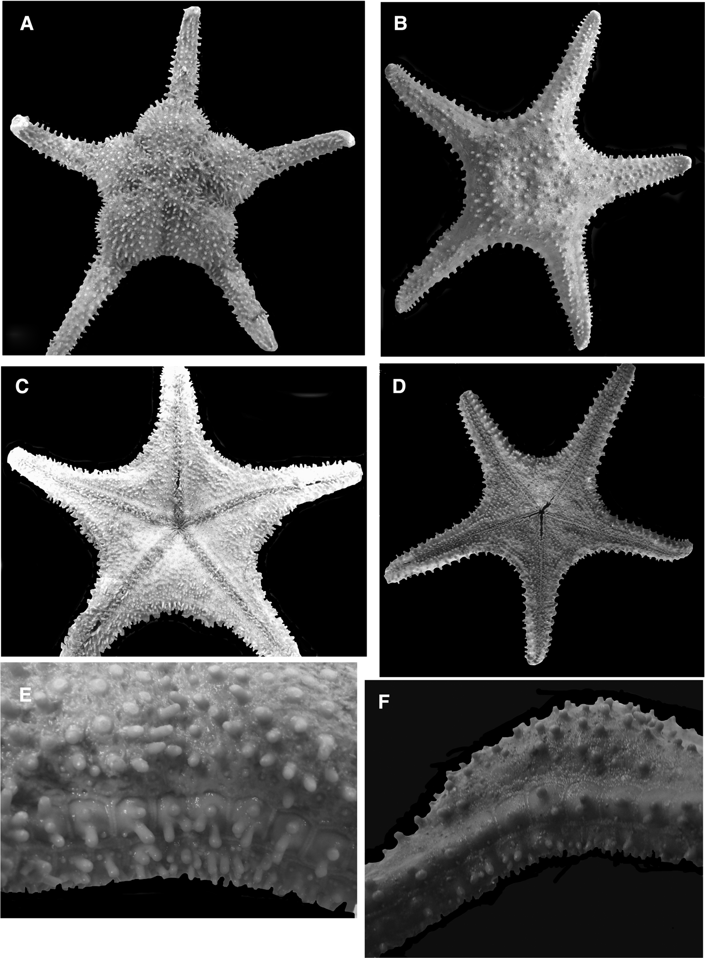
All new specimens examined displayed broad agreement with the description of Evoplosoma timorensis as outlined by Aziz & Jangoux (Reference Aziz and Jangoux1985), including the blunt, thick spines, covering the abactinal, marginal and actinal surface, pedicellariae morphology, surface texture and furrow spine number (Figure 2A–F). However, there was some additional variation between specimens. For example, the Solomon Island (MNHN IE-2007-141), the Wallis and Futuna specimen (MNHN-IE-2013-1756) and the Tahiti specimens showed larger numbers of furrow spines (7–10) versus the five to seven in the type description. For the Tahiti and Solomon Islands (Figure 2B, D, F), specimens also showed more widely dispersed and overall fewer spines across the body surface than the type R = 9.0 cm. Actinal spines were also smaller and less conspicuous.
The Wallis and Futuna Islands specimen (Figure 2A, C, E) had a highly distended disk and was the largest specimen of this species known so far with R = 10.6 cm.
In situ observations by the Okeanos Explorer's 2010 INDEX-SATAL expedition to Indonesia reveal this species to be yellowish-orange.
Evoplosoma timorensis variation. Wallis and Futuna Islands Specimen: (A, C, E). Solomons specimen: (B, D, F).
OCCURRENCE
Solomon Islands, Wallis and Futuna Islands, Tahiti/Tuamoto, Papua New Guinea, East Timor Region and Celebes Sea, Indonesia (see coordinates below for range extension), water depth 795–1279 m.
MATERIAL EXAMINED
MNHN IE-2007-141, Solomon Islands; coordinates: 8°24′S 159°22′E; water depth: 1057 m; coll. by Bouchet, Waren and Samedi; IRD on N/O Alis. SALOMON 2. 24 October 2004 (one wet specimen R = 6.9 cm, r = 2.6 cm). MNHN IE-2007-31-89, Vitiaz Strait, Papua New Guinea: coordinates: 05°59′S 147°39′E; water depth: 860–880 m; coll. by BIOPAPUA CP 3724, 10 July 2010 (one wet specimen R = 4.0 cm, r = 1.3 cm). MNHN-IE-2013-1756, Wallis and Futuna Islands; coordinates: 12°13.8′S 177°28.0′W; water depth: 795–820 m; coll. by MUSORSTOM 7, CC 554, 18 May 1992 (one wet specimen R = 10.6 cm, r = 2.6 cm). MNHN-IE-2013-4016, Tuamoto to southern Tahiti TARASOC; coordinates: 16°40′S 151°25′W; water depth: 650–800 m; 16 October 2009. DW 3440, N/O Alis. (one wet specimen R = 9.3 cm, r = 1.9 cm).
VIDEO OBSERVATIONS
Image expl 5497; 20100701_0528mm41s10_ROVHD_STAR; coordinates: 4.67218 N 125.25679 E: water depth 873.0 m.
Evoplosoma virgo Downey, Reference Downey1982
Downey, Reference Downey1982, p. 772; Clark, Reference Clark1993, p. 253; Mah et al., Reference Mah, Nizinski and Lundsten2010, p. 282.
COMMENTS
This species is known only from the holotype and was illustrated by Mah et al. (Reference Mah, Nizinski and Lundsten2010).
OCCURRENCE
Gulf of Mexico; water depth: 2056 m.
MATERIAL EXAMINED
Holotype: USNM E24285, Gulf of Mexico, North Atlantic Ocean; coordinates: 26°08′N 92°43′W; water depth: 2056 m (1124 fms); coll. by W. Pequegnat, 30 July 1971 (one dry specimen R = 11.5 cm, r = 3.3 cm).
Evoplosoma voratus Mah et al., Reference Mah, Nizinski and Lundsten2010
Mah et al., Reference Mah, Nizinski and Lundsten2010, p. 279.
OCCURRENCE
Davidson Seamount; water depth: 2669.9 m.
MATERIAL EXAMINED
Holotype: USNM 1124506, Davidson Seamount; coordinates: 35°37′N 122°49′W; water depth: 2669.9 m; coll. by J. Barry, L. Lundsten, Sta. T-947-A10, 2 February 2006 (one wet specimen R = 8.4, r = 2.7).
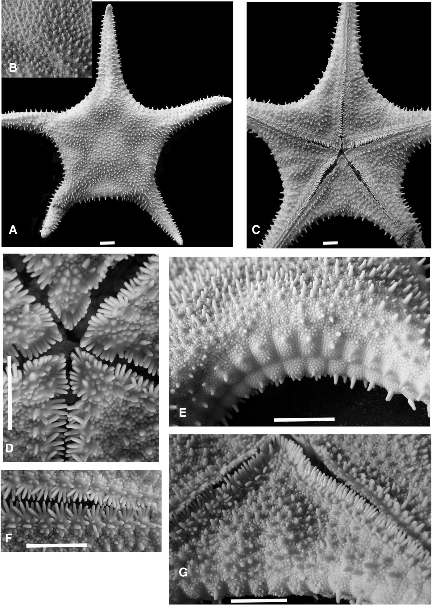
Evoplosoma watlingi n. sp. (Figure 3A–G)
ZOOBANK REGISTRATION
http://zoobank.org:pub:C991E1AF-81CE-4A07-A676-7597F463F3A2
ETYMOLOGY
This species is named in honour of its collector, Dr Les Watling, of the University of Maine and University of Hawaii.
COMMENTS
Evoplosoma watlingi n. sp. shares multiple characters with Evoplosoma scorpio Downey, Reference Downey1981 from the Gulf of Mexico, which supports the close relationship between the two. Most evident is the dense, crowded granular cover on the abactinal and marginal surfaces and to a lesser extent the actinal surface. This crowded granular covering is absent from other Evoplosoma species. Other shared characters with Evoplosoma scorpio include the presence of felipedal pedicellariae, the similar morphology and number of furrow spines (five to six), adambulacral spine/pedicellariae/granule arrangement and the overlapping numbers of furrow spines.
Evoplosoma watlingi is distinguished from Evoplosoma scorpio by the distribution of spines on the abactinal, marginal and actinal surfaces, and the differing morphology of the individual granules, which are round and smooth on Evoplosoma watlingi but spinelet-like with jagged tips on Evoplosoma scorpio. Abactinal spines in Evoplosoma watlingi are homogeneous in size and shape and occur across the complete central surface. Abactinal spines in Evoplosoma scorpio are present primarily on proximal and radial regions with spines absent or weakly present interradially. Furrow spine tips on Evoplosoma scorpio are sharply quadrate in cross-section with pointed edges as opposed to those in Evoplosoma watlingi, which are smooth and blunt. The arms in Evoplosoma scorpio are also more distinct and more tapering with an R/r = 3.33 versus an R/r = 2.8 in Evoplosoma watlingi.
Although the species is only known from its description, Evoplosoma augusti, from the Indian Ocean, displays characters that also suggest some affinity, specifically that the surface is covered by rounded, granules or tubercles. However, larger primary spines found in Evoplosoma watlingi are apparently absent in Evoplosoma virgo.
OCCURRENCE
Bear Seamount (North Atlantic); water depth: 1564 m.
DESCRIPTION
Body strongly stellate, interradial arcs rounded. Arms triangular with armtips tapering (Figure 3A, C). Disk weakly distended.
Abactinal surface covered by coarse hemispherical granules and large conical pointed spines and a distinct skin-like covering overlying abactinal plates and granules (Figure 3A, B). Abactinal plates round to irregularly round to oval in outline, weakly convex in profile. Granules, 8–20 (most have 12–18) crowded, covering each abactinal plate. Granules coarse, about 1–1.5 mm in diameter, surfaces smooth and bare, round to irregularly polygonal in outline. Granules dense and obscuring boundaries between abactinal plates and completely covering disk, arms, marginal, actinal series. Large (2–3 mm in length), conical spines present on most plates, widely distributed over abactinal surface (Figure 3B, E). Spine covering least abundant interradially closest to superomarginal contact. Madreporite triangular, shield-shaped, flanked by approximately five to six abactinal plates. Pedicellariae not observed on the abactinal surface.
Marginal plates 48 per interradius (counted from armtip to armtip), quadrate in outline, elongate with flared abactinal side, narrowing on actinal side. Superomarginal plates corresponding exactly to inferomarginal plates interradially becoming slightly offset distally near armtip. Marginal plates completely covered by coarse granules identical to those on abactinal surface and continuous with granules on abactinal surface (Figure 3E). Contact between abactinal and superomarginal plates obscured by granular covering. A large (2–3 mm), pointed, conical spine present on each superomarginal, inferomarginal plate, sometimes with smaller accessory plate. Marginal spines each in continuous series forming conspicuous peripheral fringe (Figure 3A, C, E). Central inferomarginal plates with five to 12 larger pointed spine-like granules consistent in shape with granular cover on marginal plates. Terminal plates large, triangular in shape. Felipedal pedicellariae each with five to six interclasping teeth present on interradial inferomarginal plates, one per plate, approximately one to three per interradius.
Actinal surface similar to other surfaces, covered by coarse granules and covered with large, conical spines identical to those on abactinal and marginal surfaces (Figure 3G). Actinal plates composed of approximately six to seven series with best defined proximally becoming smaller and more irregular distally but as opposed to other surfaces, individual plates are discrete. Actinal plates with two to ten round to irregularly shaped granules covering each surface. Several granules with tip spine-like to low and rounded. Majority of plates on actinal surface with large, conical spines (2–3 mm in length), widely spaced. Pedicellariae, felipedal, eight to ten in number with interclasping teeth on valves of variable sizes. Four to six pedicellariae wider (1–2.0 mm) and larger (1–2.0 mm) on proximal plates with smaller, more narrow, smaller pedicellariae on more distal plates (0.5 mm), adjacent to spines on some individual plates.
Furrow spines, five to six (mostly six) (Figure 3D, F). Spines quadrate to rounded in cross-section. Each furrow spine array splayed over one another. Subambulacrals two, short (similar to granules on actinal surface) at proximalmost, becoming much larger most distally. Large felipedal pedicellariae with 10–12 interlocking teeth adjacent to subambulacrals. Four to eight blunt granules, with variable sizes, on adambulacral plates facing actinal side. Adambulacral plates bare other than for granules, spines, and pedicellariae. Oral plates with five to six furrow spines projecting into oral region (Figure 3D). One enlarged oral spine projecting into mouth on each plate (so two per interradius). Furrow spines forming discrete boundary along adambulacral edge to end of tube foot groove. Furrow and oral spines quadrate in cross-section with blunt, rounded tips. Oral plate surfaces covered with angular granules/spines, 12–13 per plate, wide, each quadrate to triangular in cross-section.
Colour in life is bright, deep orange.
Evoplosoma watlingi n. sp. Holotype USNM 1208018. Scale Bar = 1.0 cm. (A) Abactinal surface. (B) Close up of abactinal spination. (C) Actinal surface. (D) Actinal surface-oral region showing mouth spination. (E) Marginal plate series. (F) Close up of furrow spines. (G) Actinal intermediate region showing spination, furrow spines, inferomarginals.
MATERIAL EXAMINED
USNM 1208018 holotype, Bear Seamount, North Atlantic; coordinates: 39°57.05N 67°24.78W; water depth: 1564 m. Perched on cf. Keratoisis, coll. by Les Watling, 11 May 2004 (one wet specimen R = 9.1, r = 3.2).
Hippasteria phrygiana (Parelius, Reference Parelius1768)
Full synonymy and description of this species is outlined by Mah et al., Reference Mah, Neill, Eleaume and Foltz2014, p. 441.
COMMENTS
This species has recently been subject to extensive phylogenetic review (Foltz et al., Reference Foltz, Fatland, Eléaume, Markello, Howell, Neil and Mah2013; Mah et al., Reference Mah, Neill, Eleaume and Foltz2014). Phylogeography of Hippasteria has shown that Hippasteria phrygiana is widely occurring in the Atlantic, Indian and Pacific Ocean basins. Regional descriptions of North Atlantic Hippasteria phrygiana are found in Clark & Downey (Reference Clark and Downey1992) as well as multiple accounts summarized by Mah et al. (Reference Mah, Neill, Eleaume and Foltz2014). Hippasteria phyrgiana is relatively well-described and so it is not included herein. Extensive accounts of its variation across its distribution are present in Mah et al. (Reference Mah, Neill, Eleaume and Foltz2014).
Figure 6A was compared with Hippasteria phrygiana, USNM E15785, a specimen with R = 2.5 cm. The asteroid pictured was comparable to the specimen in terms of overall body shape (i.e. R/r), abactinal plate shape and presence of spines on marginals and abactinals. However, colour and spine shape (more blunt and coarse in the specimen, finer and more pointed in the figured individual) can vary and so positive identification remains unconfirmed. Further possible identifications include a small sized Evoplosoma or possibly even an undescribed goniasterid species. Asteroids in Figure 6B–D are positively identified as Hippasteria phrygiana and based on comparisons with specimens and descriptions from Clark & Downey (Reference Clark and Downey1992).
VIDEO OBSERVATION
Northeast Atlantic, off the coast of Massachusetts; coordinates: 40.30338°N -67.67661°W; water depth: 1212.4 m; EX1304L2_IMG_20130813T143541Z_ROVHD_ASR_COR. Maine Observations, image 1369, dive 9 (one individual, abactinal surface facing left), Schoodic Ridges S1A; coordinates: 44°5.55 N 67°39.22 W; water depth: ~212 m. Image 2722 (three individuals), dive 15, Schoodic Ridges S1; coordiantes: 44°05.52 N 67°39.28 W; water depth: 200 m. Image 2703, dive 15 (one individual, surface facing forward), Schoodic Ridges S1; coordinates: 44°05.52 N 67°39.28 W; water depth: 205 m.
Sthenaster emmae Mah et al., Reference Mah, Nizinski and Lundsten2010
Mah et al., Reference Mah, Nizinski and Lundsten2010, p. 274.
OCCURRENCE
Known only from Savannah Banks and off the coast of Jacksonville, FL. 252–501 m.
TAXONOMIC SUMMARY
Note, the description herein is derived from that of Mah et al., Reference Mah, Nizinski and Lundsten2010.
Arms five, triangular in outline. Disk large, broad, swollen-especially in wet specimen. Body stout, thick.
Abactinal plates round to polygonal to oblong in outline with numerous interspersed secondary plates embedded in a thick, pulpy tissue forming heavily thickened abactinal body wall. Plates with high-aspect; mound-like in shape, forming deep but open channels between plates. Abactinal plates are covered with two to ten blunt to conical spine-like granules, usually forming a periphery around plate edge with only one or two granules/spines centrally located. Some plates bear enlarged conical, spine-like granules, surrounded by smaller blunt spinelets. Large abactinal clam-shell like pedicellariae present each with 9–12 interlocking teeth per valve. Pedicellariae located centrally on plate surface varying in size from 1/3 to 3/4 of plate diameter, flanked by spine-like granules. Pedicellariae occurring unevenly over abactinal surface, becoming densely concentrated in some areas but absent from others. Madreporite sunken, bordered by seven to eight abactinal plates. Papulae, small, four to six, interspersed between plates.
Marginal plates wide (W > L) 42–45 per interradius (from armtip to armtip), which become smaller and with more equivocal dimensions distally adjacent to terminal. Large spines absent from marginal plate series. Fasciolar channels present between marginal plates, relatively deep. Superomarginals facing laterally, correspond 1:1 to inferomarginals along most of series with one to two plates irregularly offset, possibly due to sublethal predation. Superomarginal plate surface convex, bare except for 20–35 widely spaced, sharp, conical spinelets, which are most densely concentrated ventrally on plate at contact with inferomarginal plate. One to two relatively small clam-shell like pedicellariae with interlocking teeth present on nearly every marginal plate but regularly present, adjacent to inferomarginal contact, irregularly present closer to contact with abactinal surface. Periphery of each plate with 15–40 small evenly spaced conical spinelets, which occur more densely at contact with inferomarginals. Inferomarginal plates with ventral facing and more densely covered by 10–70 irregularly sized conical to blunt spinelets. Higher number of larger, conical, more pointed spinelets present closer to superomarginal contact. Higher numbers of spinelets present interradially decreasing distally corresponding with smaller inferomarginal plate size. Pedicellariae, one to four, typically two, identical to the type on superomarginals present on inferomarginals facing ventrally. Terminal plate round, bulbous.
Actinal intermediate plates, similar in size, shape to abactinal plates with well-developed fasciolar channels running between plates. Actinal plate series adjacent to adambulacral plate series with large pedicellariae nearly equal to size of plate on which it sits, teeth poorly developed to absent relative to those on abactinal plates. Pedicellariae on actinal series adjacent to adambulacral plates occur with less frequency distally along the arm with some smaller actinal plates adjacent to armtip with pedicellariae absent, bearing only 3–6 spinelets. Actinal pedicellariae occur most heavily adjacent to mouth becoming less common to absent adjacent to inferomarginal plate contact. Pedicellariae on each actinal plate surrounded by 20–35 sharp spinelets, some round, some triagonal to polygonal in cross-section. Actinal plates adjacent to inferomarginal plate series covered with 4–30 sharp, irregularly sized, conical granular spines. Some with polygonal to triangular in cross-section. Oral cavity sunken.
Adambulacral furrow spines blunt tipped, three to four per plate, triangular to oblong ovate in cross-section in weakly convex series. Distinct grooves between adambulacral plates. Central spines longest and thickest with spines shortest on ends. Subambulacral spines three in two series. Subambulacral spine series adjacent to furrow spines somewhat shorter than furrow spines but comparable in thickness, round to oval in cross-section. Subambulacral spine series farthest away from furrow spines shortest, with central spines only slightly less thick than furrow spines with smallest spines located on ends of second subambulacral series. Distinct fasciolar channel separates adambulacral plates from actinal intermediate plates.
Oral plates with four to six furrow spines. Oral plate surface with three to four spines per plate (six to eight total on paired oral plates) with one to three spines projecting into oral opening. Spines triangular to flattened triangular in cross-section, most are comparable in length to furrow spines but sometimes with one or two that are similar to granular spines.
Colour in life is orange.
RESULTS AND DISCUSSION
The feeding events presented below were observed from a video recorded by ROVs deployed by the R/V Okeanos Explorer . With the exception of Evoplosoma watlingi n. sp., no specimens were collected for any of the video observations recorded herein. However, video observations in all instances where feeding is listed, show either the direct or indirect evidence of predation – these are discussed below. Aside from Circeaster, Evoplosoma and Hippasteria, the hippasterine Sthenaster emmae Mah et al., Reference Mah, Nizinski and Lundsten2010 is also a likely corallivore. As reported by Mah et al. (Reference Mah, Nizinski and Lundsten2010), gut contents of this species contained spicules of the co-occurring gorgonian Eunicella modesta indicating likely predation.
Circeaster americanus feeding observations
Images from Manning Seamount in the North Atlantic (1328–1333 m) taken in 2004 and 2005 show four individuals of Circeaster americanus (identified by arm and body shape) climbing over and feeding on a downed colony of the alcyonacean octocoral Paragorgia (observations herein based on unpublished data, Scott France, September 2014). The cause of the downed colony is unknown. The images taken in 2005 are shown in Figure 4A–C. The determination of feeding behaviour is based on the bare patches of tissue on the Paragorgia colony and from the hunched postures of the asteroids on the branches of the colony. The initial observation of this feeding event was made in 2004 (Figure 4A) and was revisited 15 months later in 2005 (Figure 4B, C). The subsequent visit showed only three individuals of Circeaster americanus but others could have been under the colony and out of sight of the cameras. The 2005 visit also shows a smaller fourth unidentified star, possibly a poraniid but definite identification was not possible, which was observed near the edge of the colony.
It is argued that the three individuals observed on the colony from the 2005 observation are the same as those from the 2004 observation. Overall resemblance and size and number of individuals is consistent providing a basis for this conclusion. Also, based on prior collections and in situ observations of anticipated species occurrence, it does not appear that this species is very abundant.
The 2005 observation shows larger barren patches present on the Paragorgia colony than in the first 2004 image (Figure 4B). Based on the scale taken by the lasers (=10.0 cm), a bare patch of tissue measuring approximately 30.76 cm long observed on a Paragorgia branch (Figure 4B) was devoured within a 15 month period. This suggests a conservative feeding rate of about 2 cm of tissue per month. The cause of the decay observed on the distal and peripheral branches of the Paragorgia colony was more unclear. It was uncertain if these were caused by predation by the asteroids, by decay resulting from injury of the downed coral or perhaps some other predator. It is significant to note the 15 month feeding period, which implies a relatively slow feeding rate. This assumes constant feeding over the time period, which is likely not the case. Nonetheless, this provides an estimate of the duration required by a deep-sea corallivore to feed on prey.
This provides a stark contrast to feeding rates of shallow-water taxa. Birkeland & Lucas (Reference Birkeland and Lucas1990) summarized feeding rates of the tropical reef corallivore Acanthaster planci and calculated feeding rates of approximately 0.35–0.57 m2/month/animal. Under circumstances where feeding rates were significantly higher, Birkeland & Lucas (Reference Fujikura, Okutani and Maruyama1990) also mentioned that the role of water temperature might be a possible factor in the determination of feeding rates. If that is the case, and the converse were true, then slower feeding rates would be consistent with cold-water/deep-sea habitat temperatures.
The observed feeding behaviour of Circeaster americanus is consistent with that of shallow-water species, which display non-directional movement once an abundant food source is encountered. Individuals then exploit this resource and remain in areas of high prey density (McClintock & Lawrence, Reference McClintock and Lawrence1985). However, it is unclear as to what individuals of Circeaster americanus would do following depletion of the food source. Conceivably, they could possess a ‘home’ range as some shallow-water asteroids, such as Oreaster reticulatus, are known to do (Scheibling, Reference Scheibling1980) or possibly they practice a more widespread foraging behaviour. This latter behaviour may account for the disappearance of the fourth individual from the 2005 observation, which was originally observed from the 2004 image assuming it was not out of sight.
The Okeanos Explorer images taken in the Gulf of Mexico show Circeaster americanus climbing up isidid octocorals (also known as ‘bamboo corals’) well off the bottom, clearing off polyps and leaving behind the bare skeleton (Figure 4C). Close-up images of these specimens permitted the comparison of abactinal and marginal plate morphologies, which allowed relatively certain identifications. The goniasterid Circeaster pullus from the Hawaiian Islands was also documented by Mah (Reference Mah2006) as feeding on octocorals.
Circeaster americanus in situ feeding observations. (A) Three individuals on Paragorgia, observed on 14 May 2004. (B) Subsequent observation 15 months later in 2005. White arrow points to bare surface in 2005 and that same region in 2004. Scale bar = 10.0 cm. (C) Close up on individuals from 2005 feeding observations. Scale bar = 10.0 cm. (D) Circeaster americanus individual feeding on an isidid octocoral.
Evoplosoma feeding observations
Evoplosoma scorpio feeding behaviour was observed on several occasions from Gulf of Mexico and North Atlantic localities by the R/V Okeanos Explorer video images. One individual (Figure 5A, B) was observed feeding on an unidentified isidid octocoral. The feeding behaviour of all Evoplosoma individuals was identical in that they were all observed extending their stomachs out and feeding as they climb along the stalk.
Evoplosoma watlingi was collected from Bear Seamount and was observed feeding on an isidid octocoral, provisionally identified as Keratoisis sp. (Figure 5C, D). Sclerites were collected from the mouth opening and found sequestered within the mouth cavity, where they were presumably awaiting digestion but could not be identified beyond family.
Evoplosoma in situ feeding observations. (A, B) Atlantic Evoplosoma, likely Evoplosoma scorpio feeding on isidid octocoral. (C, D) Evoplosoma watlingi n. sp. Holotype feeding on isidid octocoral. R of specimen = 9.1 cm.
Hippasteria feeding observations
The individual shown in Figure 6A has its body crouched over what appears to be Anthomastus or a similar appearing alcyonacean octocoral. The asteroid's posture suggests that its stomach is being extended over the octocoral's fleshy body. If the identification is correct, this represents the first instance of Atlantic Hippasteria phrygiana feeding on an alyconacean (cf. Anthomastus).
Further observations of H. phrygiana from the continental shelf (200–212 m) off the coast of Maine (Figure 6B–D) show it feeding on the gorgonian Primnoa resedaeformis. Gale et al. (Reference Gale, Hamel and Mercier2013) recorded H. phrygiana in the bathyal zone (304–992 m) of Newfoundland and Labrador, eastern Canada, preying on multiple cnidarian species, including sea pens, scleractinian coral, isidid octocorals (‘bamboo corals’) and sclerites in its gut representing multiple octocoral species. Gale et al. (Reference Gale, Hamel and Mercier2013) also showed the presence of sediment and amphipods in the gut, which suggests that H. phrygiana may also derive some food from infaunal sources in a minority of instances.
A revision of the genus Hippasteria, especially H. phrygiana, was recently presented by Mah et al. (Reference Mah, Neill, Eleaume and Foltz2014). An analysis of H. phrygiana from localities in the Pacific, Indian and Atlantic Oceans has showed that what were once perceived to be multiple species, are in fact one species (Foltz et al., Reference Foltz, Fatland, Eléaume, Markello, Howell, Neil and Mah2013). H. phrygiana in different ocean regions appears to be consistently focused on different types of cnidarians, including sea anemones, gorgonians and pennatulaceans (sea pens) (see Mah et al., Reference Mah, Nizinski and Lundsten2010, Reference Mah, Neill, Eleaume and Foltz2014; Foltz et al., Reference Foltz, Fatland, Eléaume, Markello, Howell, Neil and Mah2013). This suggests some ecological plasticity with regards to predator–prey item relationships, contrary to the more specific predator–prey relationship suggested by Birkeland (Reference Birkeland1974).
Hippasteria phrygiana in situ feeding observations. (A) Hippasteria cf. phrygiana feeding on alcyonacean (cf. Anthomastus). (B) Three individuals of H. phrygiana feeding on Primnoa resedaeformis. (C, D) Individual H. phrygiana feeding on P. resedaeformis.
Feeding posture and functional morphology
The feeding behaviour in all of the observed species showed that the stalk of the octocoral encloses along the tube foot groove as the stomach engulfed the polyps (e.g. Figure 5A, B). This posture has been documented in the Pacific on Evoplosoma (Fujikura et al., Reference Fujikura, Okutani and Maruyama2008; Mah et al., Reference Mah, Nizinski and Lundsten2010).
This implies a functional relevance for structures such as the furrow and subambulacral spines and the pedicellariae. Museum specimens of the observed species all display well-developed furrow spines. This includes either relatively elongated (e.g. Circeaster, Evoplosoma) or very thickened (e.g. Hippasteria) furrow and/or subambulacral spines. Pedicellariae are often oriented around the ambulacral groove, either on the adambulacral plates or adjacent to the adambulacral series. While their function is unclear, the location of these poorly understood structures could also be relevant to feeding behaviour.
CONCLUSION
In addition to a key and overview of Evoplosoma, several taxonomic changes, range extensions and behavioural observations were presented.
A new species of Evoplosoma, Evoplosoma watlingi n. sp., from Bear Seamount in the North Atlantic is presented.
Hippasteria tasmanica is transferred to Evoplosoma, establishing Evoplosoma in the New Zealand region.
Range extensions for Circeaster americanus and Evoplosoma scorpio in the North Atlantic and the Gulf of Mexico and Evoplosoma timorensis in the central Pacific are presented.
In situ feeding behaviour is presented for Circeaster americanus, Evoplosoma scorpio, Evoplosoma watlingi n. sp., Evoplosoma sp. and Hippasteria phrygiana.
The observations of Circeaster americanus provide the first example of the time duration needed for a deep-sea asteroid corallivore to feed on its prey. Four individuals participated in predation on a colony of Paragorgia, which was observed 15 months later with three individuals.
The feeding posture of octocorals presents functional morphological interpretations for furrow spines and adambulacral accessories, including pedicellariae.
ACKNOWLEDGEMENTS
I am grateful to Les Watling (University of Hawaii/University of Maine) for collecting the holotype and making it available for this study. Marc Eleaume (MNHN) and Kate Neil (NIWA) also provided additional loans of material. My thanks to Stephen Cairns (NMNH) and Les Watling (University of Hawaii) who identified the deep-sea corals observed in the figures. Thanks to Martha Nizinski (National Marine Fisheries Service) and Dave Packer (Gulf of Maine Deep Coral Science Team 2014/NURTEC-UConn/NOAA Fisheries/University of Maine) for providing the images of Hippasteria phyrigana feeding. Further thanks to Peter Auster and Rhian Waller for their involvement as part of the Gulf of Maine Deep Coral Science Team 2014. Special thanks to Scott France (University of Louisiana, Lafayette) for allowing use of his Manning Seamount observations. Special thanks to Andrea Quattrini (Temple University) for assistance with obtaining locality data for the video observations. Susan Gottfried and her staff at OE Data Management, NOAA National Coastal Data Development Centre are thanked for their assistance with image data. I gratefully acknowledge the efforts of the science team and crew of the R/V Okeanos Explorer. Three anonymous reviewers provided useful and constructive comments for the manuscript.