


Introduction
Parasitic infections pose significant threats to avian health and conservation efforts, affecting both free-ranging and captive populations. Cracid birds – including guans, chachalacas, and curassows – are among the most vulnerable taxa in the Neotropical region. They face increasing pressures from habitat loss, hunting, and naturally low reproductive rates (Brooks Reference Brooks2006; Pinilla-Buitrago et al. Reference Pinilla-Buitrago, Martínez-Morales, González-García, Enríquez, Rangel-Salazar, Guichard-Romero, Navarro-Sigüenza, Monterrubio-Rico and Escalona-Segura2014). Despite their large size and conspicuous vocalisations, which render them readily detectable in forested and adjacent human-modified landscapes, critical gaps remain in our understanding of their ecology and disease dynamics (Brooks Reference Brooks2006).
Cyclocoelidae Stossich, 1902 is a large family of digenean trematodes in the superfamily Cyclocoeloidea, comprising 6 subfamilies and nearly 128 species. The subfamily Cyclocoelinae includes 25 recognised species. The taxonomy of Cyclocoelidae has historically been unstable due to the variation in several diagnostic traits and the limited availability of molecular data. A major comparative morphological and molecular revision (Sitko et al. Reference Sitko, Bizos and Heneberg2017) helped clarify generic boundaries within the family and refined the diagnostic features of Cyclocoelinae, providing a modern framework to distinguish among those genera. The genus Circumvitellatrema (Dronen et al. Reference Dronen, Greiner, Ialeggio and Nolan2009) contains a single species, Circumvitellatrema momota, which was described from the air sacs and lungs of a captive-hatched blue-crowned motmot (Momotus momota) in Pennsylvania, USA, in 2009 (Dronen et al. Reference Dronen, Greiner, Ialeggio and Nolan2009; Dronen and Blend Reference Dronen and Blend2015). Records of C. momota are scarce. In 2012, the parasite was reported again in another blue-crowned motmot in the Montpellier Zoo in France (Libert et al. Reference Libert, Jouet, Ferté, Lemberger and Keck2012). Other reports include the United States and the Brazilian Amazon (Greiner et al. Reference Greiner, Zimmerman, Delaski, Dronen, Hammond, Stacy, Grillo, MacLean, Maurer, Okimoto, Douglass, Yu and Reavill2023). Recently, the parasite was found in a free-ranging blue-crowned motmot that was found dead in Costa Rica, which represents the first report of the species in our region and in a free-ranging bird (Chaves-Hernández et al. Reference Chaves-Hernández, Jiménez-Rocha, Quesada-Vasquez, Lizano-Zumbado, Martínez and Jiménez-Soto2025).
To our knowledge, this study reports the first documented occurrence of C. momota in cracid birds and the first record of any helminth parasite infecting Crax rubra and Penelope purpurascens (both cracid birds) in Costa Rica. In addition to expanding the known host range of this rare trematode, our findings provide a detailed pathological characterisation of C. momota infection, and we present a phylogenetic analysis to place this parasite within the broader evolutionary framework of the subfamily Cyclocoelinae. This study underscores the importance of integrating parasitology into avian conservation strategies, particularly for threatened species inhabiting rapidly changing ecosystems.
Materials and methods
An adult female great curassow (C. rubra), free-roaming within the facilities of Rescate Wildlife Rescue Centre (RWRC) in Alajuela Province, Costa Rica, was admitted as an emergency case presenting with shock and dyspnoea. As signs worsened despite treatment, the bird was euthanised and subjected to necropsy at the Pathology Laboratory at Universidad Nacional de Costa Rica, Heredia. The bird weighed 3.2 kg and measured 85.0 cm in total length. Twenty-two trematodes were extracted from the air sacs and the coelomic cavity. Specimens were collected and preserved in 70% ethanol for further processing at the Helminthology Unit, Parasitology Department, Universidad de Costa Rica, San Pedro, San José. The organs were fixed with 10% formalin and processed for standard haematoxylin and eosin staining.
One month later, an adult female crested guan P. purpurascens, also from RWRC, presented with clinical signs consistent with dyspnoea and shock. The bird was also euthanised and underwent necropsy at the same pathology facility. Trematodes were recovered from the air sacs and coelomic cavities: 18 adults and 6 juveniles were extracted and sent to the helminthology laboratory for further processing.
Specimen preparation and mounting
Four specimens from C. rubra and three from P. purpurascens were washed in phosphate-buffered saline and then relaxed in warm 70% ethanol at 29°C. The trematodes were then dehydrated through a graded ethanol series (70%, 80%, 90%, and 95%) for 20 minutes in each solution. After dehydration, the specimens were cleared in lactophenol for 2 hours, then stained with hydrochloric acid–carmine for 30 minutes. Subsequently, a 1% acetic acid solution was used to eliminate excess carmine. The stained specimens were mounted in Canada balsam on glass slides for microscopy. Six voucher specimens were deposited as reference slides in the National Helminth Collection of Costa Rica (CHCR) (three from C. rubra and three from P. purpurascens) under the catalogue numbers: CHCR-214-1, CHCR-214-2, CHCR-214-3, CHCR-214-4, CHCR-214-5, and CHCR-214-6.
Molecular analysis
DNA was extracted from four trematodes (two from each host species) using the Qiagen DNeasy Blood & Tissue Kit. Tissue samples were lysed and homogenised in a buffer containing proteinase K to digest proteins and release DNA. Contaminants such as protein residues and RNA were removed through a series of washes and centrifugation, following the manufacturer’s protocol. Purified DNA was eluted in a low-salt buffer, and its concentration and purity were assessed using spectrophotometry.
Polymerase chain reaction (PCR) was performed using a master mix containing Green Taq DNA polymerase. Primers targeting the ribosomal DNA internal transcribed spacer (ITS-2) gene of trematodes were used: A28 (5’-TTGATTGGTGGTTTTGGTAA-3’) and 3S (5’-TACAGTAAAGTGTCTTTGTTAA-3’) (Prasad et al. Reference Prasad, Goswami, Tandon and Chatterjee2011). DNA from Fasciola hepatica was used as a positive control and nuclease-free water as a negative (no-template) control. Amplicons were Sanger-sequenced bidirectionally using the BigDye Terminator protocol (Applied Biosystems) at Macrogen.
The obtained sequences were visually inspected and aligned with published ITS-2 sequences from the family Cyclocoelidae in MEGA7 (Kumar et al. Reference Kumar, Stecher and Tamura2016). The best-fit nucleotide substitution model was determined to be Kimura 2+G and a Bayesian phylogenetic tree was inferred using the BEAST package (Bouckaert et al. Reference Bouckaert, Vaughan, Barrido-Sottani, Duchêne, Fourment, Heled, Jones, Kühnert, De Maio, Matschiner, Mendes, Müller, Ogilvie, du Plessis, Popinga, Rambaut, Rasmussen, Siveroni, Suchard, Wu, Xie, Zhang, Stadler and Drummond2019). Sequences were uploaded in BEAUTi and 108 Markov Chain Monte Carlo (MCMC) chains with a sampling of every 103 and 10% burn-in were selected. Convergence of chains was checked in Tracer v1.7.2 with values larger than 300 for each effective sample size. Finally, a consensus tree was generated in TreeAnnotator v2.7.6 and depicted in FigTree v1.4.4.
Results
Specimens were recovered from the air sacs of two captive birds: P. purpurascens (18 adults and 6 juveniles) and C. rubra (22 adult parasites), both in the family Cracidae. This constitutes the first report of C. momota parasitising a member of the Cracidae family, extending the family host range for this parasite (Table 1).
Geographical distribution of all published reports and hosts of Circumvitellatrema momota

Pathology
Pathological examination of C. rubra revealed severe and extensive pulmonary involvement. Grossly, the lungs showed diffuse consolidation and marked pleural irregularity, affecting an estimated 70–80% of the pulmonary parenchyma. Lungs showed pneumonia with heterophilic infiltrate, multiple diffuse haemorrhages, severe vascular congestion, and mucoid exudate. Adult trematodes were embedded on the coelomic side of the lung, while eggs were within the parenchyma. The pleural surfaces were irregular and nodular, with multiple granulomatous lesions (Figure 1 A–C). Elongate parasites were present in the pleural tissue and were invading the lung parenchyma. The mesothelium exhibited petechial haemorrhages, particularly over the liver and air sacs. Extensive inflammation and fibrinous deposits were also present.
A General aspect of the affected lung of C. rubra. Adult parasites can be seen in the cranial portion of the lung with necrotic zones and profuse haemorrhage (blue arrowhead). B Adult parasites (see arrowheads) are observed in the coelomic portions of both lungs. C Detail of an adult into the lung’s coelom (red arrowhead). D Tissue from C. rubra’s air sacs prepared in regular haematoxylin and eosin stain showing a longitudinal section of an adult worm in situ (yellow arrowheads indicated). Tissue of the air sac (black arrowhead) shows abundant inflammatory infiltrates.

A similar pathological condition was observed in P. purpurascens, which exhibited extensive diffuse haemorrhages, inflammation, and granulomas involving the pleura, lungs, and liver with associated mesothelial changes. The histopathological examination of the air sacs and lungs revealed severe multifocal inflammation characterised by heterophilic infiltrates and interstitial thickening. Adjacent to the worm, areas of necrosis and loss of the pulmonary parenchymal architecture were observed (Figure 1D). Collectively, these findings indicate systemic pathological alterations caused by the parasites, leading to a fatal outcome.
Parasite morphology
Details on all measurements of each morphological character are available in the Supplementary Table S1. Of the trematodes examined, 49 were adults and 13 were juveniles. Specimens were dorsoventrally flattened, elongated, and tapered towards the anterior and posterior ends of the body. The mean length of adults was 9.2 ±1.2 mm (range = 6.4 to 11.2, n = 20), while the mean width was 3.1 ± 0.3 mm (range = 2.9–4.1, n = 20). Oral sucker was poorly developed (133.7 ± 8.4 μm wide, range = 112.0–143.2, n = 15), and an acetabulum was absent.
A short prepharynx was visible and measured 141.0 ± 2.2 μm (range = 137.2–144.5, n = 12), and the pharynx measured 366.6 ± 31.2 μm (range = 291.5–388.2, n = 15). The intestinal caeca were long, simple, tubular, and straight. Both caeca converged at nearly 1 mm anterior to the distal end of the body.
Testes were trapezoidal in shape. The lower testis was 839.8 ± 90.2 μm long (range 734.8–1044.0, n = 15) and 508.5 ± 57.0 μm (range = 417.1–610.6, n = 15) wide. It was located approximately 600 μm anterior to the distal end of the body. The upper testis was smaller, 669.0 ± 23.6 μm long (range = 632.0–706.0, n = 15) and 547.3 ± 52.1 μm wide (range = 417–597.4, n = 12) (fig. 2).
A Carmin–hydrochloric acid-stained specimen of C. momota found in C. rubra from Costa Rica. B Digitally hand-made descriptive drawing. Trapezoid-like testes and ovary form a triangle, each gonad separated one from the other by approximately 2 mm. Vitelline glands form a complete ellipse around the reproductive tract, confluent in both anterior and posterior portions of the body.
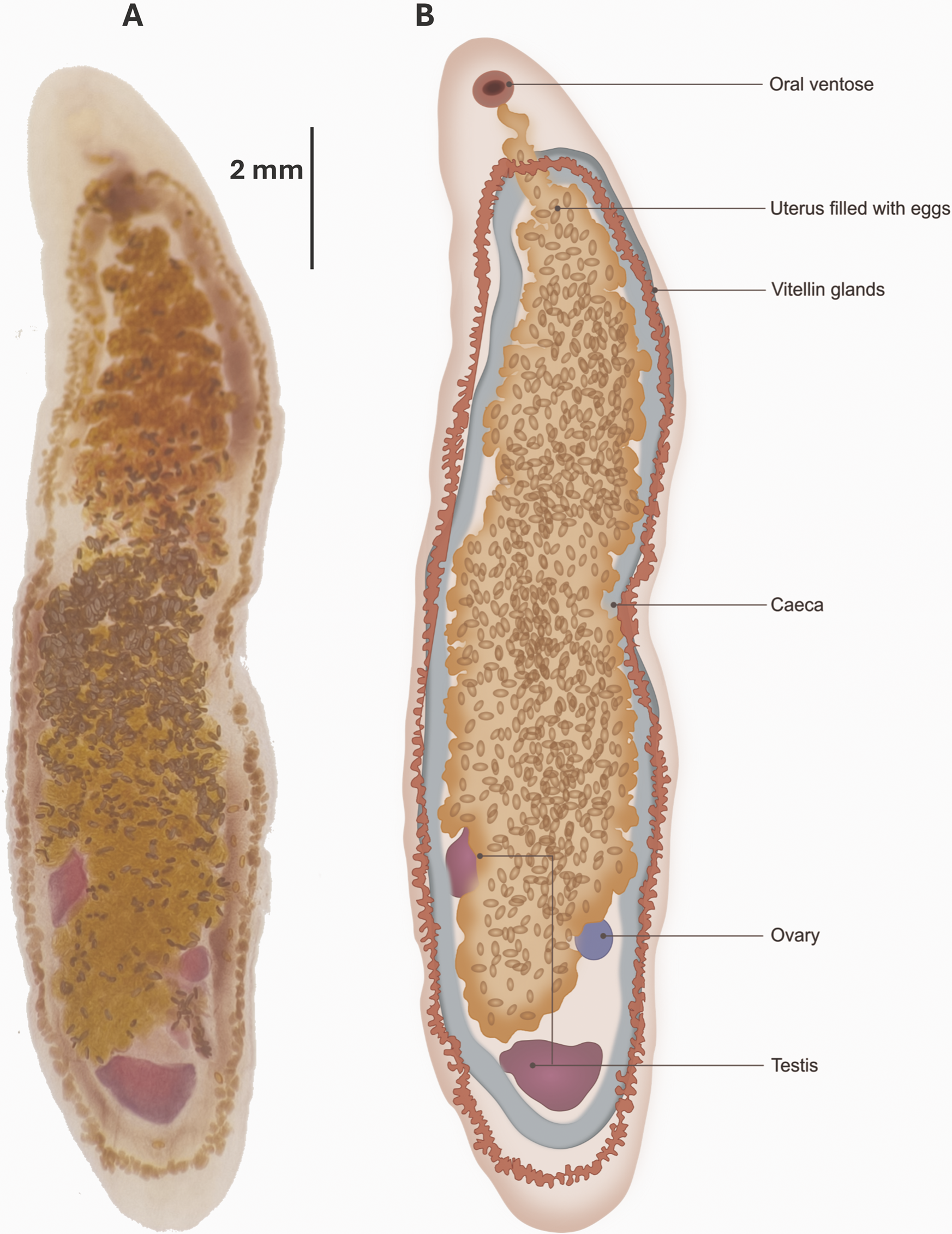
The ovary was nearly spherical and measured 571.3 ± 13.8 μm in length (range = 545.6–591.8, n = 12), and 400.2 ± 20.5 μm in width (range = 366.8–424.3, n = 12). The gonads formed a triangular arrangement, with the ovary in an intertesticular position. The gonads are separated from one another by nearly 2 mm. The genital pore was post-pharyngeal. Vitelline fields formed a complete ellipse, becoming confluent in both the anterior and the posterior portions of the body (Fig. 2).
The uterus was extremely large, occupying most of the body, located internally relative to the caeca and vitelline fields. The uterus does not extend beyond the post-testicular field (fig. 2). Eggs measured 138.1 ± 2.9 μm in length (range = 136.6–141.3, n = 20) by 68.4 ± 3.8 μm in width (range = 65.52–72.61, n = 20).
Molecular and phylogenetic analysis
DNA was extracted from trematodes recovered from C. rubra and yielded a 599 bp ITS-2 fragment, showing 100% identity with C. momota from a blue-crowned motmot kept in captivity in France (accession no. JQ886068) with a coverage of 94%. DNA from trematodes isolated from P. purpurascens amplified a 614 bp ITS-2 fragment identical to the same C. momota sequence, with 92% query coverage (JQ886068). The three ITS-2 sequences generated in this study were deposited in GenBank under accession numbers PQ753276 (from C. rubra), PQ753277, and PQ753278 (from P. purpurascens).
The Bayesian phylogenetic analysis recovered a strongly supported monophyletic clade comprising C. momota specimens from Costa Rica and France (posterior probability = 1.00). Cyclocoelum mutabile constituted a well-separated sister clade to C. momota, supported by a posterior probability of 1.00. This topology provides a clear genetic distinction between the genera Circumvitellatrema and Cyclocoelum, thereby corroborating the morphological identification of the Costa Rican isolates (Figure 3).
Bayesian phylogenetic tree based on ITS-2 sequences, showing the monophyly of major digenean families and the placement of Circumvitellatrema momota from Costa Rica and France within Cyclocoelidae. Posterior probabilities are indicated at nodes; clades corresponding to Echinostomatidae, Fasciolidae, Philophthalmidae, and Cyclocoelidae are highlighted.

Discussion
To the best of our knowledge, this is the first report of C. momota infecting a member of the Cracidae family, representing a significant host-range extension for this parasite. Most previous reports have originated from birds in rescue centres or captive wildlife facilities in Brazil, France, and the United States (Dronen et al. Reference Dronen, Greiner, Ialeggio and Nolan2009; Greiner et al. Reference Greiner, Zimmerman, Delaski, Dronen, Hammond, Stacy, Grillo, MacLean, Maurer, Okimoto, Douglass, Yu and Reavill2023; Libert et al. Reference Libert, Jouet, Ferté, Lemberger and Keck2012). Recently, C. momota was also reported in a free-ranging blue-crowned motmot (M. momota) found dead in Heredia, Costa Rica (Chaves-Hernández et al. Reference Chaves-Hernández, Jiménez-Rocha, Quesada-Vasquez, Lizano-Zumbado, Martínez and Jiménez-Soto2025), and in the grey-winged trumpeter (Psophia crepitans) in the Brazilian Amazon (Greiner et al. Reference Greiner, Zimmerman, Delaski, Dronen, Hammond, Stacy, Grillo, MacLean, Maurer, Okimoto, Douglass, Yu and Reavill2023). These findings suggest that C. momota may be a generalist parasite with a broader host range than previously recognised, particularly in captive or human-modified environments.
Knowledge of metazoan endoparasites in wild cracids is scarce. Most studies have focused on captive birds, primarily in Brazil (Cunha et al. Reference Cunha, Mendonça, Oliveira, Baratella-Evêncio, Oliveira-Filho, Simões, Simões and Evêncio-Neto2008; Marques et al. Reference Marques, Junior, de Assis, Fernandes, De Araújo, de Resende and da Silva-Martins2013). In Costa Rica, only the haemosporidian Hemoproteus sp. and Trypanosoma everetti have been previously reported in cracids, both from free-ranging C. rubra in the Área de Conservación Guanacaste (Valkiūnas et al. Reference Valkiūnas, Iezhova, Brooks, Hanelt, Brant, Sutherlin and Causey2004). Other parasites known to affect cracid birds include the stramenopile Blastocystis, and protozoans such as haemosporidians and unidentified intestinal coccidians. Helminths reported in cracids include Hymenolepis nana, Heterakis sp., Ascaridia sp., Capillaria sp., and Strongyloides sp. (Galindo and Sousa Reference Galindo and Sousa1966; Marques et al. Reference Marques, Junior, de Assis, Fernandes, De Araújo, de Resende and da Silva-Martins2013).
Morphological and molecular identification
The general shape and arrangement of testes and ovary in the trematodes recovered from both cracid hosts were typical of members in the subfamily Cyclocoelinae. These characters are consistent with the diagnostic boundaries of Cyclocoelinae revised in Sitko et al. (Reference Sitko, Bizos and Heneberg2017), who demonstrated that traits such as the configuration of the vitelline fields, the position of the genital pore, and the arrangement of the gonads are among the most reliable morphological markers for distinguishing genera within Cyclocoelidae (Sitko et al. Reference Sitko, Bizos and Heneberg2017). All key morphological characteristics – including the egg size, post-pharyngeal position of the gonopore, the extensive uterus occupying most of the body, and the vitelline fields confluent both anteriorly and posteriorly – matched the original description of C. momota by Dronen et al. (Reference Dronen, Greiner, Ialeggio and Nolan2009).
These features also enabled the exclusion of closely related species. For example, in Psophiatrema greineri, vitelline fields are not confluent anteriorly; in Cyclocoelum spp., the gonopore is prepharyngeal; and in Selfcoelum spp., posterior vitelline fields are not confluent (Dronen et al. Reference Dronen, Greiner, Ialeggio and Nolan2009; Dronen and Kinsella Reference Dronen and Kinsella2009; Greiner et al. Reference Greiner, Zimmerman, Delaski, Dronen, Hammond, Stacy, Grillo, MacLean, Maurer, Okimoto, Douglass, Yu and Reavill2023; Sitko et al. Reference Sitko, Bizos and Heneberg2017; Vainutis et al. Reference Vainutis, Voronova, Andreev and Shchelkanov2024). Morphological characters such as the relative position of the uterus, gonopore, testes, and ovary, as well as the distribution of vitelline fields, remain critical for species-level identification within the Cyclocoelidae. Such distinctions align with the findings of Sitko et al. (Reference Sitko, Bizos and Heneberg2017), as they reassessed the morphological limits of Cyclocoelidae genera using integrated molecular and morphometric criteria (Sitko et al. Reference Sitko, Bizos and Heneberg2017).
Molecular and phylogenetic analyses provided further support for the identity of the trematodes. The ITS-2 sequences obtained from Costa Rican C. momota were 100% identical to the sequence from a C. momota specimen from France (JQ886068), with 92–94% query coverage. A monophyletic clade comprising C. momota specimens from Costa Rica and France was recovered with high nodal support (posterior probability =1.00) and was clearly distinct from C. mutabile (Figure 3). This clustering aligns with our morphological identification and supports the presence of C. momota in cracid birds. It is important to note that our interpretation of these phylogenetic data is strictly limited to species-level identification within the family Cyclocoelidae, as our molecular analysis was restricted to the nuclear ITS-2 marker due to technical limitations. Although this marker provided strong support for the identification of C. momota when compared to available sequences, the absence of mitochondrial sequences (e.g., cox1) limits the depth of our phylogenetic inference. Future studies incorporating mitochondrial markers are warranted to fully elucidate the evolutionary relationships and intraspecific diversity of this trematode genus.
Pathological impact in cracid hosts
A total of 22 adult trematodes were extracted from C. rubra. Pathological analysis revealed severe multifocal pneumonia with heterophilic infiltration, diffuse haemorrhages, vascular congestion, and mucoid exudate. Trematodes adhered to the coelomic side of the lung, while eggs were embedded within the parenchyma. The pleural surfaces exhibited irregular, nodular granulomatous lesions. Elongate parasites invaded the pleural tissue and lung parenchyma, with petechial haemorrhages over the liver and air sacs, and extensive fibrinous deposits.
Penelope purpurascens exhibited a comparable pathological condition, characterised by diffuse haemorrhages, inflammation, and granulomas affecting the pleura, lungs, liver, and mesothelium. Histopathology revealed severe multifocal heterophilic inflammation, interstitial thickening, necrosis, and loss of bronchoalveolar architecture adjacent to the parasites. These findings suggest systemic alterations induced by the parasite.
Our information is not sufficient to establish this infection as the definitive cause of death in either host, also, the necropsy had not enough detail to support a strict causal association. However, the severity and extent of the lesions, together with the involvement of organs beyond the lungs, strongly suggest that the parasites played a significant contributory role in the mortality of both birds. These findings align with those reported by Chaves-Hernández et al. (Reference Chaves-Hernández, Jiménez-Rocha, Quesada-Vasquez, Lizano-Zumbado, Martínez and Jiménez-Soto2025) in a necropsy practiced on a C. momota–infected blue-crowned motmot from Heredia, Costa Rica (Chaves-Hernández et al. Reference Chaves-Hernández, Jiménez-Rocha, Quesada-Vasquez, Lizano-Zumbado, Martínez and Jiménez-Soto2025).
In C. mutabile, metacercariae are known to perforate the digestive tract to reach the abdominal cavity, then migrate towards the lungs, causing severe damage to the liver and other organs during transit (McLaughlin Reference McLaughlin1976). A similar migratory pattern can be hypothesised for C. momota, which would explain the extensive tissue damage observed in our cases – particularly the pleural and hepatic lesions not in direct proximity to the adult worms.
Transmission dynamics and potential role of wild avian visitors
Although the life cycle of C. momota remains incompletely understood, experimental studies on related cyclocoelids suggest a shortened life cycle requiring only a single intermediate host – typically a snail. Miracidia penetrate the snail and develop into a single redia, which then produces metacercariae directly (Assis and Pinto Reference Assis and Pinto2024; McKindsey and McLaughlin Reference McKindsey and McLaughlin1995; McLaughlin Reference McLaughlin1976). Transmission to the definitive host is presumed to occur via ingestion of infected snails.
Although motmots – the original described hosts of C. momota – frequently consume vertebrates, cracids are primarily frugivorous but opportunistically ingest invertebrates, including small snails present on fruits or foliage (Muñoz and Kattan Reference Muñoz and Kattan2007; Pinilla-Buitrago et al. Reference Pinilla-Buitrago, Martínez-Morales, González-García, Enríquez, Rangel-Salazar, Guichard-Romero, Navarro-Sigüenza, Monterrubio-Rico and Escalona-Segura2014; Porter and Riehl Reference Porter and Riehl2017). The snail Subulina octona, widespread in Costa Rica and common in anthropogenically altered habitats, occurs sympatrically with the infection sites of P. purpurascens and C. rubra (Barrientos-Llosa, Reference Barrientos-Llosa2010). Its congener, Subulina striatella, has been confirmed as an intermediate host for C. momota in France via molecular methods (Libert et al. Reference Libert, Jouet, Ferté, Lemberger and Keck2012), making S. octona a plausible candidate in Costa Rica. However, the involvement of S. octona in the life cycle of C. momota in Costa Rica remains unconfirmed and should be regarded as a plausible but unverified hypothesis.
Given the proximity of captive enclosures to forested habitats, environmental contamination by infected wild avian visitors may have introduced C. momota into the cracid enclosures in RWRC facilities – an ecologically plausible, albeit indirect, transmission pathway. Future screening of local snails for cyclocoelid metacercariae would be essential to test this possibility more rigorously. Wild motmots (Momotus lessonii) are frequent visitors at RWRC, and some of the cracids roam freely in parts of the facility. While quarantine protocols are in place, the movement of birds and wildlife between captivity and the wild – inherent to reintroduction programmes – creates opportunities for parasite spillover in both directions (Carrera-Játiva et al. Reference Carrera-Játiva, Morgan, Barrows, Uzcátegui and Tituaña2020; Ferreira-Junior et al. Reference Ferreira-Junior, de Angeli Dutra, Silveira, Pacheco, Witter, de Souza Ramos, Pacheco, Escalante and Braga2018). These dynamics warrant further investigation, including sampling of local snails for trematode metacercariae, as demonstrated in the successful detection of Philophthalmus gralli metacercariae in local snails at RWRC (Heneberg et al. Reference Heneberg, Rojas, Bizos, Kocková, Malá and Rojas2014). These considerations directly inform management strategies for captive facilities, where monitoring potential intermediate hosts and minimising environmental contamination should be prioritised.
Conservation and management implications
Parasitic infections, particularly air sac trematodiasis, pose a significant threat to avian conservation programmes. Captive birds are especially vulnerable due to high stocking densities and environmental conditions that may favour parasite persistence (Delaski et al. Reference Delaski, Nelson, Dronen, Craig, Pond and Gamble2015; Ombugadu et al. Reference Ombugadu, Angbalaga, Lapang, Micah, Njila, Dogo and Anzaku2019; Otegbade and Morenikeji Reference Otegbade and Morenikeji2014). If captive facilities aim to reintroduce birds into the wild, the risk of parasite spillover to naïve wild populations becomes a serious concern – especially for generalist parasites like C. momota. This risk has been demonstrated in Brazil, where chickens acted as reservoirs of Plasmodium juxtanucleare for wild passerines (Ferreira-Junior et al. Reference Ferreira-Junior, de Angeli Dutra, Silveira, Pacheco, Witter, de Souza Ramos, Pacheco, Escalante and Braga2018). Conversely, wild birds visiting captive facilities can introduce generalist parasites to confined populations, complicating disease dynamics (Carrera-Játiva et al. Reference Carrera-Játiva, Morgan, Barrows, Uzcátegui and Tituaña2020).
Treatment options for cyclocoelid infections in birds remain limited. In North America, prolonged praziquantel regimens (20 mg/kg daily for 28 days), alone or combined with clorsulon (20 mg/kg, 4 days/week for four cycles), have shown efficacy in passerine, yet failed to eliminate the infection in a motmot treated at Montpellier Zoo (Libert et al. Reference Libert, Jouet, Ferté, Lemberger and Keck2012). Given the difficulty of treatment and the severe pathological consequences observed here, prevention and risk assessment are critical – especially for institutions like RWRC, which has operated a captive breeding and reintroduction programme since the early 2000s (Fournier and Janik Reference Fournier and Janik2008). This is particularly urgent for vulnerable taxa like cracids, which already face conservation challenges due to habitat loss and low reproductive rates (Brooks Reference Brooks2006).
Supplementary material
The supplementary material for this article can be found at http://doi.org/10.1017/S0022149X25101077.
Acknowledgements
We are grateful to Cristian Fonseca Chaves for his contribution to the slide’s preparation and parasites preservation for the Costa Rica’s National Helminth Collection.
Author contribution
ASB, AR, and ARM: conceptualisation and initial conception of the idea for this paper, ASB morphological identification of the worms, morphometric measurements, and original draft writing. IH performed birds’ necropsies, was the first to recognise the parasitic nature of the worms found in the birds and their relation to their pathological findings, made photographs, extracted the parasites, and sent them to MR. IH, MR, and AAA provided key information to describe the clinical presentations, pathological, and histopathological findings. ARM made the slide preparation, parasites’ staining, DNA extraction, and PCR for parasites identification. AR worked up the obtained sequences, performed the phylogenetic analysis, and prepared figures. MR performed the histopathological analysis and histopathology figures preparation. All authors made a significant contribution to writing and provided insights for the final reviews of the manuscript.
Funding source
Funds for this research were provided by Vicerrectoría de Investigación, Universidad de Costa Rica, as part of the funding obtained by the project B7-733-21, ‘Colección Helmintológica de Costa Rica’.
Competing interests
Authors declare they have no competing interests.
Ethics statement
No experimental use of animals was made, but medical veterinary management of animals was done, which followed all the ethical standards that veterinary practice must accomplish in Costa Rica. Genetic access to biological resources is granted for our project through Resolution No. 420 of the Biodiversity Committee of Universidad de Costa Rica, further explained in the official document CBio-73-2023.



