Introduction
Intrauterine or fetal growth restriction (IUGR/FGR) is a principal risk factor for neonatal brain injury, including white and gray matter injury, as well as intraventricular hemorrhage, which may all lead to long-term neurological impairments in motor function, cognition and behavior.Reference Barker 1 – Reference Hack, Breslau and Weissman 3 Placental insufficiency is the most common cause of FGR, mediated via reduced oxygen and nutrient transfer to the developing fetus. Accordingly, the growth restricted fetus develops in a chronically hypoxic environment which leads to oxidative stress, inflammation and redistribution of blood flow to vital organs including the brain, termed brain sparing. The developing brain is vulnerable to oxidative stress due to its high metabolic rate, abundance of iron and unsaturated fatty acids as well as it is low antioxidant capacity.Reference Ferriero 4 Indeed we have recently described the complex structural/microstructural alterations observed in the brain of FGR infants, and functional outcomes associated with FGR.Reference Miller, Huppi and Mallard 5 Further, in the brain of FGR lambs, we have observed reduced density of white matter (WM), disorganized fiber tracts, reduced WM blood vessel coverage, and compromise of the neurovascular unit, particularly reduced interactions between blood vessels, astrocytes and pericytes.Reference Castillo-Melendez, Yawno and Allison 6 , Reference Tolcos, Bateman and O’Dowd 7 Dissociation of the cells comprising the neurovascular unit can decrease blood–brain barrier (BBB) integrity and the ability of the fetus to regulate the supply of oxygen to the brain. Taken together, these structural differences may compromise cerebral oxygen delivery, further increasing the risk of hypoxic WM injury in the FGR infant. Despite the acknowledgment that the FGR fetus is vulnerable to injury, very few studies have investigated how FGR infants differ from their appropriately grown (AG) counterparts in the vital hours after birth, particularly as these infants are also often premature and requiring medical care.
Most FGR infants are born preterm, particularly in moderate to severe cases of growth restriction, and preterm birth exacerbates neuropathology in FGR infants.Reference Kok, Lya den Ouden, Verloove-Vanhorick and Brand 8 It is not known how the interaction between FGR and preterm birth acts to increase brain injury. Preterm infants often require resuscitation and mechanical ventilation after birth, and mechanical ventilation, while life saving, is a causal factor to lung injury in preterm infants.Reference Jobe, Hillman and Polglase 9 Similarly, preterm infants are susceptible to ventilator-induced brain injury.Reference Polglase, Miller and Barton 10 It is likely that the relative immaturity of the preterm brain, and particularly the developing WM, contributes to brain injury in preterm infants.Reference Khwaja and Volpe 11 In preterm lambs, we have previously demonstrated that ventilation with high tidal volumes leads to cerebral hemodynamic instability, increased brain inflammation and oxidative stress.Reference Polglase, Miller and Barton 10 FGR preterm offspring have an increased risk of intraventricular hemorrhage. Therefore, given the fundamental structural differences in the neurovascular unit between the FGR and appropriately grown (AG) brain, we wondered whether this contributed to increased brain injury in preterm FGR lambs that received ventilation soon after birth.
There are no studies to date that have assessed whether FGR further exacerbates the risk of ventilation-induced brain injury in infants born preterm. This is important, given that clinicians must balance the risks associated between preterm birth, with declining placental function and increasing hypoxia in utero. Accordingly, in this study we examined the effects of preterm birth and 2 h of mechanical ventilation on the brain of FGR and AG (control) lambs. We hypothesized that FGR preterm lambs would be more vulnerable to acute brain injury imposed by mechanical ventilation.
Methods
Ethics statement
The experimental protocol was performed in accordance with guidelines established by the National Health and Medical Research Council of Australia and was approved by the Monash Medical Centre animal ethics committee at Monash University.
Animals
Twin bearing Border-Leicester pregnant ewes (n=27) underwent surgery on days 103–105 of pregnancy (term approximately 148 days) to induce FGR via single umbilical artery ligation (SUAL), as previously described.Reference Sutherland, Crossley and Allison 12 Briefly, anesthetized ewes underwent surgery to instrument each fetus with a femoral artery catheter and an amniotic catheter for determination of blood gases and to administer antibiotics, respectively. In one fetus (randomly selected), the SUAL procedure was performed with ligation of one of the two umbilical arteries (FGR); in the other fetus the umbilical cord was manipulated but not ligated (AG). The fetuses were returned to the uterus and a maternal jugular vein catheter was inserted for antibiotic administration before recovery of the ewe.
Experimental procedures
For 3 days after surgery, antibiotics were administered to both the fetus (Ampicillin, 1 g via the amniotic catheter) and the ewe (Engemycin 5 ml i.v.). The maternal and fetal catheters were flushed daily with heparinized saline and a fetal blood sample (3 ml) was taken every second day for assessment of fetal well-being. The partial pressure of arterial oxygen (PaO2) and carbon dioxide (PaCO2), oxygen saturation (SaO2), pH, hematocrit (Hct), glucose and lactate were measured (ABL 700 blood gas analyzer; Radiometer, Copenhagen, Denmark).
Delivery of preterm lambs
At 123–127 days gestation, ewes were anaesthetized as above and fetuses were either immediately culled (unventilated controls; UVC, AGUVC and FGRUVC) or delivered and ventilated (VENT, AGVENT and FGRVENT) as previously described.Reference Allison, Hooper and Coia 13 Briefly, the VENT groups of lambs were ventilated (Babylog 8000+; Dräger, Lüberk, Germany) with an initial peak inspiratory pressure (PIP) of 40 cmH2O and positive end-expiratory pressure (PEEP) of 5 cmH2O for 20 min. The inspired oxygen fraction (FiO2) commenced at 0.21 and was adjusted to maintain SpO2 between 85 and 95%. All lambs received prophylactic surfactant (100 mg/kg, Curosurf; Chiesi Farmaceutici S.p.A., Italy) 15 min after birth. Lambs were subsequently ventilated with a target tidal volume (V t ) of 7 ml/kg, which is the normal V t for lambs at this gestation.Reference Polglase, Hooper and Gill 14 Throughout ventilation, lambs were sedated by continuous infusion of Alfaxan (3 mg/kg/min). Lamb well-being was regularly assessed (ABL30; Radiometer, Copenhagen, Denmark). At the end of the experiment, lambs were humanely euthanized by a pentobarbital sodium overdose (100 mg/kg i.v.; Valabarb, Jurox, Rutherford, NSW, Australia) and the brain removed, weighed and collected for later analysis. Cerebral spinal fluid (CSF) was also removed carefully with a 3 ml syringe and 18 G needle. CSF was aliquoted and immediately frozen for later analysis. The brains were dissected into the left and right half, and further divided at 0.5 cm coronal intervals for snap freezing and storage at −70°C, or fixed in formalin for light microscopy and immunohistochemical analysis.
Molecular assessment
Portions of the fetal brain white matter were immediately frozen in liquid nitrogen and subsequently stored at −80°C to be assessed for pro-inflammatory cytokines (IL-1β, IL-6 and IL-8) and early brain injury marker connective tissue growth factor (CTGF). WM from the left hemisphere were separately homogenized, and total messenger RNA (mRNA) was isolated from each region (RNeasy Midi Kit; Qiagen) and reverse-transcribed into complementary DNA (cDNA) (SuperScript III reverse transcriptase; Invitrogen). Genes of interest were measured by qRT-PCR using an Applied Biosystems 7900HT Fast Real-Time PCR system; 10 μl reactions containing 0.5 μl of cDNA template, 0.5 μl of each forward and reverse primer and 5 μl of SYBR green (Power SYBR® Green PCR Master Mix; Applied Biosystems) and 3.5 μl of nuclease-free water. The thermal profile used to amplify the polymerase chain reaction (PCR) produces included an initial 10 min incubation at 95°C, followed by 40 cycles of; denaturation at 95°C for 15 s, annealing at 60°C for 1 min and elongation at 72°C for 20 s. The fluorescence was recorded after each 72°C step. Dissociation curves were performed to ensure that a single PCR product had been amplified for each primer pair. Each sample was measured in triplicate and a control sample, containing no template, was included in each run. A threshold value (C
t
value) for each sample was determined. Minor differences in the amount of cDNA template added to each reaction were adjusted by subtracting the C
t
value for 18S from the C
t
value for the gene of interest (ΔC
t
). To enable comparisons between assays, a calibrator sample (in quadruplicate) was run in each assay. The average C
t
value for the calibrator sample was subtracted from the ΔC
t
of each sample (ΔΔC
t
). The mRNA level of genes of interest were normalized using the equation
 $${\rm 2}^{{{\minus}\Delta \Delta C_{t} }} $$
and the results were expressed relative to the mean mRNA levels of the gene of interest in AGUVC. Relative mRNA expression of key pro-inflammatory interleukins (IL-1β, IL-6 and IL-8) and early brain injury marker CTGF were measured. Primer details have previously been described.Reference Polglase, Hooper and Gill
15
–
Reference Wallace, Probyn and Zahra
17
The expression of 18S was used to normalize data.
Immunohistochemistry
Single label immunohistochemistry
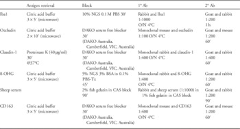
Immunohistochemistry for Iba1 (activated microglia), occludin (tight junction protein), claudin-1 (tight junction protein), 8-hydroxy-2'-deoxyguanosine (8-OHG; marker of DNA damage induced via oxidative stress), sheep serum (marker of BBB integrity) and CD163 (peripheral macrophages) was performed on 10 µM paraffin-embedded brain tissue sections cut at the level of the mid-striatum (caudate nucleus).
In brief, sections were dewaxed and rehydrated in serial alcohols. Antigen retrieval, blocking as well as primary and secondary antibody incubations are outlined in Table 1. Biotinylated secondary antibody detected with streptavidin horseradish peroxidase (HRP; 1:200 Amersham Bioscience, UK). Sections were washed, counterstained lightly with nissl stain, dehydrated, mounted, and viewed using light microscopy for assessment.
Immunohistological protocols

Ab, antibody; ′, minute; microwave, microwave on high; O/N, overnight.
S100B Quantification
S100B ELISA was performed as per manufacturer’s instructions (DiaSorin, MN, USA) on CSF (50 μl) obtained at postmortem to further assess BBB integrity. Assay sensitivity was 0.03 μg/l with a run imprecision of <10% and total imprecision of <15%.
Quantification
Single label immunohistochemistry
All slides were coded, with counting and qualitative assessment performed by a single examiner (B.J.A.) blinded to the experimental group. Images were taken under light microscopy using an Olympus BX41; DP25 Camera (Olympus, Australia). Photographs were taken at 200× magnification (sheep serum) or 400× (Iba1, occludin, claudin-1, CD163 and 8-OHG). Specific magnifications were chosen to allow for maximal regional specificity. Five fields of view were chosen, at random, from the cortical gray matter (GM), periventricular white matter (PVWM) and subcortical white matter (SCWM) from each section for analysis. Cells positive for Iba1, CD163 and 8-OHG were manually counted. Iba1 positive cells were deemed to be ‘activated’ or ‘not-activated’ based on morphological appearance of their processes. All other analysis was measured in pixels and converted to μm. Measurement of the vascular density of claudin-1 and occludin was assessed by densitometry as previously described.Reference Baburamani, Lo, Castillo-Melendez and Walker 18 Total number of positive staining in pixels was assessed and corrected for total percentage area occupied by blood vessels. Presence or absence of microbleeds (sheep serum) were assessed qualitatively by a single observer (B.J.A.) blinded to the experimental group.
Statistical analysis
Data are expressed as mean and s.e.m. Two-way analysis of variance (SigmaStat 3.5) was used to compare histological and morphological, molecular and biochemical indices. P<0.05 was accepted as statistically significant.
Results
Baseline characteristics and physiological parameters
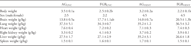
In the current study, we used an established ovine model of FGR, which has been previously described.Reference Allison, Hooper and Coia 13 Baseline characteristics are described in Table 2. Two fetal FGRUVC animals were found to be recently deceased at postmortem, despite having healthy arterial blood gases throughout the experimental period, and were therefore excluded from further analysis. FGRUVC and FGRVENT animals weighed less than AGUVC and AGVENT, but brain weight/body weight ratio was increased in FGRUVC and FGRVENT animals compared with AGUVC and AGVENT animals, indicative of brain sparing. FGR had no effect on corrected lung or heart weights.
Cohort properties

AG, appropriately grown; UVC, unventilated controls; FGR, fetal growth restriction; VENT, ventilation.
Different letters indicate significant difference P<0.05.
Brain structure
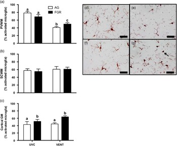
In the PVWM, FGRVENT lambs (49.8±3.4%) had significantly greater activated microglia compared with AGVENT lambs (40.9±2.7%, Fig. 1a). Interestingly, there was significantly less activated microglia in the AGVENT and FGRVENT lambs compared to UVC groups. Within cortical gray matter (GM), both unventilated FGR fetuses (FGRUVC, 51.7±5.3%, Fig. 1e) and ventilated FGRVENT lambs (63.9±3.0%, Fig. 1g), had significantly greater (P<0.01) microglia activation, compared with AGUVC (43.0±8.1%, Fig. 1d) and AGVENT (44.7±3.0%, Fig. 1g) animals (Fig. 1c). Microglia activation was not different within SCWM (Fig. 1b) between any groups.
Microglia activation. Microglial activation presented as a percent (%) of total microglia in the periventricular (PVWM) (a) and subcortical (SCWM) (b) white matter as well as the cortical gray matter (GM) (c). Differing letters indicate statistical difference between groups P<0.01. Representative micrographs from the cortical GM are shown in AGUVC (d), FGRUVC (e), AGVENT (f) and FGRVENT (g), activated microglia are indicated (black arrow). Scale bar=50 μm.

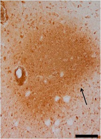
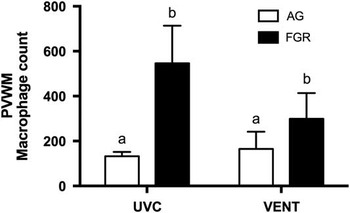
We assessed the integrity of the BBB by examining the infiltration of large protein (sheep serum, Fig. 2) and macrophages into the brain. We observed significantly more macrophages (P<0.01) within the PVWM of FGRUVC and FGRVENT animals compared with AGUVC and AGVENT animals (Fig. 3). No difference in macrophage infiltration was observed in the SCWM or the cortical GM (data not shown). Sheep serum extravasation was observed within the PVWM (Fig. 2) and SCWM regions of the FGRUVC and FGRVENT brains (four of six) relative to AGUVC and AGVENT (one of four) (data not shown).
Sheep serum extravasation. A representative image of sheep serum extravasation (arrow) in the PVWM within a FGRVENT animal is shown. Image taken at 20×. Scale bar=100 μm.

Macrophage density. Data mean±s.e.m. macrophages (CD163) in appropriately grown (AG) and growth restricted (FGR) unventilated controls (AGUVC, FGRUVC) and following ventilation (AGVENT, FGRVENT). Values are mean number of positive cells per field of view. Differing letters indicate statistical difference between groups (P<0.01) using a two-way analysis of variance.

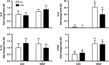
Within the PVWM, IL-8 mRNA levels were significantly increased in response to ventilation in both FGRVENT and AGVENT lambs, relative to FGRUVC and AGUVC fetuses (P<0.01, Fig. 4c). Early injury marker CTGF mRNA was similarly increased (P<0.002) in FGRVENT and AGVENT lambs following ventilation, compared with FGRUVC and AGUVC fetuses (Fig. 4d). Neither IL-6 nor IL-1β mRNA was different between groups within the white matter.
Inflammatory markers messenger RNA (mRNA). Data are interleukin (IL)-1β, IL-6, IL-8 and connective tissue growth factor (CTGF) mRNA levels in white matter of appropriately grown (AG) and growth restricted (FGR) unventilated controls (AGUVC, FGRUVC) and following ventilation (AGVENT, FGRVENT). Values are expressed as a fold change relative to values in appropriately grown unventilated controls. Differing letters indicate statistical difference between groups (P<0.01) using a two-way analysis of variance.

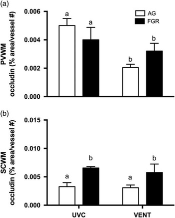
Occludin protein density was significantly decreased (P<0.005) in the PVWM of both FGRVENT (0.003±0.0005) and AGVENT (0.002±0.0002) lambs compared with unventilated AGUVC (0.005±0.0004) and FGRUVC (0.004±0.0008) fetuses. However, in the SCWM occludin density was significantly increased (P<0.05) in both FGRUVC (0.0066±0.0002) and FGRVENT (0.0058±0.0001) animals compared with both AGUVC (0.0031±0.0005) and AGVENT animals (0.0031±0.0005, Fig. 5b). Claudin-1 protein density was not significantly different between groups (data not shown).
Tight junction marker occludin protein density. Vascular occludin density in the periventricular (PVWM) and subcortical (SCWM) white matter. Differing letters indicate statistical difference between groups, P<0.005.

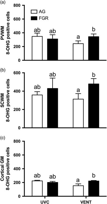
Oxidative stress was assessed by measurement of 8-hydroxyguanasine (8-OHG, Fig. 6). There was a significant interaction between growth status and ventilation resulting in significantly increased oxidative stress marker 8-OHG in FGRVENT animals in all regions measured compared to AGVENT animals. 8-OHG positive cells were increased 74% in the SCWM (Fig. 6a), 42% in the PVWM (Fig. 6b) and 44% in the cortical GM (Fig. 6c) in FGRVENT animals compared with controls.
Oxidative stress marker 8-hydroxyguanasine (8-OHG). Data shown are 8-OHG abundance (%) in appropriately grown (AG) and growth restricted (FGR) unventilated controls (AGUVC, FGRUVC) and following ventilation (AGVENT, FGRVENT). Values are mean percentage (%) of positive cells per field of view. Differing letters indicate statistical difference between groups (P<0.05) using a two-way analysis of variance.

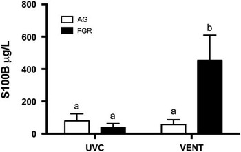
We measured astroglial protein S100B as an early marker of brain injury in the CSF (Fig. 7). S100B concentration was significantly (P<0.05) increased in FGRVENT lambs (454.0±156.0 μg/l) compared with AGVENT (57.0±31.0 μg/l) lambs and both unventilated control groups (AGUVC 79.6±44.2 μg/l, FGRUVC 40.0±23.0 μg/l).
S100B. Data mean±s.e.m. S100B levels in appropriately grown (AG) and growth restricted (FGR) unventilated controls (AGUVC, FGRUVC) and following ventilation (AGVENT, FGRVENT). Differing letters indicate statistical difference between groups (P<0.05) using a two-way analysis of variance.

Discussion
This is the first study to examine the susceptibility of growth restricted (FGR) preterm lambs to brain injury induced by the onset of mechanical ventilation. Our results have implications for the long-term development of neurological deficits associated with FGR, and the increased susceptibility of infants born preterm and with FGR. Although FGR infants are described as having a brain sparing phenotype, this nomenclature is deceptive as the FGR brain does not develop normally.Reference Castillo-Melendez, Yawno and Allison 6 The abnormalities of the FGR brain are not homogenous. FGR brain injury is highly dependent on the timing and severity of in utero insult, as well as the timing of delivery.Reference Baschat 19 This study demonstrates that late preterm FGR lambs have regions of the brain susceptible to ventilation-induced injury after birth. We show that 2 h of ventilation in FGR lambs increases oxidative cell damage, and levels of the brain injury marker S100B compared with other groups. Ventilation per se, regardless of growth status, increased inflammatory marker IL-8 within the brain of late premature lambs. The differences between the response of FGR and AG lambs to ventilation are likely to be mediated by abnormalities in the neurovascular unit, and increased BBB permeability. These changes appear to have been developmentally programmed in utero, as we noted fundamental differences in the neurovascular unit of the FGR brain before ventilation, compared with the AG brain.
The brain of FGRVENT lambs had significantly increased levels of cellular DNA fragmentation, a precursor to cell death. We used 8-hydroxyguanosine (8-OHG), a well-established marker of oxidative DNA damage, to indicate cell damage caused by oxidative stress.Reference Miller, Yan, Castillo-Melendez, Jenkin and Walker 20 The fetal brain has a high metabolic rateReference Chong, Li and Maiese 21 which increases free radical production. Excess free radicals are inadequately scavenged by immature antioxidant defensesReference Rogers, Witz, Anwar, Hiatt and Hegyi 22 hence, the preterm brain is highly susceptible to oxidative stress.Reference Volpe 23 In particular, developing oligodendrocytes have been found to be highly vulnerable to lipid peroxidation. The FGR fetus experiences chronic hypoxia caused by placental insufficiency, which we have shown previously to be associated with high levels of lipid peroxidation products within the brain.Reference Miller and Yawno 24 , Reference Miller, Chai and Loose 25 Our study was not designed to examine the long-term implications of ventilation on the FGR and AG brain, and we do not know if all cells that are positive for DNA damage will die. However, it is likely that endogenous repair mechanisms will be more limited in the FGR brain than in the AG late preterm brain, as is the case for endogenous antioxidant enzyme capacity.Reference Biri, Bozkurt and Turp 26 Further supporting the susceptibility of the FGR brain to injury after late preterm birth and ventilation is the four-fold increase in circulating concentration of S100B seen in FGRVENT lambs, when compared with all other groups. S100B is an established biomarker of brain injury, where S100B is derived predominantly from reactive astrocytes.Reference Aridas, Yawno and Sutherland 27 , Reference Gonçalves, Leite and Nardin 28 This protein is released by injured astrocytes and is found in the circulation after passage through a disintegrated BBB. Our lack of increase in S100B levels in FGRUVC fetuses is consistent with findings in human newborns.Reference Boutsikou, Mastorakos and Kyriakakou 29 In the clinical situation, an acute hypoxic-ischemic event at birth is associated with significantly increased serum concentrations of S100B at 24 h and is also correlated with long-term neurodevelopmental outcomes.Reference Nagdyman, Kömen, Ko, Müller and Obladen 30 , Reference Nagdyman, Grimmer, Scholz, Müller and Obladen 31 In addition to S100B acting as an effective biomarker for acute brain injury, elevated S100B in the peripheral circulation is also indicative of increased permeability and leakiness of the BBB.
In the current study, FGR fetuses and ventilated lambs had increased macrophage density within the brain. Hypoxia upregulates hypoxia-inducible factors (HIF-1α, HIF-2) leading to recruitment of macrophages. Macrophages are recruited to the brain in response to injury and infiltrate following BBB breakdown.Reference Denieffe, Kelly, McDonald, Lyons and Lynch 32 We determined macrophage content by the macrophage marker CD163, which specifically binds to the M2 macrophages. One of the key upregulators of M2 macrophages are glucocorticoids.Reference Martinez 33 We have previously shown that plasma cortisol is increased in both the FGRUVC and FGRVENT animals.Reference Allison, Hooper and Coia 13 Hence, in the current study, increased cortisol associated with placental insufficiency may lead to macrophage upregulation, indicative of brain tissue repair following injury.
Initiation of mechanical ventilation following premature birth induces a cerebral inflammatory cascade. We have previously demonstrated that the initiation of ventilation leads to an upregulation of pro-inflammatory cytokines in preterm lambs.Reference Barton, McDougall and Melville 16 , Reference Barton, Melville and Tolcos 34 This study is the first to demonstrate that being born FGR does not exacerbate this inflammatory response within the brain. Pro-inflammatory cytokine IL-8 mRNA in brain white matter is not different before ventilation (FGRUVC v. AGUVC) and we show that 2 h of ventilation upregulates IL-8 mRNA levels to a similar degree, irrespective of whether the newborn was growth restricted or not (FGRVENT and AGVENT). Systemic inflammation is strongly linked to brain injury, and inflammation-induced brain injury is described as an initial white matter injury, acute BBB dysfunction, and neurovascular unit injury associated with behavioral alterations.Reference Stolp, Johansson and Habgood 35 When followed to adulthood, the behavioral changes persisted, as did BBB changes.Reference Stolp, Johansson and Habgood 35 In the current study IL-6 mRNA and IL-1β mRNA were not different between groups. The detection of inflammation is correlated with both short-term injury, such as enhanced risk of intraventricular hemorrhage and cerebral white matter injury, and long-term injury such as cerebral palsy.Reference Khwaja and Volpe 11 Acute ventilation has been shown previously to increase the size and density of microglial aggregations.Reference Barton, McDougall and Melville 16 , Reference Barton, Melville and Tolcos 34 We observed increased microglial aggregation within the PVWM and cortical GM in FGRVENT compared with AGVENT. Although we have previously shown that ventilation increased activated microglia in the SCWM in AG preterm lambs,Reference Barton, Melville and Tolcos 34 we did not find any change in the SCWM in the current study. Further, both VENT groups had decreased microglial activation compared with UVC groups. It is known that the PVWM is a highly dynamic region of the brain with a high level of proliferation progenitor cells resulting in inflammatory processes. The increased microglia activation in the cortical GM is representative of established cells of the brain. Indeed it is increasingly being recognized that established white matter injury impacts on the overlaying cerebral cortex.Reference Miller, Huppi and Mallard 5 Further, mature FGR offspring have reduced gray matter volume compared with term counterparts.
In the last decade, CTGF has emerged as an early marker of lung injury.Reference Wallace, Probyn and Zahra 17 CTGF regulates cell proliferation and has been found to be rapidly upregulated in the lung of preterm lambs following ventilation-induced lung injury.Reference Wallace, Probyn and Zahra 17 CTGF also has a role in mediating brain injury, it is involved in fibroblast activation following injury, and the subsequent processes of wound healing and scar formationReference Schwab, Postler and Nguyen 36 and increases rapidly in response to traumatic brain injuryReference Liu, Liu and Li 37 and chronic neurodegeneration.Reference Ueberham, Ueberham, Gruschka and Arendt 38 This is the first study to report CTGF as an early marker for ventilation-induced brain injury in late preterm lambs and may have implications for long-term neurodegeneration.
The neurovascular unit describes the cellular components of the cerebral vasculature, and close association of vascular endothelial cells covered by a basal lamina membrane, the tight junction proteins between these cells, and the astrocytes, which provide the communication link between blood vessels and the brain.Reference Castillo-Melendez, Yawno and Allison 6 Tight junctions are vital for normal functioning of the BBB and are located between the endothelial cells of cerebral blood vessels. The BBB is a highly complex system comprising the endothelial cells, basement membranes (both endothelial and parenchymal), pericytes, perivascular antigen-presenting cells, and astrocytic endfeet. Alteration to the neurovascular unit or the BBB can alter cerebral homeostasis leading to edema and inflammatory cell recruitment. Interestingly, occludin density was increased in the SCWM but decreased in the PVWM. This is of particular importance as the PVWM is highly vulnerable to injury in both growth restriction and in prematurity. Our results suggest regional changes to tight junction integrity, which may underpin BBB permeability. Previous studies have demonstrated upregulation of occludin in response to glucocorticoids.Reference Förster, Silwedel and Golenhofen 39 We have shown that our model of FGR results in increased cortisolReference Sutherland, Crossley and Allison 12 , Reference Allison, Hooper and Coia 13 which may lead to the increased occludin observed in the SCWM. Although the alterations were not seen ubiquitously in all brain regions studied, any alteration to BBB function can have dramatic impacts on brain permeability. Indeed, we did find evidence of increased protein extravasation in FGRUVC and FGRVENT relative to the AGUVC and AGVENT. Future studies are required to determine the regionality and functionality of the tight junction changes within the FGR brain.
The mechanisms of developmental programming of neurological sequelae in FGR offspring remain poorly understood. We have shown in this study, and previously, that antenatal compromise leads to altered BBB function and impaired neurovascular unit composition.Reference Castillo-Melendez, Yawno and Allison 6 Both of these factors undoubtedly increase vulnerability to a second hit, such as mechanical ventilation. In support of this we show that FGRVENT lambs have an increased oxidative stress response compared with AGVENT lambs. Taken together, these data confirm our previous findings that FGR offspring require tailored care to minimize detrimental long-term neurological outcomes. Altered BBB function has been previously hypothesized to contribute to long-term behavioral changes, via altered protein permeability and ultimately white matter volume reduction. Breakdown of the BBB is closely related to increased risk of long-term injury.Reference Stolp, Johansson and Habgood 35 Hormonal imbalances have been implicatedReference Thompson, Syddall, Rodin, Osmond and Barker 40 with increased hypothalamic–pituitary–adrenal (HPA) axis activation associated with anxiety. We did not examine the HPA axis in the current study, however we have previously shown that cortisol is increased in our model of FGR.Reference Sutherland, Crossley and Allison 12 , Reference Allison, Hooper and Coia 13 It has been suggested that there is a ‘critical window’ during the second and third trimester during which neuroimpairment occurs. This timing suggests that placental insufficiency, rather than other pathology, and therefore hypoxia and nutrient deficiencies, programs neurological consequences. As a decline in cognitive state occurs with time, rather than being immediately evident at birth, there is an implication that additional environmental factors may contribute. We demonstrate that many markers of long-term neurological deficit (IL-8 and S100B) are increased following 2 h of ventilation (FGRVENT) but not in the unventilated FGR group (FGRUVC). These data may suggest that a ‘second hit,’ in this case injurious mechanical ventilation, increases the risk of brain injury and long-term neurological outcome.
Limitations
A limitation of this study is that we examined brain injury over a relatively short-time period, 2 h after birth. It would now be interesting to examine whether the initial response to injury we observe in this study persists long term. However, at this early gestation it is extremely difficult to keep lambs alive without additional neonatal support, which confounds results. This study determined the brain pathology of an acute mechanical ventilation insult and, while long-term implications have been extrapolated from this data, future studies should examine long-term neurodevelopmental outcomes following mechanical ventilation-induced brain injury. It would also be of interest to examine the components of the basement membrane, smooth muscle/endothelial cells, astrocytes, pericytes and gap junctions to determine how these critical cell interactions are altered in the FGR brain.
Our findings indicate that FGR and AG premature lambs have altered pathways of injury in response to an initial injurious mechanical ventilation. Further investigation of these pathways may allow therapeutic options to minimize long-term neurological deficits known to be associated with late preterm FGR birth.
Acknowledgments
The authors would like to acknowledge the assistance of Dalibor Stanojkovic and Karyn Rodgers.
Financial Support
National Health and Medical Research Council (NH&MRC) Project Grant (APP1083520), The Cerebral Palsy Alliance (PG0414), NH&MRC Research Fellowships (GRP: 1026890 and SBH: 545921), a Rebecca L. Cooper Medical Research Foundation Fellowship (GRP), Australian Research Council Future Fellowship (SLM) and the Victorian Government’s Operational Infrastructure Support Program.
Conflicts of Interest
The authors do not have any potential, perceived or real conflicts of interest, especially no financial arrangement with a company whose products are discussed in the manuscript. Chiesi Farmaceutici S.p.A. generously donated the surfactant used in this study. The funders had no role in study design, data collection and analysis, decision to publish, or preparation of the manuscript.
Ethical Standards
The authors assert that all procedures contributing to this work comply with the ethical standards of the relevant national guides on the care and use of laboratory animals (National Health and Medical Research Council of Australia) and has been approved by the institutional committee (Monash Medical Centre animal ethics committee at Monash University).