Introduction
Nasal septal perforations are anatomical defects of the nasal septum. They can alter nasal function, which may in turn cause various symptoms.Reference Pedroza, Patrocinio and Arevalo1
Nasal septal perforation may occur due to local trauma, nasal surgery or cocaine inhalation, or in association with granulomatous diseases such as Wegener's granulomatosis. The condition has also been reported following intranasal steroid therapy.Reference Williams2
Direct communication between the two nasal cavities leads to impairment of air flow and pressure, often accompanied by a wide variety of symptoms such as crusting, epistaxis, cacosmia and impaired nasal respiration.Reference Re, Paolucci, Romeo and Mallardi3 Whistling may be noted (usually in the case of smaller perforations) due to air flow through the small opening. Dried blood and crusts may lead to obstruction; however, even in a clean nasal perforation turbulence may produce a definite sense of nasal obstruction.Reference Døsen and Haye4 Large and anterior septal perforations are more symptomatic, while posterior perforations tend to be less so due to humidification from the turbinates.
Perforation size is classified as small (diameter less than 0.5 cm), medium (diameter 0.5–2 cm) or large (diameter more than 2 cm).Reference Re, Paolucci, Romeo and Mallardi3
Surgical repair is indicated for patients with symptomatic perforations, but is not usually recommended for perforations without subjective and/or functional derangement. Contraindications to surgery include: comorbidity posing a severe general anaesthesia risk; chronic nasal inflammatory processes (including sarcoidosis and Wegener's granulomatosis), and ongoing cocaine usage.Reference Kridel5
In the early 1950s, some authors proposed the use of prosthetic devices to aid patients with nasal septal perforation. Although the composition of such devices has improved over time, they are invariably ill-tolerated as they act as foreign bodies.Reference Facer and Kern6 However, silicone prosthetic devices can be used in those patients with contraindications to surgery.Reference Re, Paolucci, Romeo and Mallardi3
Many surgical procedures for nasal septal perforation repair have been described. Surgical approaches include endonasal, open external, sublabial and midfacial degloving techniques. The great variety of published techniques is evidence that no single technique is currently recognised as being uniformly reliable in closing all perforations.Reference Pedroza, Patrocinio and Arevalo1
The use of an inferior turbinate flap, aloneReference Ayshford, Shykhon, Uppal and Wake7 or with an interposition graft comprising acellular human dermal allograftReference Friedman, Ibrahim and Ramakrishnan8 or bioactive glass,Reference Stoor and Grenman9 has been described.
The use of inferior turbinate grafts with a middle layer composed of tragal cartilageReference Hussain and Kay10 or bovine cartilaginous sheetingReference Re, Paolucci, Romeo and Mallardi3 has also been described.
Other authors have described the use of bipedicled mucoperichondrium advancement flaps with an interposition supporting graft, created via a closed (with or without endoscopy) or external rhinoplasty approach. The interposition support may be formed of temporalis fascia, mastoid periosteum, acellular human dermal allograft, dermal autograft, porcine small intestinal submucosa, conchal cartilage, mastoid cortical bone, allograft dermal matrix, and/or tragal cartilage with perichondrium.Reference Pedroza, Patrocinio and Arevalo1, Reference Re, Paolucci, Romeo and Mallardi3, Reference Fairbanks11–Reference Lee, Lee, Ban and Jin16
The use of buccal flaps pedicled on the labial arteryReference Karlan, Ossoz and Sisson17 or facial arteryReference Heller, Gabbay, Trussler, Heller and Bradley18 has also been proposed.
For large perforations, various authors have suggested a sublabial midfacial degloving approach,Reference Sachs, Foster and Corovin19 intranasal tissue expansion,Reference Romo, Jablonski, Shapiro and McCormick20 pericranial rotation flapReference Paloma, Samper and Cernera-Paz21 and radial free flap.Reference Mobley, Boyd and Astor22
In the present study, we undertook endoscopic, endonasal closure of nasal septal perforations, using an inferior turbinate graft with no advancement flaps.
Method
Six patients with symptomatic nasal septal perforations were included. These patients presented (or were referred) to Fakeeh Hospital and United Doctors Hospital, Saudi Arabia, between January 2007 and July 2009. We included in the study all consecutive symptomatic patients with no contraindications who agreed to undergo the procedure and provided written consent.
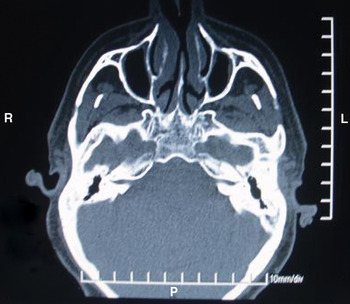
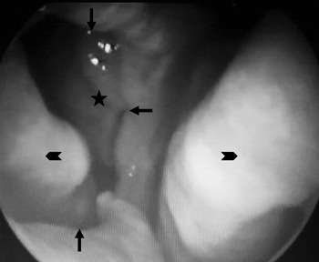
These patients' septal perforations were iatrogenic (following septal surgery) in five cases and due to non-surgical trauma in one case. Patients comprised four men (67 per cent) and two women (33 per cent), with an age range of 23 to 41 years (mean age, 33 years). The perforation size ranged from 1 to 2.5 cm in horizontal diameter and 0.5 to 1.5 cm in vertical diameter (Figures 1 and 2). Six patients complained of nasal crusts, five of nasal obstruction, three of cacosmia and three of recurrent epistaxis (Table I).
Axial computed tomography scan showing nasal septal perforation 2 cm in horizontal diameter. L = left; R = right; P = posterior

Endoscopic view of left nasal cavity showing septal perforation, with the right inferior and middle turbinates seen through the perforation. Arrows = edges of perforation; chevrons = right and left inferior turbinates; star = right middle turbinate

Patient Data

Pt no = patient number; y = years; perf = perforation; mth = months; M = male; F = female
Prospective surgical candidates were excluded due to the following contraindications: ongoing inflammatory disease; previous turbinectomy or atrophic changes; perforations larger than 2.5 cm horizontally or 1.5 cm vertically; and ongoing cocaine usage.
Surgical repair was performed via a closed, endoscopic, endonasal approach. The procedure was performed under general anaesthesia with endotracheal intubation.
A caudal septal incision (hemitranfixion) was made on one side and subperichondrial dissection commenced from anterior to posterior until the septal perforation was reached. The anterior aspect of the perforation was entered via incision of its anterior edge. A 0°, 4 mm Hopkins rod endoscope was used from this point. The edge of the perforation was incised superiorly, posteriorly and inferiorly using a number 12 scalpel blade, and a tunnel was created by dissecting between the mucoperichondrium of both sides. On one side, this tunnel was extended between the mucoperichondrium and the septal cartilage and between the mucoperiosteum and the vomer or perpendicular plate of the ethmoid.
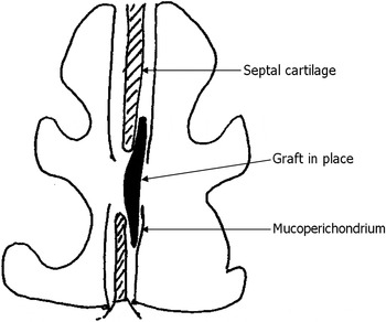
On the other side, a partial inferior turbinectomy was performed. The inferior turbinate graft was then dissected from any bony remnants and flattened, taking care not to disturb its continuity. In two patients with large perforations, part of the contralateral inferior turbinate was used to cover the centre of the raw side of the graft, to facilitate healing. Vicryl 4/0 sutures were used to fix the graft in place within the tunnel described above. The first stitch was made posteriorly by taking one bite 5 mm posterior to the posterior edge of the perforation and the other bite into the graft. The graft was then approximated to the perforation and tucked into place as the stitch was tightened. The graft was positioned between the mucoperichondrium of both sides and between the mucoperichondrium (or mucoperiosteum posteriorly) and the cartilage (or bone) of one side (Figure 3). The graft was fixed by three more stitches positioned superiorly, inferiorly and anteriorly. The anterior septal incision was then closed.
Diagram showing graft placement between the mucoperichondrium of both sides and between the mucoperichondrium and the cartilage of one side.

Silastic® splints were applied, to be removed one week post-operatively. The nasal cavities were packed with Merocel® (Medtronic Xomed, Jacksonville, Florida, USA), to be removed after two days.
Patients were followed up one, two and four weeks post-operatively, then every month for three months, and then every three months for one year.
Results
Five of the six patients (83 per cent) had complete closure of their perforation.
The remaining patient had partial closure, with a small, 2 mm, posterior residual perforation. This patient's original perforation measured 2.5 × 1.5 cm.
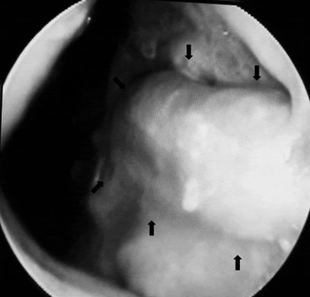
None of the patients (including the patient with residual perforation) had any post-operative complaints related to septal perforation. It was noted that the side of the septum with no turbinate mucosa healed within three to five weeks post-operatively, after which crusting ceased (Figure 4). No significant difference in healing time was noted in the two patients in whom the central part of the raw side of the graft was covered by part of the contralateral inferior turbinate.
Endoscopic view five weeks post-operatively, showing the side previously containing the raw surface of the graft, now completely healed by septal mucosa. Arrows = original perforation site

Patient follow up ranged from six months to two years.
Discussion
Many techniques have been reported for the surgical repair of nasal septal perforation, suggesting that it represents a significant surgical challenge. Various approaches have been described depending on the size and site of the perforation; these include a closed endonasal approach with or without endoscopy, external rhinoplasty, sublabial and midfacial degloving.
Reported surgical procedures have included septal rotation flaps, inferior turbinate pedicle flaps, sublabial buccal mucosal flaps, radial forearm free flaps, pericranial flaps, tissue expansion and facial artery musculomucosal flaps.
Several types of autologous interposition graft have been described for septal perforation repair, including temporalis muscle fascia, septal cartilage, nasal turbinate, conchal cartilage, mastoid periosteum, ear tragus cartilage with perichondrium, pericranium, ethmoid bone, and acellular human dermal allograft.Reference Pedroza, Patrocinio and Arevalo1
The size of the nasal septal perforation is an important factor in determining the need for surgical repair, the type of repair and the surgical outcome. The larger the vertical height of the perforation, the more difficult the repair. Larger septal perforations require greater tissue mobilisation and can result in greater tension on closure.Reference Re, Paolucci, Romeo and Mallardi3 When the vertical diameter of a defect is greater than 50 per cent of the septal height, there is insufficient remaining mucosa to achieve closure. Closure options for large, symptomatic perforations are limited, and include radial forearm free flaps, pericranial flaps, nasal tissue expansion and the facial artery musculomucosal flap.Reference Heller, Gabbay, Trussler, Heller and Bradley18
The endonasal approach to perforation repair is less invasive and leaves no external scars; however, it is a more difficult technique due to the limited exposure of the operative field.Reference Lee, Ahn, Park, Kim, Sin and Youn23
The use of a nasal endoscope provides excellent direct visualisation of smaller septal perforations, but other techniques may be needed for large perforations. The open rhinoplasty approach with an external columellar incision can achieve wide exposure of the septum, but this procedure leaves an external scar. Alternatively, the midfacial degloving approach also allows excellent visualisation of the septal perforation and is a good procedure for larger perforations.Reference Hier, Yoskovitch and Panje24
Repair using a skin or buccal mucosal graft or flap may leave the patient with a dry nose with crusts, because the normal respiratory epithelium is absent.Reference Lee, Lee, Ban and Jin16
The advantages of an inferior turbinate flap are abundant vascularity, combined skeletal and epithelial support, and ease of development and insertion. Another important advantage is that it uses respiratory tract mucosa, enabling physiological repair. The disadvantage of this technique is that repair with an inferior turbinate flap (rather than a free graft) requires a second-stage procedure to release the pedicle; furthermore, airway obstruction may be caused by the turbinate flap itself.Reference Friedman, Ibrahim and Ramakrishnan8
In contrast, the above-described technique using a free turbinate graft has the same advantages as inferior turbinate flap rotation without the need for second-stage surgery or the possibility of nasal obstruction by the flap. Although one surface of the graft is not epithelialised, mucosal healing occurs by secondary intention within three to five weeks. In two of the presented patients, the raw surface in the centre of the graft area was partially covered by part of the contralateral inferior turbinate in order to facilitate healing, but no significant difference in healing time was noted.
Some patients who seek perforation repair have already failed local advancement flaps from the septum. Other patients have had extensive previous septal surgery with removal of cartilage, and are not candidates for local advancement flaps. The inferior turbinate provides a valuable source of respiratory tract mucosa, which may still be healthy even when the septal mucosa had been previously damaged.Reference Friedman, Ibrahim and Ramakrishnan8
The described technique of septal perforation repair using an inferior turbinate graft is contraindicated in patients with septal perforations larger than 2.5 cm horizontally or 1.5 cm vertically. It is also contraindicated if the patient has atrophic changes or has previously undergone turbinectomy.
• Many nasal septal perforation repair techniques have been described
• This study treated six patients with small to medium perforations, using an endoscopic repair technique with an inferior turbinate graft
• Five patients (83 per cent) had complete perforation closure; one had partial closure
• All patients were symptom-free after surgery
The described procedure requires only minimal undermining, with no release incisions, and enables tension-free closure. This makes it a relatively simple, easily reproducible procedure with good results when used in appropriately selected patients.
Conclusion
An endoscopic endonasal approach using inferior turbinate free graft technique is effective in the closure of small and medium sized nasal septal perforations.