Introduction
Tomato (Lycopersicon esculentum Mill [Solanaceae]) is one of the key vegetables produced by smallholder farmers usually for the domestic market in sub-Saharan Africa. About 54 million metric tonnes are produced worldwide and more than 10% of this in Africa (Wang et al., Reference Wang, Liu and Mahson2006). Tomato is an integral part of an everyday diet for millions of people worldwide and has an important dietary value in respect of carotene, thiamine, niacin and vitamin C. Lycopene, a carotenoid, is a powerful antioxidant found in tomatoes and used in the management of cardiovascular diseases, urinary tract diseases and diabetes (Atessahin et al., Reference Atessahin, Türk, Karahan, Yilmaz, Ceribasi and Bulmus2006).
Tomato is one of the most important local market vegetables in Kenya. The crop is mainly grown by small-scale farmers in most arable areas of the country. The main production areas in Central Province are Kirinyaga, Nyeri, Kiambu, Mwea and Gikambura. In 2004, an estimated 75,101 tonnes of tomato valued at over Kshs 1 billion (approximately $12 million) were produced in Central Province, surpassing all the other vegetables in value (KARI, 2005). The major tomato production constraints reported in Kenya include diseases (bacterial wilt, early and late blight, leaf curl, tomato spotted wilt virus, leaf spot and powdery mildew), insect pests and other arthropods (spider mites, thrips, whiteflies, African bollworm) and nematodes, leading to high economic losses (Varela et al., Reference Varela, Seif, Löhr and Ng'eny-Mengech2003; KARI, 2005).
Root-knot nematodes (RKN) are one of the major pathogens of tomatoes worldwide and limit fruit production (Sikora and Fernandez, Reference Sikora, Fernandez, Luc, Sikora and Bridge2005; Mehdi, Reference Mehdi2009). There are more than 90 described species in the genus Meloidogyne (Tylenchida: Heteroderidae), but the four most commonly occurring species are Meloidogyne incognita, M. arenaria, M. javanica and M. hapla (Hunt et al., Reference Hunt, Luc, Manzanilla-Lopez, Luc, Sikora and Bridge2005). They have a wide host range and are hence difficult to control as they can survive and reproduce on other host crops, including weeds. RKN infection is characterized by galled roots, yield losses, collapse of plants and stunted growth (Jacquet et al., Reference Jacquet, Bongiovanni, Martinez, Verschave, Wajnberg and Castagnone-Sereno2005).
Recommended control measures for RKN include use of nematicides, soil solarization and crop rotation. However, use of these methods by small-scale growers in Kenya is greatly hampered due to high cost of using nematicides and diminishing size of land holdings. Plant resistance is one of the most environmentally safe and economically viable means for controlling RKN. Several cultivars of tomatoes such as Montelle, Sun6082, Pik Red, Celebrity and Beefmaster have been developed in an attempt to produce RKN-resistant cultivars (Milligan et al., Reference Milligan, Bodeau, Yaghoobi, Kaloshian, Zabel and Willamson1998; Jacquet et al., Reference Jacquet, Bongiovanni, Martinez, Verschave, Wajnberg and Castagnone-Sereno2005). Resistance mechanism in response to invasion by RKN involves the formation of necrotic cells at the infection site to prevent the juveniles from developing any further. However, a high level of genetic variability of RKN has led to the development of races and virulent populations which can reproduce even on plants carrying the resistance genes (Castagnone-Sereno, Reference Castagnone-Sereno2006). Successful use of tomato cultivars resistant to RKN in Kenya depends on the accurate identification of RKN species found associated with tomatoes.
In Kenya, identification of RKN has primarily relied upon morphological characteristics. This technique suffers major limitations. Morphological characterization relies on variability of the perineal patterns in the natural populations of the Meloidogyne spp. that may be indistinct (Baum et al., Reference Baum, Gresshoff, Lewis and Dean1994) but which has proved to be useful and remains a tool in diagnostics. It also relies on the variability of the stylet length, which values do overlap, making species identification difficult. Isozyme phenotype analysis used in RKN identification is more accurate and less subjective (Carneiro et al., Reference Carneiro, Almeida and Quénéhervé2000). To complement isozyme phenotype technique, DNA markers have been useful for the identification and phylogenetic comparison of plant-parasitic nematodes (Curran, Reference Curran and Nickle1991; Jones et al., Reference Jones, Phillips and Armstong1997; Floyd et al., Reference Floyd, Abebe, Papert and Blaxter2002). The objective of the present study was to assess the prevalence and identify RKN infesting tomatoes using isozyme phenotypes and restriction fragment length polymorphism (PCR-RFLP).
Materials and methods
Study sites and sampling
Surveys were conducted in three major tomato-growing districts in Kenya that are hot spots for nematodes: Mwea (1°24.0′S; 37°25.0′E), Gikambura (1°16.6′S; 36°37.6′E) and Athi River (1°26.60′S; 36°58.6′E). Ten fields per district were selected and 30 young tomato plants (total of 900) were uprooted at random and inspected for occurrence of RKN symptoms, which were characterized by galled roots. Root samples were kept in polyethylene bags, brought to the laboratory and thoroughly examined for the presence of galls. Debris on the roots was washed off with distilled water and then immersed in an aqueous solution of phloxin B (0.15 g/l) for 15 min to stain egg masses. The number of root galls per root system was counted and prevalence determined on the following scale: 0 = 0, 1 = 1–2, 2 = 3–10, 3 = 11–30, 4 = 31–100 and 5 = greater than 100 galls. The number of egg masses per root system was counted and prevalence determined on the following scale: 0 = 0, 1 = 1–2, 2 = 3–10, 3 = 11–30, 4 = 31–100 and 5 = greater than 100 egg masses (Taylor and Sasser, Reference Taylor and Sasser1978). RKN infestation load in each locality was calculated using the formula:

Maintenance of inoculum
The inoculum of selected field populations from each locality was maintained on RKN-susceptible tomato var. Money Maker in a greenhouse at 25 ± 3°C, 90 ± 2% relative humidity with a photoperiod of 12 h light:12 h dark by inoculating each seedling in a pot containing autoclaved soils with chopped infested roots collected from the field. To make pure cultures of field populations maintained on tomato in the greenhouse, a single egg mass inoculation was made (Mehdi, Reference Mehdi2009). Single mature egg masses were inoculated in pots around the roots of a young tomato seedling for each population and the collection maintained separately. To maintain sufficient inoculum for further studies, sub-culturing was done. Each new seedling was inoculated with at least 15 egg masses obtained from a pure culture.
Identification of RKN using isozyme profiles
Sample preparation
Six females per plant were isolated under a dissecting microscope. After the isolation, they were rinsed in reagent-grade water and then transferred to an ice-cold 1.5 ml Eppendorf tube containing 60 μl extraction buffer (20% sucrose, 2% Triton X-100 and 0.01% Bromophenol Blue) (Esbenshade and Triantaphyllou, Reference Esbenshade and Triantaphyllou1985) and squashed to release body contents. Samples were centrifuged at 12,000 g at 4°C for 1 min.
Native PAGE electrophoresis and enzyme staining
Electrophoresis was done on native polyacrylamide gels (8–25%) at 4°C as described by Karssen (Reference Karssen2002). Meloidogyne javanica (laboratory reared) obtained from the botanical farm at Kenyatta University in Nairobi, Kenya, was used as a reference standard. After electrophoresis, gels were stained for enzymatic activity in a Petri dish at 37°C. Malate dehydrogenase (MDH) activity was measured using a staining solution containing 0.05 g β-NAD, 0.03 g nitroblue tetrazolium, 0.02 g phenazine methosulphate, 50 ml of 0.5 m Tris pH 7.1 and 7.5 ml stock (10.6 g Na2CO3+1.34 g l-malic acid in 100 ml H2O) dissolved in 70 ml reagent grade water. Incubation of MDH lasted for 5 min. Following this, the gel was washed twice with distilled water and further stained for esterase (EST) activity for 30 min. EST was measured using a staining solution containing 100 ml of 0.1 m phosphate buffer pH 7.3, 0.06 g fast blue RR salt, 0.03 g EDTA and 3 ml of 0.1 g α- and 0.1 g β-naphthyl acetate dissolved in 10 ml acetone (Karssen, Reference Karssen2002). The reaction was terminated at 30 min. This was achieved by rinsing the gel with distilled water and fixing for 5 min in a solution of 10% acetic acid, 10% glycerol and 80% distilled water. This experiment was conducted four times.
Identification of RKN using PCR-RFLP
DNA extraction and purification
Six female nematodes were handpicked from the infected root systems under a dissecting microscope and second-stage juveniles (J2) were allowed to hatch in 2% sucrose at 25°C. Three J2 were used for the subsequent analysis. The respective J2 were frozen in a pre-cooled mortar and ground to fine powder in liquid nitrogen. A 50-μl lysis buffer (1–10 mg/ml proteinase K, 100 mm Tris–HCl pH 8.0, 5 mm EDTA, 200 mm NaCl and 1% w/v of SDS buffer) was added to the fine powder, mixed gently and incubated at 37°C overnight. The liberated DNA was extracted against an equal volume of phenol saturated with TE (100 mm Tris–Cl pH 8.0 and 1 mm EDTA pH 8.0). The aqueous phase was transferred to a clean 1.5 ml microcentrifuge tube using a wide-bore pipette and mixed with an equal volume of chloroform isoamyl alcohol (24:1). DNA was then precipitated from the final aqueous phase by addition of an equal amount of ice-cold 95% ethanol with an incubation period of 5 h at − 20°C. DNA was pelleted by centrifugation at 12,000 g for 10 min, washed with 500 μl of 70% ethanol, air-dried and re-suspended in 20 μl of 10 mm Tris and 1 mm EDTA at pH 7.5. DNA was quantified using spectrophotometer at 280/260 nm.
Mitochondrial DNA COII PCR amplification
Primers C2F3 (5′-GGTCAATGTTCAGAAATTTGTGG-3′) (Powers and Harris, Reference Powers and Harris1993) and MRH106 (5′-AATTTTCTAAAGACTTTTCTTAGT-3′) (Stanton et al., Reference Stanton, Hugall and Moritz1997) were used to amplify the region between (MtDNA COII-LSUrRNA) genes. Amplifications were performed using 25-μl volumes containing 12 ng DNA, 200 μm dNTPs (MBI Fermentas®), 0.4 μm primers, 1 U Taq DNA polymerase (MBI Fermentas®) and 1 × Taq DNA polymerase reaction buffer. The temperature profile for all reactions was 94°C for 2 min, followed by 35 cycles of 94°C for 30 s, 50°C for 30 s and 72°C for 2 min, with a final extension at 72°C for 10 min. Confirmation of the amplified DNA fragments was conducted on 1% agarose gel in 1 × TAE buffer at 70 V for 1.5 h.
Restriction digestion of mitochondrial DNA COII PCR products
The region between mtDNA COII-LSUrRNA PCR products was digested using HinfI (Fermentas®). The restriction digestion with Hinf1 was carried out on 1 μl buffer, 1 μl (10 U) of Hinf1, 4 μl water and 4 μl of PCR product. Digestion was performed for 4 h at 37°C. Restricted products (5 μl) were separated on a 2% agarose gel in 1 × TAE at 70 V for 1.5 h.
Results
Prevalence and severity of RKN in tomato fields
Tomato plants in all the three districts were found infested with RKN, but the severity of infestation varied. Highest prevalence per plant (62.6%) was found in Athi River, closely followed by Mwea (47.6%) and Gikambura (28.6%). The disease severity on tomato was estimated on the basis of average gall and egg mass indices. Both gall and egg mass indices (averages) ranged from 2.5 to 5.3. The greatest egg mass and gall indices were found in Athi River, the area in which the prevalence of RKN was also greatest (Table 1). Meloidogyne incognita was found in all the three districts and accounted for 59.1% of all the samples analysed, closely followed by M. javanica (35.7%). Meloidogyne arenaria (EST phenotypes A1 and A2) occurred only in Athi River and accounted for 5.2% of the total samples.
Prevalence and distribution of root-knot nematodes on tomatoes in three districts in Kenya

GI, gall index; EMI, egg mass index on Taylor and Sassers' scale.
RKN identification by isozyme profiles
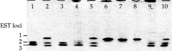
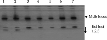
On the basis of isozyme profiles (N = 900), three species of RKN (M. incognita, M. javanica and M. arenaria) were identified in the three districts. Five EST phenotypes were detected in this study. Meloidogyne javanica produced three bands at EST loci 1, 2 and 3, while M. incognita occurred in two EST phenotypes; M. incognita (EST phenotype I1) with one strong band at EST locus one and M. incognita (EST phenotype I2) with a strong band at locus one and an allele at the same band. Two EST phenotypes of M. arenaria were detected in this study: M. arenaria (EST phenotype A1), which had two strong bands at second and third EST locus, and M. arenaria (EST phenotype A2), which had a strong band at the second locus (Fig. 1). One MDH phenotype designated N1 was observed for all the species (M. javanica, M. incognita and M. arenaria) identified in this study (Fig. 2).
Esterase (EST) phenotypes of representative specimens of Meloidogyne incognita, M. javanica and M. arenaria from the three locations. Lane 1: M. arenaria (EST phenotype A2); 2: M. javanica (J3); 3: M. arenaria (EST phenotype A2); 4: M. arenaria (EST phenotype A2); 5: M. javanica EST phenotype (J3); 6: M. incognita (EST phenotype I1); 7: M. incognita (EST phenotype I1); 8: M. incognita (EST phenotype I1); 9: M. arenaria (EST phenotype A2); 10: Standard (M. javanica).

Malate dehydrogenase (MDH) and esterase (EST) phenotypes of the representative populations of Meloidogyne incognita and M. javanica in tomato production systems (Athi River samples). Lane 1: M. javanica A13 (MDH-EST phenotype) (N1-J3); 2: M. javanica A14 (N1-J3); 3: M. incognita A5 (NI-I1); 4: M. incognita A6 (N1-I1); 5: M. incognita A10 (N1-I1); 6: M. incognita A16 (I1); 7: M. incognita A18 (N1-I1).

RKN identification by mitochondrial DNA (mtDNA) COII PCR-RFLP
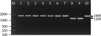
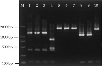
Using mtDNA COII-LSUrRNA primers (C2F3 and MRH106), an amplification product was obtained from purified DNA fragments of two different sizes from the same samples used for isozyme identification. Meloidogyne incognita, M. javanica and M. arenaria (EST phenotype A1) had a PCR product of approximately 1800 bp, while M. arenaria (EST phenotype A2) produced smaller products of approximately 1200 bp (Fig. 3). When the mtDNA COII-LSUrRNA PCR products were digested using restriction enzyme HinfI, different restriction profiles on the 1800 bp fragment of M. incognita, M. javanica and M. arenaria (EST phenotype A1) were observed. Meloidogyne incognita produced three fragments of approximately 1300, 400 and 100 bp, except one specimen that had an additional restriction site in the 1300 bp fragment producing four fragments of approximately 900, 420, 380 and 100 bp (Fig. 4, lane 4). Two fragments were generated in M. arenaria (EST phenotype A1) of 1700 and 100 bp, while M. arenaria (EST phenotype A2) 1200 bp PCR products was cleaved into two fragments of 1100 and 100 bp. The 1800 bp PCR product of M. javanica had no restriction sites with HinfI (Fig. 4).
PCR products amplified using mtDNA COII-LSUrRNA primers from representative specimens of Meloidogyne incognita, M. javanica and M. arenaria. Lane 1: M. incognita (EST phenotype I1); 2: M. incognita (EST phenotype I1); 3: M. incognita (EST phenotype 12); 4: M. arenaria (EST phenotype A2); 5: M. arenaria (EST phenotype A1); 6: M. javanica (EST phenotype J3); 7: M. javanica (EST phenotype J3); 8: M. arenaria (EST phenotype A2); 9: M. arenaria (EST phenotype A2); 10: M. javanica (EST phenotype J3); M: 2000 bp ladder.

HinfI restriction patterns of the PCR products amplified using mtDNA COII primers from the representative specimens of Meloidogyne incognita, M. arenaria and M. javanica. Lane 1: M. incognita (EST phenotype Mwea); 2: M. incognita (EST phenotype I1); 3: M. incognita (EST phenotype I2); 4: M. incognita (EST phenotype I1); 5: undigested PCR product (M. incognita EST phenotype I2); 6: M. javanica (EST phenotype J3); 7: M. javanica (EST phenotype J3); 8: M. arenaria (EST phenotype A2); 9: M. arenaria (EST phenotype A2); 10: M. arenaria (EST phenotype A1); M: 2000 bp ladder.

Discussion
RKN were found infesting tomatoes in all the three districts with a high prevalence. The occurrence of M. incognita, M. javanica and M. arenaria from 900 samples collected clearly demonstrated the widespread distribution of these species. The high prevalence of these nematodes shows the significance of RKN as a threat to tomato production. These three species were associated with yield loss of tomato ranging from 24 to 38% in Brazil (Sikora and Fernandez, Reference Sikora, Fernandez, Luc, Sikora and Bridge2005). Meloidogyne incognita was the most common and widely spread species in all tomato production areas compared with M. javanica and M. arenaria, which showed that this species is significant and a damaging tomato pest. Meloidogyne incognita is reported to be widely distributed in the vegetable-growing regions of Pakistan (Anwar et al., Reference Anwar, Zia, Hussain and Kamran2007; Shahid et al., Reference Shahid, Rehman, Khan and Mahmood2007). Soil type is a primary factor that may influence the damage potential of Meloidogyne spp (Siddiqui and Mahmood, Reference Siddiqui and Mahmood1998; Desaeger and Rao, Reference Desaeger and Rao2000). In Athi River, sandy-loamy soils are mostly found while in Gikambura and Mwea, black cotton soils are predominant (Thomas, Reference Thomas1997). The sandy-loamy soils in Athi River correlate with high magnitude of damage by nematodes due to easier movement and reproduction rate of nematodes.
Morphological characters have been the basis for nematode identification for many years in East Africa (Nono-Womdim et al., Reference Nono-Womdim, Swai, Mrosso, Chadha and Opeña2002). Species identification based on differences in morphological characters requires a lot of skill and is often inconclusive at species level (Hartman and Sasser, Reference Hartman, Sasser, Barker, Carter and Sasser1985). Isozyme analysis used in the current study was a relatively fast and accurate method to identify species of the genus Meloidogyne as earlier reported by Esbenshade and Triantaphyllou (Reference Esbenshade and Triantaphyllou1990). Five EST phenotypes detected in this study were species-specific, which is in agreement with a previous study (Karssen, Reference Karssen2002). These were recognized on the basis of a single band or a combination of several bands. As a result of such associations, these phenotypes were designated a letter suggesting a particular nematode species, followed by a number indicating the number of major bands of enzymatic activity. The following phenotype nematode species associations were recognized: I1 and I2 for M. incognita, A1 and A2 for M. arenaria and the phenotype J3 for M. javanica (Ibrahim et al., Reference Ibrahim, Perry and Webb1995). Differences in activities of the isozymes were indicated by the colour intensity of the bands of the different alleles from the same locus. The three species (M. javanica, M. incognita and M. arenaria) found in the three districts had one MDH phenotype designated as N1 that reconfirms their identity. MDH was found to be polymorphic with M. hapla species (Ibrahim et al., Reference Ibrahim, Perry and Webb1995); however, this species was not found in the current study. A limitation of isozyme analysis is that it can only be done on fresh, healthy, young females (Esbenshade and Triantaphyllou, Reference Esbenshade and Triantaphyllou1985).
Genomic DNA-based techniques do not rely on the expressed products of the genome and are independent of environmental influence or developmental stage of an organism. In our analysis, all isolates of M. incognita, M. javanica, M. arenaria (EST phenotype A1) produced 1800 bp fragment, which is different from the isolates of M. arenaria (EST phenotype A2) that produced a 1200 bp fragment. Hence M. arenaria (EST phenotype A2) could be distinguished on the basis of the size of the PCR amplicon alone. The PCR-RFLP results of the current study agree with those of Powers and Harris (Reference Powers and Harris1993), which indicate different size classes of amplification products in reactions with five Meloidogyne species using mitochondrial DNA. The different restriction profiles of RKN isolates studied corroborate earlier studies (Castagnone-Sereno et al., Reference Castagnone-Sereno, Vanlerberghe-Masutti and Leroy1994; Blok et al., Reference Blok, Wishart, Fargette, Berthier and Phillips2002) except for the variant M. incognita (Fig. 4, lane 4), which showed an additional HinfI restriction site on the amplification product. The size variation in this M. incognita is as a result of intraspecific variation in the cytochrome oxidase subunit II region for this species. The absence of the HinfI restriction site for M. javanica in this study is a result similar to what was reported for the same species by Hugall et al. (Reference Hugall, Moritz, Stanton and Wolstenholme1994).
The J2 stage of plant-parasitic RKN has been difficult to identify because of their small size and lack of distinguishing morphological features. Identification of this stage is important, since they include the infective and overwintering stage of RKN species. Whereas the isozymes used were successful in identifying the enlarged female stages, they lack the sensitivity to detect the relatively smaller vermiform and this was resolved by the use of PCR-RFLP. This study confirms the identity of the prominent and prevalent species of RKN in three tomato-growing districts in Kenya.
Acknowledgements
The present work was funded by the Gatsby Charitable Trust, and Rael Birithia received a training fellowship from the World Federation of Scientists. We thank Dr Bonaventure Aman for valuable comments on an earlier version of the manuscript.