Implications
Goat milk has a high content of short and medium-chain fatty acids, they are mainly produced by fatty acid synthase (FASN) in mammary gland. In this study, inhibition of FASN suppresses the expression of acetyl-CoA carboxylase α (ACCα), lipoprotein lipase and heart-type fatty acid binding protein (H-FABP). Furthermore, the reduction of FASN expression level decreases the abundance of medium-chain fatty acids (C10:0 and C12:0). Taken together, our study provides a direct evidence for inhibition of FASN reduces cellular medium-chain fatty acids synthesis in goat mammary gland epithelial cells.
Introduction
Goat milk is one of the important nutrient-rich foods produced in the world. High contents of short and medium-chain fatty acids endow goat milk a high nutritional value (Haenlein, Reference Haenlein2004), which also prevents some human metabolic illness (Williams, Reference Williams2000; Haenlein, Reference Haenlein2004). Most of the short and medium-chain fatty acids in milk are produced via de novo fatty acid synthesis catalyzed by fatty acid synthase (FASN) in ruminant mammary gland.
FASN is the key enzyme of milk lipogenesis and secretion (Smith, Reference Smith1994). It participates in the metabolic regulation of cell life and activities, such as energy storage, formation of biological membrane structures, cell signaling transduction and protein acetylation (Olsen, Reference Olsen2011). Comparison of FASN sequences of caprine (DQ 223929), ovie (XM 004013447), bos (NM 001012669), rattus (NM 017332), homo (NM 004104), and gallus (NM 205155) have demonstrated that the FASN gene is comprised of 43 exons with the exception of the bovine sequence where exon 42 and 43 are not separated by an intron (Roy et al., Reference Roy, Taourit, Zaragoza, Eggen and Rodellar2005).
Fatty acid synthesis differs considerably between mammary gland of mono-gastric and ruminant species (Roy et al., Reference Roy, Taourit, Zaragoza, Eggen and Rodellar2005). The predominant product of FASN is a 16-carbon long-chain palmitate in non-ruminants. Palmitate is a significant material for triglyceride synthesis and fatty acid elongation, which is believed to be the main source of mono-unsaturated fatty acids in rat insulinoma cell line (Green et al., Reference Green, Ozguden-Akkoc, Wang, Jump and Olson2010). However, FASN can also produce smaller amounts of myristate, laureate, and even shorter-chain fatty acids in lactation mammary gland (Hillgartner et al., Reference Hillgartner, Salati and Goodridge1995) and rat liver (Hansen et al., Reference Hansen, Carey and Dils1970). Although FASN in lactating non-ruminant mammary glands and waterfowl uropygial glands produce medium-chain fatty acids (C8, C10, C12) with the help of thioesteraseII (TEII) (Grunnet and Knudsen, Reference Grunnet and Knudsen1978), an independent enzyme which is not a part of the FASN complex and only be detected in the mammary glands of non-ruminants as a tissue-specific protein (Thompson and Smith, Reference Thompson and Smith1985). Purified FASN proteins, without the presence of TEII, from rabbit, bovine, rat and guinea pigs produce only long-chain and short-chain but not medium-chain fatty acids (Carey et al., Reference Carey, Dils and Hanse1970). In contrast, FASN from goat mammary gland could release all three types of fatty acids (long-chain, medium-chain and short-chain fatty acids) (Grunnet and Knudsen, Reference Grunnet and Knudsen1978). Therefore, it has been proposed that, there are different mechanisms of fatty acid synthesis at work between non-ruminants and ruminants. The expressed and purified goat MAT protein from Escherichia coli showed medium-chain thioesterase activity in ruminants (Rangan and Smith, Reference Rangan and Smith1996). Moreover, some studies also showed that short-chain acyl-CoA and medium-chain acyl-CoA shared the same enzyme for fatty acids release in ruminants (Mikkelsen et al., Reference Mikkelsen, Højrup, Hansen, Hansen and Knudsen1985), suggesting that MAT may be the dominant regulatory factor for short and medium-chain fatty acids synthesis in ruminants. Therefore, we hypothesize that FASN may play a crucial role in regulating the specific fatty acids composition synthesis of ruminants.
In order to complete the sequence and expression information of Xinong Saanen dairy goat, and reveal the effect of FASN expression on the cellular fatty acid compositions, we cloned the FASN gene and characterized its expression in fat tissue, small intestine and mammary gland of Xinong Saanen dairy goats. We provided an evidence that inhibition of FASN induce significant suppression of the crucial genes in the process of fatty acid metabolism, including acetyl-CoA carboxylase α (ACCα), lipoprotein lipase (LPL) and heart-type fatty acid binding protein (H-FABP), by orlistat treatment and shRNA mediated silence of FASN. Furthermore, reduction of FASN expression level changed the composition of fatty acid in goat mammary gland epithelial cells (GMEC). Together, our study provides direct evidence for inhibition of FASN reduces cellular medium-chain fatty acids synthesis in GMEC.
Material and methods
Animals and samples collection
The Animal Care and Use Committee of the Northwest A&F University approved all procedures and experiments. Three healthy, lactating (60 days) Xinong Saanen dairy goats with similar physiological condition (4 years old in the 3rd parity) were raised in the experimental farm of Northwest A&F University, Shaanxi, China. After slaughtering, tissue samples (subcutaneous fat, small intestine, mammary gland, lungs, rumen, muscle, spleen, liver, kidney and heart) were immediately collected. Besides, mammary gland tissue samples from three healthy, 3-year-old goats of similar weight were surgically collected at mid-lactation and dry-off period. All samples were rinsed with DEPC (Sigma, St. Louis, USA) sterile water then immediately frozen in liquid nitrogen.
Total RNA extraction and real-time PCR
Total RNA were extracted from tissues (listed above) with Trizol reagent (Invitrogen, New York, USA). First-strand cDNA of various tissues were synthesized from 0.1 to 5 µg of purified total RNA, using the PrimeScriptTM RT kit (for perfect real-time) (Takara, Osaka, Japan) according to the manufacturer’s instructions. SYBR® Premix Ex Taq™ II (Takara) was used for measuring the relative expression level of FASN in various tissues by real-time quantitative PCR (RT-qPCR) fluorescent technology according to the manufacturer’s manual on a CFX96 (BioRad, Hercules, USA). Glyceraldehyde-3-phosphate dehydrogenase (GAPDH) was used as an endogenous reference for normalization of targeted mRNA profiles and three technical replicates were analyzed per sample (Lin et al., Reference Lin, Luo, Zhang, Wang, Shi and Zhu2013; Shi et al., Reference Shi, Luo, Zhu, Li, Sun, Lin, Zhang, Yao and Shi2013).
Full-length cDNA cloning of FASN from the mammary glands
The primers for FASN cloning, as described in Supplementary Table S1, were designed based on the multiple alignment of the conserved sequence in the GenBank database, capra (DQ 223929), bovine (Bos taurus, NM 001012669), human (Homo sapiens, NM 004104), mouse (Mus musculus, NM 007988), rattus (Rattus norvegicus, NM 017332), pig (Sus scrofa, NM 001099930), chicken (Gallus, NM 205155) and dog (Canis lupus, XM 540497.3). Briefly, annealing temperatures for open reading frame (ORF) cloning were shown in Supplementary Table S1. The cDNA cloning of the 5′- and 3′-UTR were performed according to the manufacture’s protocol of 5' RACE system Ver.2.0 kit (Invitrogen) and 3′-full RACE core set Ver.2.0 kit (Clontech, Mountain View, USA), respectively. All the PCR fragments were cloned into pMD® 19-T (Takara) vectors and sequenced by Invitrogen.
Sequence analysis
The core fragment, 3′ end and 5′ end sequences were assembled using the SeqMan II software in the DNAStar Package to obtain full-length cDNA of FASN. The cross-species similarities of both nucleotide and putative amino acid sequences were then calculated for homologyon ClustalW (http://www.ebi.ac.uk/Tools/clustalw2/index.html). Exon information was predicted by the NCBI online tool (http://www.ncbi.nlm.nih.gov/sutils/splign/splign.cgi?textpage=online&level=form). Putative translation of the goat FASN protein potential functional domains were predicted using a server of online accessible bioinformatics tools (http://myhits.isb-sib.ch/cgi-bin/motif_scan) including Protparam (http://www.expasy.org/tools/protparam.html) for protein chylomicron (CM) and isoelectric point (PI) value, ProtScale (http://us.expasy.org/cgi-bin/protscale.pl) for hydrophobicity structure prediction TMHMM (http://www.cbs.dtu.dk/services/TMHMM/) for trans-membrane structure prediction, SignalP (http://www.cbs.dtu.dk/services/SignalP/) for signal peptide sequence prediction, conserved domain database search (http://www.ncbi.nlm.nih.gov/Structure/cdd/cdd.shtml) for conserved domains and CPH models, 3.0 Server (http://www.cbs.dtu.dk/services/CPHmodels/) for tertiary structure prediction (Nomura et al., Reference Nomura, Long, Niessen, Hoover, Ng and Cravatt2010). The phylogenetic tree was computed using the ClustalW and MEGA 5.05 programs (http://www.megasoftware.net) with standard parameters.
Cell culture and treatments
A total of 293A cells were cultured in Dulbecco’s modified Eagle medium (DMEM) (Hyclone, Massachusetts, USA) compensated with 10% FBS (Hyclone). GMEC were cultured following our previously described protocol (Wang et al., Reference Wang, Luo, Zhong, Lin, Shi, Zhu, Li, Sun and Zhao2012; Shi et al., Reference Shi, Luo, Zhu, Li, Sun, Lin, Zhang, Yao and Shi2013). For the orlistat (Roche, Basilea, USA) treatment experiments, GMEC were seeded in 12-well cell culture clusters (NUNC, Massachusetts, Denmark) for 24 h (about growing to 90% confluence) before treated with different concentration of orlistat (0, 5, 15, 25, 35 μM) for 48 h in DMEM/F-12 complete medium. Before experiments, cells were washed twice with phosphate buffered saline (PBS) and once with serum-free DMEM/F-12 without antibiotics. All analyses were conducted on three independent GMEC culture preparations.
Cellular fatty acid synthesis activity assay
Cellular FASN activity was assessed as described previously, with some modifications (Chuang et al., Reference Chuang, Chang and Hwang2011). In brief, cells in 6-well plate were washed with cold PBS and then collected. Ultrasonic disruption was achieved in an assay buffer with 0.6 mM phenylmethyl sulfonylfluoride (PMSF) (Pierce, Massachusetts, USA) on ice and then centrifuged at 18 000×g for 30 min at 4°C. The assay buffer contained a 100 mM potassium phosphate buffer, 1 mM ethylenediaminetetraacetic acid (Sigma), and 1 mM dithiolthreitol (Sigma) at pH 7. The supernatant was used to evaluate the FASN activity at 340 nm (before/after malonyl-CoA). The assay solution without malonyl-CoA served as a background for the assay. Each sample was assayed in triplicate.
Adenovirus generation and RNA interference
shRNA sequences were designed and synthesized from Invitrogen, according to the sequences reported previously (sense: 5′-GATCCGAGACGCCATGCTGGATAAGAGTACTGT TATCCAGCATGGCGTCTCTTTTTTC-3′, antisense: 5′-TCGAGAAAAAAGAGACGCCATG CTGGATAACAGTACTCTTATCCAGCATGGCGTCTCG-3′), with the negative shRNAs as control (sense: 5′-GATCCACTACCGTTGTTATAGGTGGAGTACTGCACCTATAACAACGG TAGTTTTTTTC-3′, antisense: 5′-TCGAGAAAAAAACTACCGTTGTTATAGGTGCAGTACT CCACCTATAACAACGGTAGTG-3′). The recombinant adenovirus containing the screened shRNA sequences were constructed using BLOCK-iTTM system according to manufacturer’s protocol using 293A cells (Lin et al., Reference Lin, Luo, Zhang, Wang, Shi and Zhu2013). GMEC were seeded in 12-well cell culture clusters, and grown to 70% to 80% confluence. Before experiments, cells were washed twice with PBS and once with serum-free DMEM/F-12 without antibiotics. This was followed by 72 h incubation in DMEM/F-12 medium, containing the adenovirus constructed previously. Three independent experiments were conducted for all analyses.
Western blot
The protein samples were extracted with RIPA (Solarbio, Beijing, China) supplemented with PMSF. Western blot was assessed following the protocol described previously (Shi and Shen, Reference Shi and Shen2010). Polyclonal rabbit anti-FASN (Bioss, bs-1498 R, USA, 1 : 500) and monoclonal mouse anti-GAPDH (CWBIO, CW0266, China, 1 : 1000) were used as the primary antibodies. Polyclonal goat anti-mouse IgG coupled to HRP (TIANGEN, AB101, China, 1 : 1000) was used as secondary antibody. Signals were detected using the chemiluminescent ECL Western blot detection system (Pierce).
Fatty acid analysis
After 72 h incubation with virus containing shRNA in 60 mm culture plates, cells were washed twice with PBS, followed by 5 min of shaking and 5 min of centrifugation at 1200×g. Total fatty acids were extracted from approximately 100 mg cells, using 2 ml of vitriol/methanol (2.5 : 1, v/v) in a clean tube according to the method described by Folch et al. (Reference Folch, Lees and Sloane Stanley1957). Methylated lipid samples were analyzed using a gas chromatography-mass spectrometry device (GC-MS) (Agilent, Santa Clara, USA) equipped with an HP-5 column (60 m×0.25 mm. i.d.×0.25 µm df) according to published protocol with some modifications (Wang et al., Reference Wang, Luo, Zhong, Lin, Shi, Zhu, Li, Sun and Zhao2012). 2 μl sample, with a 1 : 10 split ratio, and 0.8 mL/min He gas was provided for 2 min at 40°C, and the temperature was then increased to 240°C for 15 min at a rate of 8°C/min. For analysis, the temperature of injector and detector was 250°C and 280°C, respectively, and the ionization energy was 70 eV with 35 to 500 amu mass number.
Statistical analysis
SPSS 19.0 was used for statistical calculations. The RT-qPCR data was analyzed using transcript C t values normalized with GAPDH as the endogenous control gene (ΔC t). Group data for multiple comparisons were analyzed by ANOVA using a general linear model procedure followed by Tukey’s test. Significance was established at a P<0.05.
Results
Characterization of the full-length FASN cDNA from goat mammary gland
To study the function of FASN, we cloned FASN mRNA sequence from a Xinong Saanen dairy goat mammary gland (GenBank ID: DQ915966). The full-length of goat FASN is 8271 bp and contained 42 exons, including 88 bp of 5′UTR, 7545 bp of ORF and 584 bp of 3′UTR and encodes a 3514-amino acids polypeptide. Sequence analysis revealed that the start codon (ATG) is located in the 2nd exon, while the stop codon is in the 42nd exon (Supplementary Figure S1). In addition, the 3′UTR contains one AATAAA motifs that represents putative poly-adenylation signals. We subsequently compared the ORF to the sequences of goat, bovine, mice, rat, pig and human using BLAST and found that goat shares the highest similarity with bovine, with an extra amino acid in the MAT domain of bovine FASN (data is not shown).
The theoretical CM of FASN was estimated to be 273.8 kDa and the PI is 5.99 with a 45.05 instability index, predicted by the Protparam program. The signal peptide prediction performed by SignalP 4.0 revealed that this polypeptide contains no signal peptide in its amino acids region and is a non-secretory protein. Hydrophobicity analysis using the Protscale program showed no significant hydropathicity or hydrophobicity. Prediction of transmembrane domain made by the TMHMM program indicated that the protein is not a potential membrane protein.
The conserved domain database alignment illustrated that the FASN protein containes seven activity sites, which are located in 7 domains respectively. These are Cys161 of KS domain (Met1…Pro414), Ser580 of MAT domain (Arg429…Pro816), His877...Gly881...Pro886 of DH domain, Gly1679 XGly1681XXGly1684 of ER domain (Asp1638…Ala1866), Gly1895XGly1897XXGly1900 of KR domain (Val1867…Ser2120), S2159 of ACP domain and (S2311... DH2484) of TE domain (Leu2210…Glu2514), respectively.
To analyze the phylogenetic relationship between goat FASN and that of other species, a neighbor-joining phylogenetic tree was constructed, based on sequences of nine representative animals. Tree topology revealed that the deduced amino acid sequence of the FASN from Xinong Saanen dairy goat is in a same clade with sheep (XM 004013447), which is followed by bovine, dog and pig, while it is most distant from that of chicken (NM 205155). The distances and dendogram showed the evolutionary relationship of various species and also validate the current classification of these proteins (Supplementary Figure S2).
Tissues expression analysis of FASN
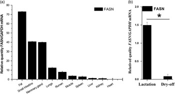
We found that FASN gene expresses in every tissue examined including fat, small intestine, mammary gland, lungs, rumen, muscle, spleen, liver, kidney and heart. However, fat, intestine and mammary gland have higher expression levels, followed by lungs, stomach, muscle, spleen, liver and kidney. Negligible level of expression was detected in heart (Figure 1a). Relative to the dry period, the expression level of FASN increased significantly (17.9-fold) (P<0.05) at lactation period (Figure 1b).
Fatty acid synthase (FASN) expression correlates with lactation. (a) The mRNA expression of FASN gene in various tissues from dairy goats. (b) The mRNA expression of FASN gene in mid-lactation and dry-off period in mammary glands of dairy goats. Relative quantities of mRNA were determined in each sample using the ΔC t method of RT-qPCR data analysis with GAPDH as an endogenous control. The data of each experimental group represents the mean±s.d. of three independent experiments where RT-qPCR samples were measured in triplicates.

Inhibition of FASN suppresses the expression of genes related to fatty acid metabolism in GMEC
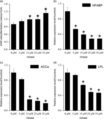
To investigate the effect of FASN inhibition on the expression of key genes involved in fatty acid metabolism, we therefore used orlistat to inhibit FASN activity. We found that there was a significant suppression of FASN activity by orlistat at concentrations higher than 15 μM (P<0.05) (Figure 2a), in accordance with previously published data (Chuang et al., Reference Chuang, Chang and Hwang2011). Additionally, the effect appeared to be concentration dependent with the maximal concentration, 35 μM orlistat, having the greatest inhibition effect in GMEC (Figure 2a). To investigate the effect of FASN inhibition on regulating fatty acid metabolism, we assayed the expression of genes related to fatty acid metabolism by RT-qPCR. The expression level of LPL was also dose-dependently downregulated in orlistat-treated GMEC (Figure 2d). Orlistat treatment also inhibited the expression of ACCα substantially in GMEC (Figure 2c). In addition, H-FABP, a crucial carrier protein for fatty acids transportation, was also significantly downregulated by FASN activity suppression (Figure 2b).
Inhibition of fatty acid synthase (FASN) by orlistat suppresses the expression of genes related to fatty acid metabolism in goat mammary gland epithelial cells (GMEC). (a) Concentration dependent inhibition of orlistat on cell fatty acid synthesis activity. GMEC were treated with different concentration orlistat (0, 5, 15, 25, 35 μM) for 48 h for malonyl-CoA detection at A340. (b, c, d) Effect of different concentrations orlistat (0, 5, 15, 25, 35 μM) on mRNA of HFABP, ACCα and LPL, respectively. The data represents the mean±s.d. of three independent experiments. *P<0.05 v. the control.

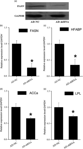
Western blotting revealed that the protein level of FASN was reduced after treatment with shRNA targeting FASN sequence (MOI=200) (Figure 3a). RT-qPCR showed that shRNA mediated gene knockdown decreased FASN expression significantly, while the control shRNA had no significant influence on FASN mRNA (Figure 3b). Additionally, similar to the orlistat treatment in GMEC, Silencing of FASN also caused significant decrease in the expression of H-FABP, ACCα and LPL, respectively (Figure 3c, d and e).
Silence of fatty acid synthase (FASN) inhibits the expression of genes related to fatty acid metabolism in goat mammary gland epithelial cells (GMEC). GMEC were treated with virus containing shRNA targeting FASN mRNA (AD-shRNA) and a negative control shRNA sequence (AD-NC) for 72 h, respectively. (a) Western blot detection of FASN protein with GAPDH as control. (b, c, d, e) RT-qPCR detection for the mRNA of FASN, HFABP, ACCα and LPL, respectively. The data represents the mean±s.d. of three independent experiments. *P<0.05 v. the control.

Effect of silence of FASN on cellular fatty acid composition
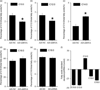
Absolute content of each fatty acid species was calculated for fatty acid synthesis activity analysis. The results showed that the total fatty acids content was downregulated significantly by the loss of FASN. Furthermore, we observed significant decrease in decanoic acid (C10:0), lauric acid (C12:0), palmitic acid (C16:0) and stearic acid (C18:0) absolute content, and an increase in myristic acid (C14:0) (Figure 4f), which collectively suggests that fatty acid synthesis activity was decreased. In addition, the analysis of relative content indicated that there was a significant increase in C14:0 fatty acid fraction (Figure 4c) with a substantial decrease in C10:0 (Figure 4a) and C12:0 (Figure 4b) fatty acids. However, no apparent changes were observed in C16:0 (Figure 4d) and C18:0 (Figure 4e) relative fatty acid content. Overall, the total relative content of medium-chain fatty acids (C10:0, C12:0, C14:0) were decreased substantially.
Silence of fatty acid synthase (FASN) regulates cellular fatty acids contents in goat mammary gland epithelial cells (GMEC). The data showed relative changes (a, b, c, d and e) and absolute changes (f) AD-NC and AD-shRNA represented the adenovirus vectors contained negative shRNA and shRNA target FASN mRNA, respectively. The data represents the mean±s.d. of three independent experiments. *P<0.05 v. the control (AD-NC).

Discussion
Sequence and expression analysis of FASN gene
The FASN sequence of various species (caprine, ovie, rattus, homo, anser, gallus, bos) have been cloned (Kameda and Goodridge, Reference Kameda and Goodridge1991; Amy et al., Reference Amy, Williams-Ahlf, Naggert and Smith1992; Jayakumar et al., Reference Jayakumar, Tai, Huang, al-Feel, Hsu, Abu-Elheiga, Chirala and Wakil1995), but little is known about goat FASN. In this study, we cloned the full-length cDNA of goat FASN gene sequence. The result of homology alignment revealed that there is an extra amino acid found in the goat MAT domain of FASN, compared with that of bovine. Although ovie FASN protein contains 250 amino acids less than capra FASN protein (contained 2514 amino acids and 2264 amino acids, respectively), capra FASN shares the most similarity (97.08%) with ovie FASN, much higher than the similarity between capra and bos (contained 2514 amino acids and 2513 amino acids, respectively). Cpara also shares a same clade with ovie. These results had shown that there is a closest evolutionary relationship between capra and ovie. We compared the conversed sites among various species mentioned above, and found that all the activity sites described in results section are highly conversed, except there is a Gly1900 deletion in ovie FASN protein. Whether these differences of the sequences contribute to the functional discrepancy is still unknown.
Previous studies showed that FASN expressed higher levels in the brain, testis and adipose tissues of bovine (Bionaz and Loor, Reference Bionaz and Loor2008). To investigate how FASN is expressed in goat, we performed a comparative gene expression study in various tissues of goat using RT-qPCR (different from Bernard et al., Reference Bernard, Leroux, Rouel, Bonnet and Chilliard2012). Similar to the studies in bovine (Roy et al., Reference Roy, Taourit, Zaragoza, Eggen and Rodellar2005), we found that FASN is expressed at higher levels in energy-intensive tissues in the goat (e.g. adipose tissue, small intestines and the mammary gland). A higher FASN expression could provide more energy for maintaining the normal metabolism during lactation, including mammary cell proliferation and the development of the mammary gland. This may explain the higher expression level in adipose tissue, mammary gland and small intestines. Additionally, all the species mentioned above share the same activity sites. In addition, we also found higher expression level of FASN at mid-lactation. All these data showed that FASN may play an important role in lactation period.
Inhibition of FASN suppresses the expression of genes related to fatty acid metabolism
Fatty acid metabolism in mammary gland is controlled by a complex gene network. The fatty acid synthesis pathway involves two crucial enzymatic reactions. First, acetyl-CoA is carboxylated into malonyl-CoA catalyzed by ACCα in the cytosol, a key regulatory step in the fatty acid synthesis pathway. Acetyl-CoA and malonyl-CoA are then sequentially used by FASN, the subsequent enzyme in the pathway, as a donor of acetate moieties to the growing acyl chain, resulting in the synthesis of fatty acids (Chajès et al., Reference Chajès, Cambot, Moreau, Lenoir and Joulin2006). Depletion of FASN in both human MCF-7 and MDA-MB-231 cells caused a reduction in the levels of ACCα (Chajès et al., Reference Chajès, Cambot, Moreau, Lenoir and Joulin2006). The synthesized fatty acids then bind with H-FABP and are transferred into endoplasmic reticulum for triglyceride synthesis (Chajès et al., Reference Chajès, Cambot, Moreau, Lenoir and Joulin2006). As a natural inhibitor of FASN, orlistat was reported to have a significant inhibition effect on FASN activity by curbing the binding of TE domain between FASN subunits (Chuang et al., Reference Chuang, Chang and Hwang2011). FASN inhibition by orlistat induces multiple adaptive changes of the genes in fatty acid synthetic pathway and associated metabolic pathways (Chuang et al., Reference Chuang, Chang and Hwang2011; Sankaranarayanapillai et al., Reference Sankaranarayanapillai, Zhang, Baggerly and Gelovani2012), including a significant up-regulation of ACCα in human non-small cell lung carcinoma cells (Sankaranarayanapillai et al., Reference Sankaranarayanapillai, Zhang, Baggerly and Gelovani2012). However, depletion of FASN by siRNA in both human MCF-7 and MDA-MB-231 cells were found to reduce the levels of ACCα (Chajès et al., Reference Chajès, Cambot, Moreau, Lenoir and Joulin2006). Although the different cell types may contribute to the difference, the reason for this discrepancy is still unknown. Orlistat is also believed as a drug for obesity therapy partly because of its thioesterase inhibition activity even in normal cells. Therefore, it may provide us a tool to study the effect of FASN inhibition on the expression of genes related to fatty acid and fatty acid composition in GMEC. In fact, in our study, orlistat suppressed the fatty acid synthesis activity significantly in GMEC when the concentration was over 15 μM. In addition, it has been previously described that other esterases may also be responsive to orlistat (Olsen, Reference Olsen2011). To verify the results of orlistat treatment, recombinant adenovirus vectors containing the shRNA sequences targeting FASN were constructed to determine the regulatory function of FASN on expression of the genes related to fatty acid metabolism. Consistent with Chajès et al., we found a significant suppression effect on ACCα expression by FASN inhibition in GMEC, indicating that the responses may be similar among different types of mammary gland cells (GMEC, MCF-7 and MDA-MB-231) and the difference may be due to differences between cell types.
Previously, it has been shown that inhibition of FASN activity suppresses the expression of peroxisome proliferator-activated receptor γ (PPARγ) in mice 3T3 cells (Schmid et al., Reference Schmid, Rippmann, Tadayyon and Hamilton2005). Decreased PPARγ suppresses the expression of ACCα in human THP-1 cells (Chawla et al., Reference Chawla, Boisvert, Lee, Laffitte, Barak, Joseph, Liao, Nagy, Edwards, Curtiss, Evans and Tontonoz2001). Our previous study has shown a significant inhibition on ACCα expression by PPARγ knockdown (Shi et al., Reference Shi, Luo, Zhu, Li, Sun, Lin, Zhang, Yao and Shi2013). It is generally believed that SREPB1 plays a key role in regulation of mammary lipogenesis including mice (Rudolph et al., Reference Rudolph, McManaman, Phang, Russell, Kominsky, Serkova, Stein, Anderson and Neville2007), sheep (Hussein et al., Reference Hussein, Harvatine, Weerasinghe, Sinclair and Bauman2013) and dairy cows (Harvatine et al., Reference Harvatine, Boisclair and Bauman2009). In general, the expression of SREBP1 is decreased to even a greater extent than the expression of several lipogenic genes, including the ones measured above, along with those associated with triglyceride assembly and desaturation (Harvatine et al., Reference Harvatine, Boisclair and Bauman2009). Thus, it is possible that inhibition of FASN might suppress ACCα, other related genes as well, expression through decreasing the expression of PPARγ and SREBP1. This is also consistent with the results of Bernard et al. (Reference Bernard, Leroux, Rouel, Bonnet and Chilliard2012), in which FASN and ACCα was demonstrated to be a positive expression associations in goats. In addition, excess malonyl-CoA, which can be induced by inhibition of FASN, is predicted to be the inhibitor of fatty acid synthesis (McGarry et al., Reference McGarry, Mannaerts and Foster1977). Thus, it is possible that a feedback effect of malonyl-CoA accumulation may be involved in this process. However, the mechanism still needs more study in the future.
Decreased expression of ACCα and FASN, accompanied by a decreased de novo fatty acid synthesis activity, also induced the downregulation of H-FABP in this study. H-FABP is involved in lipid metabolism by intracellular transport of long-chain fatty acids that bind to fatty acyl-CoA and acyl-l-carnitines (Cho et al., Reference Cho, Kim, Jeon and Chung2011), and it is also involved in palmitate-induced single pathway (Kusudo et al., Reference Kusudo, Kontani, Kataoka, Ando, Shimokata and Yamashita2011). Additionally, PPARγ can also enhance the expression of H-FABP (Chawla et al., Reference Chawla, Boisvert, Lee, Laffitte, Barak, Joseph, Liao, Nagy, Edwards, Curtiss, Evans and Tontonoz2001), ACCα and FASN (Shi et al., Reference Shi, Luo, Zhu, Li, Sun, Lin, Zhang, Yao and Shi2013). Thus, PPARγ may also mediate the decreased expression of H-FABP in GMEC. Therefore, it is possible that both of decreased fatty acid synthesis (especially for the decreased palmitate synthesis) and PPARγ pathway contribute to the downregulation of H-FABP induced by FASN inhibition.
LPL plays a key role in lipoprotein metabolism by catalyzing the hydrolysis of triglycerides of very low density lipoproteins (VLDL) and CMs, thereby releasing free fatty acids (Yang et al., Reference Yang, Nevin, Peng, Brunzell and Deeb1995), which provides another source of cellular fatty acids. Similar to the result of Caenorhabditis elegans (Olsen, Reference Olsen2011), we found a significant downregulation of LPL expression when FASN activity was inhibited. However, the mechanism underlying this regulation is still unclear. It is possible that decreased FASN expression induces a reduction in triglyceride synthesis, which may result in suppressed LPL expression.
Downregulation of FASN alters the fatty acid composition profile in GMEC
To determine whether FASN contribute to the regulation of cellular short and medium-chain fatty acid synthesis in GMEC, we performed cell fatty acid composition analysis after the loss of FASN. Knudsen and Grunnet (1980) found that FASN possessed a transferase activity for both butyryl-CoA and C12 fatty acids in ruminants (Mikkelsen et al., Reference Mikkelsen, Højrup, Hansen, Hansen and Knudsen1985). Competitive binding experiments with malonyl-CoA and [1–14C] acetyl-CoA, [1–14C] butyryl-CoA or [1–14C] decanoyl-CoA indicate that all these substrates are transferred to the synthesis loop of lactating-goat mammary gland fatty acid by the same transferase (Mikkelsen et al., Reference Mikkelsen, Højrup, Hansen, Hansen and Knudsen1985). Isolation and determination of the amino acid sequence of [1–14C] decanoyl-labeled CNBr-cleavage peptide from the decanoyl transferase site showed that this transferase shares the same transferase with acetyl/malonyl transferase (Mikkelsen et al., Reference Mikkelsen, Højrup, Hansen, Hansen and Knudsen1985). Hansen’s research about the effect of substrates on the chain-length of fatty acids synthesized by rat liver fatty acid synthase also resulted in a similar conclusion (Hansen et al., Reference Hansen, Carey and Dils1970). In order to identify the MAT’s role as a shorter-chain fatty acids transferase, Rangan and Smith (Reference Rangan and Smith1996) overexpressed and purified recombinant goat MAT protein in E. coli. Their results suggested that MAT possessed not only a transferase activity but also a thioesterase activity. It is proposed that MAT may be an important regulatory factor for fatty acid composition. In our study, decreased MAT (due to reduced FASN expression), reduced the relative amount of medium-chain fatty acids (C10:0 and C12:0). This is also consistent with the results of Grunnet et al. (Rangan and Smith, Reference Rangan and Smith1996). An alternative hypothesis is that inhibited FASN increased malonyl-CoA content which was reported to inhibit the increasing of C10:0 fatty acid content induced by extra albumin (Green et al., Reference Green, Ozguden-Akkoc, Wang, Jump and Olson2010). Although decreased ACCα may partly alleviate the increasing trend of malonyl-CoA, it is just through as a feedback regulation to alleviate the effect of FASN reducing. For LPL, it was already reported to play an important role in hydrolyzing VLDL and CM (Yang et al., Reference Yang, Nevin, Peng, Brunzell and Deeb1995) to release free fatty acids especially for C10:0 in goats (Kim Ha and Lindsay, Reference Ha and Lindsay1993). Thus, it seems that both of ACCα and LPL are involved in C10:0 fatty acid content regulation. In this study, silencing of FASN led to significant decrease in the expression of ACCα and LPL. Either MAT or ACCα and LPL may provide alternative mechanism for decreased FASN expression reducing cellular fatty acid composition.
Additionally, our data suggests that inhibition of FASN increased the relative content of C14:0. Although, downregulation of FASN could increase the content of malnoyl-CoA that was reported to be able to decrease the content of C10:0 (Green et al., Reference Green, Ozguden-Akkoc, Wang, Jump and Olson2010), the mechanism of how malnoyl-CoA affected the content of C10:0 is still unknown. It is possible that increased malnoyl-CoA enhanced the elongation of C10:0 to C14:0, and suppressed oxidation of C14:0 (Foster, Reference Foster2012). This may also be an explanation for the increase of C14:0. A deep mechanism for the issue still needs more research.
Acknowledgments
This research was jointly supported by the ‘National Natural Science Foundation of China (31072013)’, the ‘Transgenic New Species Breeding Program of China (2014ZX08009-051B)’, and ‘Special Fund for Agro-scientific Research in the Public Interest (201103038)’.