Introduction
A cholesteatoma is a non-neoplastic but destructive lesion containing layers of keratin in a cavity lined by squamous epithelium and subepithelial connective tissue. Cholesteatoma occurs mainly in the middle-ear cleft but has also been reported in other areas of the skull, intracranially, and in the external auditory canal (where it has been confused with keratosis obturans).Reference Olszewska, Wagner, Bernal-Sprekelsen, Ebmeyer, Dazert and Hildmann1–Reference Persaud, Hajioff, Thevasagayam, Wareing and Wright3 This review focuses on middle-ear cleft cholesteatoma, which can cause serious complications by eroding nearby structures or by precipitating infection, either within the temporal bone or the brain. Complex surgery is often required, and there is currently no effective medical therapy.
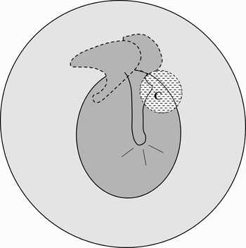
Cholesteatoma can occur in the middle ear with a normal tympanic membrane and external auditory canal. This is termed ‘congenital’ or ‘primary’ cholesteatoma, and is most commonly found in the anterosuperior mesotympanum in children (Figure 1). In contrast, acquired cholesteatoma is associated with a defect in the tympanic membrane, usually posterosuperiorly in adults. ‘Primary acquired’ cholesteatoma refers to a limited diverticulum of the pars flaccida, whilst ‘secondary acquired’ cholesteatoma appears with a posterosuperior tympanic membrane perforation. However, this approach to classification is difficult to apply clinically. Tos therefore proposed an alternative classification, based on otoscopic findings, which divided the disease into two main forms: attic cholesteatoma and cholesteatoma arising from retraction of the pars tensa.Reference Tos4 The latter is further divided into lesions of the posterosuperior quadrant of the tympanic membrane (marginal disease) and those of the central region (central disease). Mills and Padgham added a fourth category to the Tos classification in order to include squamous disease occurring behind an intact tympanic membrane.Reference Mills and Padgham5
Diagram of congenital cholesteatoma (C) in the anterosuperior region of the right middle ear, behind a virginal tympanic membrane.

The incidence of cholesteatoma is reported as 3 per 100 000 in children and 9.2 per 100 000 in adults, and is 1.4 times more common in males compared with females.Reference Olszewska, Wagner, Bernal-Sprekelsen, Ebmeyer, Dazert and Hildmann1 Congenital cholesteatoma was considered rare until the publication of large case series.Reference Levenson, Parisier, Chute, Wenig and Juarbe6–Reference Potsic, Korman, Samadi and Wetmore8 The estimated incidence of congenital cholesteatoma is 0.12 per 100 000,Reference Persaud, Hajioff, Thevasagayam, Wareing and Wright3 but the true incidence may be higher as the disease can perforate the tympanic membrane and be mislabelled as acquired cholesteatoma.
The aetiopathogenesis of cholesteatoma remains unclear. Both types of cholesteatoma are derived from keratinising stratified squamous epithelium, but the mechanisms by which this epidermal tissue reaches the middle ear and forms a lesion are still debated. Here, we review the most relevant evidence for the main aetiopathological theories of congenital and acquired cholesteatoma, in the light of recent studies. We hope to clarify some aspects of this long-standing debate.
Method
An Ovid Medline search was performed from inception to July 2006. The medical subject heading ‘cholesteatoma’ was used in combination with ‘aetiology’, ‘pathology’ or ‘pathophysiology’. We also screened the bibliographies of collected papers and our personal archives for references pertaining to the principal aetiopathogenic hypotheses of cholesteatoma. In addition, hand-searches were carried out following communication with four internationally recognised otopathologists. Only articles written in English were scrutinised.
Theories of congenital cholesteatoma aetiopathogenesis
The many aetiopathogenic theories for cholesteatoma formation behind a normal tympanic membrane have been the subject of intense discussion for over a century.
Migration theory
A topographical study of 33 cases of congenital cholesteatoma revealed that the majority of these lesions occurred near the tympanic isthmus of the middle ear (i.e. the junction between the first and second branchial arches).Reference Aimi9 This, coupled with a study of four clinical cases and histopathological examinations of three human temporal bones (from patients aged 15, 16.5 and 20 weeks), led AimiReference Aimi9 to suggest that the tympanic annulus plays a major role in controlling the proliferation and migration of skin from the external ear canal during fetal development. This author hypothesised that the absence of a ‘barrier’ (viz, connective tissue of the tympanic ring) allows ectodermal tissue to migrate from the auditory canal into the middle ear to form congenital cholesteatoma. However, the evidence for this theory was rather weak, consisting only of a histological section from a 16.5-week fetus showing ectodermal projections into the mesenchyme where the tympanic ring was missing. Moreover, this observation has never been confirmed, and the migration theory does not explain the frequent location of congenital cholesteatoma in the anterosuperior mesotympanum.
Amniotic fluid contamination theory
After examining the amniotic fluid cellular content of 63 temporal bones from neonates, aged 10 minutes to 70 days, Northrop et al. Reference Northrop, Piza and Eavey10 postulated that congenital cholesteatoma develops from innoculation of the middle ear with epidermal cells present in amniotic fluid, which enter the anterosuperior mesotympanum via the eustachian tube. However, these authors did not provide any evidence to show how the cells could implant into the mucosa to form cholesteatoma. Furthermore, the number of viable ectodermal cells found in amniotic fluid is small (compared with mesenchymal cells) and it is difficult to imagine how they could embed in the middle ear and give rise to cholesteatoma.Reference Eavey11, Reference Michaels12 Other areas, such as the nose and bronchus, are exposed to a larger number of epidermal cells during fetal life, yet cholesteatoma-like lesions are not a feature of these regions.Reference Michaels12 Finally, there is also a natural biological inhibition preventing (normal) cells of one epithelial type from invading those of a different type.
Inclusion theory
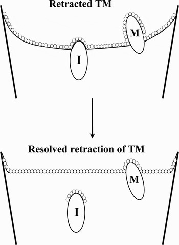
In a subsequent paper, TosReference Tos13 reviewed the literature and stated that the majority of cholesteatomas occurred close to the malleus handle and neck, whilst the minority developed near the long process of the incus. This author proposed that, under frequently observed inflammatory conditions, there is an increased risk of retraction, adherence and loosening of the tympanic membrane on these ossicles. During the loosening process, some of the cells of the tympanic membrane (presumably from a monomeric squamous segment) may become trapped in the middle-ear cavity (Figure 2) and subsequently form a cholesteatoma. Tos concluded that this conjecture neither fits with the origin of congenital cholesteatoma nor is proven for the aetiopathogenesis of acquired cholesteatoma, but justifies further research in order to test the proposition. So far, the only supporting evidence is a single temporal bone showing histopathological features of a cholesteatoma behind an intact tympanic membrane, following a resolved retraction.Reference Sudhoff and Linthicum14
Diagram of the concepts of the ‘inclusion’ theory; note the cells trapped on the incus following resolution of a retracted eardrum. I = incus; M = malleus; TM = tympanic membrane

Epidermoid formation theory
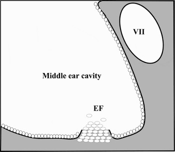
In 1936, formation of an epidermoid (i.e. a distinct squamous cell nest with unknown function) was first identified in the anterosuperior mesotympanum of a 22-week-old human fetus.Reference Teed15 Fifty years later, the lesion was independently rediscovered in fetal middle ears between the 10th and 33rd gestational week.Reference Michaels16 Interestingly, epidermoid formation originates from ectodermal tissue of the first pharyngeal cleft during development of the external auditory canal. After examining stained sections of 76 ears from 62 aborted, still- or live-born infants (gestational age 10 weeks to term), MichaelsReference Michaels16 proposed that the postpartum persistence of the epidermoid formation, for unknown reasons, could be the potential precursor of congenital cholesteatoma. This theory was strengthened by the presence of the embryonic rests always found in the anterosuperior mesotympanumReference Michaels16 – the commonest site of the lesion. However, this prompted the question of whether the epidermoid formation could explain the occasional occurrence of congenital cholesteatoma elsewhere in the middle ear.Reference Mueller, Schwetschenau and Isaacson17 Recently, however, Liang et al. Reference Liang, Michaels and Wright18 identified varying numbers of CK14 immuno-positive, novel cell rests in the posterosuperior (Figure 3), posteroinferior and anteroinferior regions of the middle ear. These were among 116 epidermoid formations in 22 temporal bones, aged 16 gestational weeks to eight months postpartum (the majority of which were in the anterosuperior mesotympanic area). Also of great importance were the independent discoveries of keratinising epidermoid formations (i.e. intermediate forms of congenital cholesteatomas),Reference Liang, Michaels and Wright18, Reference Karmody, Byahatti, Blevins, Valtonen and Northrop19 and further confirmation of four epidermal rests in a histological survey of 30 post-natal human temporal bones (the oldest aged two years and seven months).Reference Levine, Wright, Pawlowski and Meyerhoff20 Hence, there is powerful evidence to support epidermoid formation as the anlage for congenital cholesteatoma. Conclusive ‘proof’ would require a ‘real time’ demonstration of an epithelial rest forming a lesion in vivo, but this is likely to prove very difficult.
Diagram of epidermoid formation (EF) in the posterosuperior region of the middle-ear cavity in a human fetus between 20 and 22 weeks. VII = facial nerve

Theories of acquired cholesteatoma aetiopathogenesis
The various theories on the aetiopathogenesis of acquired cholesteatoma, apart from metaplasia, can account for the presence of the lesion in different situations.
Metaplasia theory
Using immunohistochemistry, Sadé et al. Reference Sadé, Babiacki and Pinkus21 identified aggregates of keratin fibres surrounded by ‘special cells’ within subepithelial, inflamed connective tissue in 19 out of 101 biopsies taken from cholesteatomatous ears. In addition, all 19 biopsies contained mucous glands. These authors interpreted these findings as representing metaplasia, and suggested that this could explain the origins of both congenital and acquired cholesteatomas.Reference Sadé, Babiacki and Pinkus21 However, whilst islands of squamous epithelium may be present in middle ears with chronic inflammation,Reference Sadé, Babiacki and Pinkus21, Reference Palva, Palva and Darmmert22 there is no histological or experimental evidence that metaplasia results in cholesteatoma formation or even in an incipient lesion. Furthermore, histopathological examination of four human temporal bones with middle-ear cholesteatomas did not reveal any areas of squamous metaplasia, despite a meticulous search.Reference Wells and Michaels23 In studies involving propylene glycol-induced cholesteatomas in 17 middle ears of chinchillas, it was noted that the disease originated from the epidermis of the tympanic membrane.Reference Imamura, Nozawa, Imamura and Murakami24 Additionally, tissue culture experiments revealed that cholesteatoma epithelium possesses the same unique locomotor properties as the epidermis of the tympanic membrane and deep canal.Reference Boxall, Proops and Michaels25 Moreover, immunocytochemical studies have indicated that cytokeratins (i.e. epithelium-specific cellular proteins) expressed in cholesteatoma are similar to those found in the external ear, but different from those present in middle-ear mucosa.Reference Broekaert, Coucke, Leperque, Ramaekers, Van Muijen and Boedts26, Reference Lee, Mackenzie, Hall and Gantz27 Finally, ultrastructural studies have shown a remarkable resemblance between cholesteatoma and the epidermis of the tympanic membrane and adjacent canal wall.Reference Youngs and Rowles28 These findings are highly indicative, if not conclusive, that acquired cholesteatoma originates from ectodermal tissue, rather than from metaplasia of the normal endodermal cuboidal epithelium of the middle ear.
Immigration theory
This theory (also know as the migration, emigration or invasion theory) invokes squamous epithelium progressing from the margin of a tympanic membrane perforation into the middle ear and then forming a cholesteatoma.Reference Bezold29 In an experimental model involving otitis media induced by injecting bacteria into the middle ears of 266 guinea pigs, FreidmannReference Freidmann30 elegantly demonstrated frequent epithelial movements from the outer auditory meatus or tympanic membrane into the middle ear, with subsequent cholesteatoma formation. Similar results were obtained in two separate studies involving 30 and 32 chinchillas with perforations caused by propylene glycol (with or without antibiotics) applied into the middle ear.Reference Masaki, Wright and Lee31, Reference Wright, Meyerhoff and Burns32 Migration of epithelium has been observed in patients with central perforations from time to time, and has also been seen in temporal bone examinations (S Merchant, personal communication). The intriguing question is why this does not happen more often, considering the incidence of eardrum perforation in the general population.
Basal hyperplasia theory
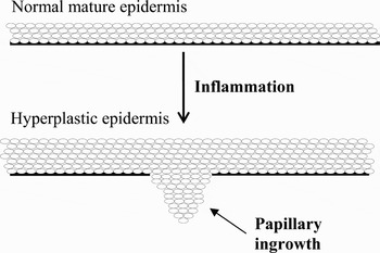
RuediReference Ruedi33 found that the application of talc and fibrin behind intact tympanic membranes of guinea pigs generated cholesteatomas from the active proliferation of basal cells breaking through the basement membrane; inflammation appeared to be important in the process (Figure 4). This was also evident in the chinchilla model,Reference Masaki, Wright and Lee31 in which some cholesteatomas appeared to form due to invasion of epidermal hyperplastic pseudopodia through an intact tympanic membrane. Immunohistochemical and ultrastructural studies, involving 30 cholesteatoma specimens, seven samples of canal skin and five samples of middle-ear mucosa from patients undergoing surgery, have also confirmed the presence of squamous pegs extending into the subepithelial region.Reference Sudhoff, Bujia, Borkowski, Hildmann, Koc and Holly34 More recently (2005), inflammation-associated epidermal hyperplasia was described as fetal-type epithelium, based on pilot histopathological studies of stained step sections of 428 temporal bones.Reference Michaels, Persaud, Liang, Soucek and Wright35, Reference Persaud, Michaels, Liang, Soucek and Wright36 The authors suggested that chronic inflammation may lead to transformation of the normal, thin, mature epidermis of the eardrum and adjacent canal into a thicker form resembling that of the fetus. They further posited that the fetal-type epithelium may have invasive pathogenic properties. However, whilst basal hyperplasia could explain some cases of acquired cholesteatoma, it is still unclear whether inflammation is the cause or an effect of hyperplasia. Moreover, if chronic inflammation is responsible for the development of invasive hyperplastic epidermis, one would expect a higher incidence of its sequelae, considering the prevalence of otological inflammatory conditions. The concept of fetal-type epithelium requires further investigation, including identification of relevant molecular, cellular and genetic markers, in order to verify that the epithelium is indeed fetal-like.
Diagram of epidermal hyperplasia with a papillary ingrowth through a breach in the basement membrane. Chronic inflammation may play a vital role in the pathological changes.

Iatrogenic, post-traumatic or implantation theory
Based on a literature review and 25 years' experience of tympanomastoidectomies, Wullstein and WullsteinReference Wullstein and Wullstein37 suggested that some cases of acquired cholesteatoma may occur due to surgery for non-cholesteatomatous conditions (e.g. myringoplasty) or injury to the temporal bone. One year later, Plester and PusalkarReference Plester and Pusalkar38 documented 48 cases of anterior annular cholesteatomas secondary to onlay surgical repair of tympanic membrane perforations. In 27 cases, the lesions were found in the substance of blunted anterior annular sulci, and in 12 cases the cholesteatomas had entered the middle ear. In 1989, McKennan and CholeReference McKennan and Chole39 presented three cases of post-traumatic cholesteatomas in which one case had undergone previous oto-surgery, and they conceded the possibility of iatrogenic implantation of skin into the middle ear.
Retraction pocket theory
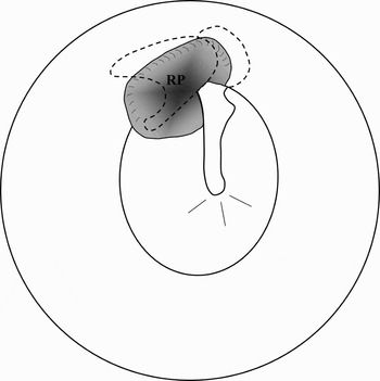
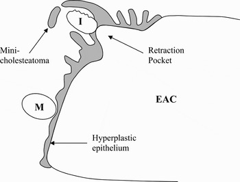
In this theory, part of the tympanic membrane, usually the pars flaccida, invaginates towards the middle ear (Figure 5) prior to cholesteatoma formation.Reference Portman and Sadé40 The main precipitating factor is functional obstruction of the eustachian tube, resulting in poor middle-ear and mastoid ventilation.Reference Youngs, Ludman and Wright41 This is particularly relevant to cleft palate patients, who appear to have a propensity for acquiring cholesteatoma on the side of the cleft, amongst other middle-ear conditions.Reference Dominguez and Harker42, Reference Goldman, Martinez and Ganzel43 In 1986, Wolfman and CholeReference Wolfman and Chole44 induced eustachian tube dysfunction (by cauterisation) in 16 gerbils and showed that cholesteatoma developed from the resulting retraction pockets in 75 per cent of the animals within 16 weeks. A previous histological inspection of four human temporal bones had shown that chronic retraction pockets were associated with a loss of the fibrous component of the eardrum, facilitating hyperplastic epidermal ingrowths into the middle ear.Reference Wells and Michael45 In the recent preliminary histopathological study by Persaud et al., Reference Persaud, Michaels, Liang, Soucek and Wright36 it was noted that epidermal hyperplasia with invasive cones was prominent in atelectatic pockets associated with inflammation (Figure 6). These findings corroborate those of an ultrastructural examination of specimens from retraction pockets in 10 human cases of chronic otitis media.Reference Akyildiz, Akbay, Ozgirgin, Bayramoglu and Sayin46 A cholesteatoma formed by such a mechanism might then cause a secondary perforation in the tympanic membrane due to expansion, infection and/or inflammation. This challenges the notion of cholesteatoma development from epithelial migration directly through a primary defect of the tympanic membrane within a retraction pocket. However, whilst cholesteatomas associated with atelectatic pockets are frequently seen in clinics, some pockets can remain free from cholesteatoma indefinitely. The reason for this is unclear, but the absence of inflammation may be significant.
Diagram of a retraction pocket (RP) in the posterosuperior region of the right middle ear.

Diagram of a posterosuperiorly retracted tympanic membrane with papillary ingrowths and the formation of a small epidermoid cyst (or ‘mini-cholesteatoma’) in the middle ear. EAC = external auditory canal; I = incus (eroded); M = malleus

Conclusions
A thorough understanding of cholesteatoma biology is crucial because of the potentially dangerous effects of the lesion, the lack of effective non-surgical treatment and the complexity of surgery often required for its management. Currently, none of the proposed aetiopathogenic theories are unequivocally proven (or involve genetics, which is an essential feature of neoplasia).
It appears that the post-natal persistence of the fetal epidermoid formation is the most robust explanation for the origin of congenital cholesteatoma; conclusive ‘proof’ awaits direct, in vivo observation of the transition of an epidermoid nidus into a lesion in humans. In contrast, acquired cholesteatoma can be explained by multiple aetiopathogenic suppositions: immigration, basal hyperplasia, retraction pocket and/or trauma (iatrogenic or non-iatrogenic); however, metaplasia within the middle ear seems highly unlikely. Chronic inflammation may play a key role in many of these patho-mechanisms.
Whilst the methodologies of the reported investigations are generally sound, one should note that most temporal bone studies present a static ‘snap-shot’ view of a dynamic process. Therefore, any derived evidence is inevitably subject to varying degrees of speculation (the best evidence would be obtained by following a disease in real time and through to autopsy). Caution is also necessary when extrapolating the findings from animal and tissue culture experiments to humans, because of the intricate bio-physiological differences.
Continued otopathological, cellular and molecular research would enhance our limited understanding of cholesteatoma and may even lead to new therapeutic strategies for this erosive disease, which often defies surgical treatment. Meanwhile, if inflammation is a trigger of, rather than just a response to, acquired cholestatoma formation, one might suggest that early treatment of otological inflammatory disorders (e.g. chronic otitis media) could reduce their sequelae, probably by preventing the development of hyperplastic epidermis with invasive pathogenic projections.
Acknowledgements
Part of the work in this paper was supported by a grant (KT13) to the first author from the Royal National Institute for Deaf People. The award facilitated a visit to the Otopathology Laboratory at the Massachusetts Eye & Ear Infirmary and to the Department of Otolaryngology, Harvard Medical School, Boston, USA. Many thanks to all those who commented favourably and constructively on various drafts of the manuscript, especially Liam Flood FRCS (UK), Sunil N Dutt MS, FRCS, PhD (UAE), and expert otologist and otopathologist Fred H Linthicum Jr MD (USA).