Introduction
The family Notocotylidae Lühe, 1909 is one of the taxonomically diverse trematode families. This group comprises parasites of birds and mammals. Until 2019, the taxonomy of representatives of Notocotylidae was based on morphological characteristics of sexually mature individuals (Gonchar et al. Reference Gonchar, Jouet, Skírnisson, Krupenko and Galaktionov2019; Izrailskaia et al. Reference Izrailskaia, Tatonova and Besprozvannykh2018, Reference Izrailskaia, Besprozvannykh, Tatonova, Nguyen and Ngo2019). In particular, the genera were differentiated on the basis of the structure of the ventral glands (Barton and Blair Reference Barton, Blair, Jones, Bray and Gibson2005; Skrjabin Reference Skrjabin1953). Subsequently, new genera of the Notocotylidae were identified using consolidated morphological and molecular data, and the use of gland structure as the only characteristic became inappropriate for generic differentiation (Gonchar et al. Reference Gonchar, Jouet, Skírnisson, Krupenko and Galaktionov2019; Izrailskaia et al. Reference Izrailskaia, Besprozvannykh, Tatonova, Nguyen and Ngo2019, Reference Izrailskaia, Tatonova and Belousova2024). To date, sufficient molecular data have been accumulated for various genera of the family. The NCBI (GenBank) database contains information on more than 20 species from eight genera of the Notocotylidae. However, in a number of cases, e.g., for the most speciose genera of the family such as Notocotylus Diesing, 1839, and Catatropis Odhner, 1905, molecular characteristics of the type species from the type localities of discovery are not available. This significantly complicates resolution of both taxonomic and systematic issues in the structure of the family. The type species of these genera, Notocotylus triserialis Diesing, 1839 (=Notocotylus attenuatus (Rudolphi, 1809)) (Vlasenkov et al. Reference Vlasenkov, Yakovleva and Sokolov2025) (Ireland) and Catatropis verrucosa (Frölich, 1789) Odhner, 1905 (Western Europe), were first discovered in the European region. These species that have been recorded not only from western but also from eastern Eurasia and also from America are currently considered cosmopolitan (Aguilar-Morales et al. Reference Aguilar-Morales, Sánchez-Nava, Ortigoza and Rodríguez-Romero2022; Besprozvannykh et al. Reference Besprozvannykh, Ermolenko and Nadtochy2012; Filimonova Reference Filimonova1985; Le et al. Reference Le, Ngo and Ermolenco2013). However, without molecular confirmation, the question of whether N. triserialis and C. verrucosa are truly cosmopolitan remains open.
In the works of Izrailskaia et al. (Reference Izrailskaia, Besprozvannykh, Tatonova, Nguyen and Ngo2019, Reference Izrailskaia, Tatonova and Belousova2024) and Gonchar et al. (Reference Gonchar, Jouet, Skírnisson, Krupenko and Galaktionov2019), polyphyly was shown for the genus Notocotylus; however, the lack of molecular data for the type species did not allow us to ask the question of which clade the true representatives of the genus belong to. Vlasenkov et al. (Reference Vlasenkov, Yakovleva and Sokolov2025) obtained morphological and molecular data for N. triserialis from Karelia (Russia), which made it possible to isolate trematodes belonging to the genus Notocotylus.
Currently, it is known that the morphologically similar N. triserialis and Notocotylus ikutai Sasaki, Kobayashi, Yoshino, Asakawa, & Nakao, 2021 (https://www.marinespecies.org/aphia.php?p=taxlist&tName=Notocotylus) are the only representatives of the Notocotylidae that use snails from the family Lymnaeidae Rafinesque, 1815 as the first intermediate host. Catatropis verrucosa involves snails from the Bithyniidae J. E. Gray, 1857 in its life cycle (Filimonova Reference Filimonova1985; Kanev et al. Reference Kanev, Vassilev, Dimitrov and Radev1994; https://www.marinespecies.org/aphia.php?p=taxdetails&id=18269).
In one of the water bodies in the southern Russian Far East, we found individuals of the snails Radix auricularia (Linnaeus, 1758) (Lymnaeidae Rafinesque, 1815) and Boreoelona ussuriensis (Büttner & Ehrmann, 1927) (Bithyniidae Gray, 1857) that emitted cercariae of the family Notocotylidae. As a result of experimental studies, we obtained metacercariae from the cercariae and then sexually mature flukes. Based on the morphology and molecular markers of these trematodes, we determined their taxonomic positions and resolved their phylogenetic relationships with other Notocotylidae representatives. The results of these studies are presented below.
Materials and methods
Morphological data
During the examination of freshwater gastropods from an abandoned channel of the Komissarovka River (south of the Russian Far East), we found one snail of R. auricularia and one snail of B. ussuriensis that emitted Notocotylidae cercariae. We obtained metacercariae from those cercariae and fed them to a duckling and three chickens, respectively. Each of them was fed a total of 50 metacercariae. Mature flukes were found in the blind appendages of the duckling’s intestine on day 21 post infection (a total of 35 individuals); in all chickens, on day 14 post infection (15 to 30 individuals).
The morphology of live cercariae was examined, and metric data were obtained from cercariae fixed in hot 4% formalin. Adult flukes were fixed in 70% ethanol and stored in 96% ethanol. Whole mounts of adult flukes were prepared by staining with alum carmine, dehydrating in a graded ethanol series, clearing in clove oil, and mounting in Canada balsam. All measurements are in micrometres (μm).
DNA extraction, amplification, and sequencing
Genomic DNA was extracted from the fluke specimens using the HotSHOT technique (Truett et al. Reference Truett, Heeger, Mynatt, Truett, Walker and Warman2000). Partial sequences of the 28S rRNA gene (28S) nuclear DNA and partial sequences of the cox1 gene (cox1) of mitochondrial DNA were amplified by polymerase chain reaction (PCR) using specific primers (Table S1, Supplementary Materials). Both external and internal primers were used for sequencing (Table S1, Supplementary Materials). The annealing temperature was 57°C for the 28S marker and 50°C for the cox1 marker. The efficiency and contamination of PCR were tested by setting positive and negative controls, respectively. The PCR products were sequenced by the Sanger method using a BigDye Terminator Cycle Sequencing kit (Applied Biosystems, USA). Nucleotide sequences were determined on an Applied Biosystems 3500 Genetic Analyzer at the Instrumental Center for Biotechnology and Gene Engineering, Federal Scientific Center of East Asia Terrestrial Biodiversity, Far Eastern Branch, Russian Academy of Sciences (FSCEATB FEB RAS) (Vladivostok, Russia).
Analysis of genetic data
Processing and alignment of consensus sequences were carried out using the FinchTV 1.4 and MEGA 5.0 programs (Tamura et al. Reference Tamura, Peterson, Peterson, Stecher, Nei and Kumar2011). Sequences for the 28S marker that were less than 1,000 bp in length were removed from the analysis. The p-distances between and within the species were analysed in the MEGA software without including indels. Phylogenetic relationships within the family Notocotylidae were assessed independently for both markers using newly obtained sequences and data for all species accessed from GenBank. The members of the trematode families located left (basal) of the Notocotylidae in the phylogenetic tree constructed by Olson et al. (Reference Olson, Cribb, Tkach, Bray and Littlewood2003) using nucleotide sequences of the rRNA genes were selected as an outgroup in our study. The list of specimens used in the study is provided in Supplementary Materials, Table S2.
Phylogenetic relationships were reconstructed using the Bayesian Inference (BI) algorithm in the MrBayes 31.2. (Ronquist and Huelsenbeck Reference Ronquist and Huelsenbeck2003) and the maximum likelihood algorithm in the PhyML 3.1 program by applying a model selected as optimal on the basis of the Akaike information criterion (AIC) in the jModeltest 2.1.7 program (Darriba et al. Reference Darriba, Taboada, Doallo and Posada2012): TVM+I+G for 28S and TVM+G for cox1. The statistical significance of phylogenetic connections for trees constructed using the maximum likelihood algorithm was established using the bootstrap method. The method of posterior probabilities was used for the trees constructed by the BI algorithm. In the BI analysis, 1,300,000 and 1,100,000 generations of the Markov chain Monte Carlo (MCMC) posterior were simulated for 28S and cox1, respectively. The number of generations was determined as sufficient, since the SD value calculated was ˂0.01. The chain was sampled every 100 generations. Of the samples assessed, 25% were excluded to permit the construction of consensus trees.
Results
Morphological description
Notocotylus ikutai Sasaki & Nakao, Reference Sasaki, Kobayashi, Yoshino, Asakawa and Nakao2021
Host: Anas platyrhynchos Linnaeus, 1758 dom. (experimentally infected).
Site: Caeca of intestine.
Intermediate host: Radix auricularia.
Type locality: an abandoned channel of the Komissarovka River (44.89°N, 131.57°E), Primorsky Krai, Russia.
Deposition: This material was deposited in the parasitological collection of the zoological collection (FSCEATB FEB RAS, Vladivostok, Russia) (Email: petrova@ibss.dvo.ru) on August 2, 2025. ZooBank accession no. 58D469F2-EB2A-413F-B1C1-F9DD2089B6A3.
Adult fluke (material examined: 5 specimens) (Fig. 1a, b; Table 1). Body flat, elongated, ventrally concave, with tapered anterior and rounded posterior ends. Ventral surface of anterior half of body covered by scale-like spines. Ventral papillae arranged in three rows: 15 papillae in lateral rows and 14 in median row. Two papillae in median row located at level of anterior half of cirrus sac, in front of anterior lateral papillae. Posterior papilla of median row located immediately posteriorly to ovary. Posterior papillae of lateral rows at level of posterior ends of testes. Oral sucker subterminal. Oesophagus short. Caeca extending laterally to uterine coils, between ovary and testes, and ending blindly at level of posterior ends of testes. Testes at posterior end of body symmetrical, slightly lobed. External seminal vesicle tubular and coiled. Loops of seminal vesicle located immediately posteriorly to cirrus sac. Cirrus sac elongated, narrowed anteriorly and expanded posteriorly, containing curved seminal vesicle, with well-expressed oval prostatic surrounded by numerous prostatic cells. Cirrus with minute scale-like spines. Genital pore median, located immediately posteriorly to intestinal bifurcation. Ovary intertesticular, consisting of 3–4 lobes. Mehlis’ gland preovarian. Uterus forms 18–19 coils. Metraterm thick-walled, with a widened posterior end surrounded by numerous gland cells, reaching half of cirrus sac length. Vitellarium consisting of irregular follicles, located in posterior half of body. Vitellarium anteriorly reaching level of 13–14th uterine coils, with posterior part located at level of anterior margins of testes. Eggs oval, operculate, with two polar filaments. Excretory vesicle saccular. Excretory pore located dorsally.
Notocotylus ikutai: (a) adult, ventral view; (b) terminal genitalia; (c–d) cercaria. Neocatatropis primorskii n. g. n. sp.: (e) adult, ventral view; (f) terminal genitalia; (g–h) cercaria. Scale bars in μm.

Measurements of adult Notocotylus specimens (μm)

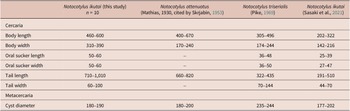
Cercaria (material examined: 10 specimens) (Fig. 1c, d; Table 2). Cercariae belonging to ‘Monostomi’ group. Body oval, brown pigmented, tri-oculate. Lateral eyespots longitudinally elongated, median eyespot transversely elongated. Body filled with numerous cystogenous glands. Postero-lateral body margin bearing adhesive pockets. Oral sucker subterminal. Oesophagus short. Intestinal bifurcation posteriorly to median eyespot. Caeca reaching level of excretory vesicle. Excretory vesicle sac-like, opening as excretory pore near posterior body end. Main excretory ducts fused anteriorly to intestinal bifurcation. Main excretory ducts filled with excretory granules. Tail almost twice length of body.
Measurements of cercariae and metacercariae of Notocotylus (μm)

Metacercaria (material examined: 10 specimens) (Table 2). Cyst dome-shaped.
Neocatatropis primorskii n. g. n. sp. Izrailskaia & Besprozvannykh
Host: Gallus gallus (Linnaeus, 1758) dom. (experimentally infected).
Site: Caeca of intestine.
Intermediate host: Boreoelona ussuriensis.
Type locality: an abandoned channel of the Komissarovka River (44.89°N, 131.57°E), Primorsky Krai, Russia.
Type deposition: holotype No. 281-Tr; paratype No. 282–285-Tr. This material was deposited in the parasitological collection of the zoological collection (FSCEATB FEB RAS, Vladivostok, Russia) (Email: petrova@ibss.dvo.ru) on August 2, 2025. ZooBank accession no. 0E39676B-BF68-46F7-BC6E-5227571CD3DA.
Etymology: due to the morphological similarity of adult flukes from the above-described genus with those from the genus Catatropis, the new genus was given the name Neocatatropis n. gen. The species was named after the region of discovery.
Adult fluke (material examined: 5 specimens) (Fig. 1e, f; Table 3). Body flat, elongated, ventrally concave, with tapered anterior and rounded posterior ends. Ventral surface of anterior half of body covered by scale-like spines. Ventral papillae in median row merged into continuous ridge and ridge from level of middle anterior half of body to level middle of ovary. Two lateral rows with 13 papillae in each. First pair of papillae in lateral rows located posteriorly to anterior margin of ridge. Posterior papillae in lateral rows located at level of posterior ends of testes. Oral sucker subterminal. Oesophagus short. Caeca extending laterally to uterine coils, between ovary and testes, and ending blindly at level of posterior edge of testes. Testes at posterior end of body symmetrical, with numerous lobes on external margin. External seminal vesicle tubular and coiled. Loops of seminal vesicle located immediately posteriorly to cirrus sac. Cirrus sac elongated, narrowed anteriorly and expanded posteriorly, containing curved seminal vesicle, with oval prostatic surrounded by numerous prostatic cells. Cirrus with minute scale-like spines. Genital pore median, immediately posteriorly to intestinal bifurcation. Ovary intertesticular, consisting of 4–5 lobes. Mehlis’ gland preovarian. Uterus forming 14–19 coils. Metraterm thick-walled, reaching 1/3 length of cirrus sac. Vitellarium consisting of irregular follicles, located in posterior half of body. Vitellarium anteriorly reaching level of 14–18th uterine coils, with posterior part located at level of anterior margins of testes or slightly laterally to testes. Eggs oval, operculate, with two polar filaments. Excretory vesicle saccular. Excretory pore located dorsally.
Measurements of adult Neocatatropis primorskii n. g. n. sp. and Catatropis specimens (μm)

Cercaria (material examined: 10 specimens) (Fig. 1g, h). Cercariae belonging to ‘Yenchingensis’ group. Body 420–460 × 200–260, oval, brown pigmented, tri-oculate. Eyespot irregular in shape. Median eyespot less developed than lateral ones. Body filled with numerous cystogenous glands. Postero-lateral body margin bearing adhesive pockets. Oral sucker 50 in diameter, subterminal. Oesophagus short. Intestinal bifurcation located at level of lateral eyespots. Caeca reaching level of excretory vesicle. Excretory vesicle sac-like, opening as excretory pore near posterior body end, between dorsal adhesive pockets. Main excretory ducts fused anteriorly at level of intestinal bifurcation. Diverticulum of anterior arc reaching level of median eyespot. Main excretory ducts filled with excretory granules. Tail 610–690 × 40–70.
Metacercaria (material examined: 10 specimens). Cyst dome-shaped, 200–240.
Molecular data
For the reconstruction of phylogenetic trees, we used aligned sequences of 1,014 and 303 bp for 28S and cox1, respectively. In the phylogenetic reconstruction based on nuclear marker (28S), the flukes experimentally grown in the duckling appeared in a cluster with representatives of the genus Notocotylus (Notocotylus malhamensis Boyce, Hide, Craig, Harris, Reynolds, Pickles, & Rogan, 2012, N. triserialis, Notocotylus multipapillus Izrailskaia, Tatonova, & Belousova, Reference Izrailskaia, Tatonova and Belousova2024, N. ikutai, MW358653, EU712725, OQ672264, AY222219, KY513158, LC599518 Notocotylus sp.), whereas the flukes grown in the chickens formed a branch externally to representatives of the genera Hippocrepis Travassos, 1922, Notocotylus, Pseudocatatropis Kanev & Vassilev, 1986, and Quinqueserialis Skvortsov, 1935 (Fig. 2). In the phylogenetic reconstruction based on the cox1 marker, the flukes experimentally grown in the duckling appeared in a cluster with representatives of the genus Notocotylus from Branch 2 and flukes grown in the chickens formed a branch located externally to the clade comprising the genera Pseudonotocotylus Izrailskaia, Tatonova, & Belousova, Reference Izrailskaia, Tatonova and Belousova2024, Ogmogaster Jägerskiöld, 1891, and Catatropis (Fig. 3).
Phylogeny of the family Notocotylidae based on 28S sequences using the Bayesian Inference (BI) and Maximum Likelihood (ML) algorithms. Posterior probability for BI and nodal support for ML are shown in the nodes, respectively. The scale bar indicates number of substitutions per site.

Phylogeny of the family Notocotylidae based on cox1 sequences using the Bayesian Inference and Maximum Likelihood (ML) algorithms. Posterior probability for BI and nodal support for ML are shown in the nodes, respectively. The scale bar indicates number of substitutions per site.

Discussion
To establish the species identity of the detected trematodes, we used a comprehensive approach, namely, we analysed both morphological and molecular data.
An analysis of the morphological characteristics of the fluke that we obtained by infecting the duckling showed the greatest similarity with the N. triserialis from the regions of finding: Ireland (Skrjabin Reference Skrjabin1953), South Wales (Pike Reference Pike1969), from South Korea, Mexico (Eom et al. Reference Eom, Rim and Jang1984; Aguilar-Morales et al. Reference Aguilar-Morales, Sánchez-Nava, Ortigoza and Rodríguez-Romero2022), Karelia (Russia) (Vlasenkov et al. Reference Vlasenkov, Yakovleva and Sokolov2025), and with the N. ikutai from Japan (Sasaki et al. Reference Sasaki, Kobayashi, Yoshino, Asakawa and Nakao2021). The sexually mature worms in our material have the most similar metric characteristics to those from Ireland and Karelia (Table 1). In terms of papilla arrangement, they are similar to specimens published by Pike (Reference Pike1969): two papillae of the median row occur anterior to the first papilla of the lateral rows. The remaining N. triserialis specimens have a single median papilla anterior to the first lateral papillae. Thus, there are no significant morphological differences between the specimens from different geographic locations.
The only difference between the worms we discovered and N. triserialis from Pike (Reference Pike1969) and N. ikutai (Sasaki et al. Reference Sasaki, Kobayashi, Yoshino, Asakawa and Nakao2021) is the difference in cercariae size (Table 2). In both cases, this may indicate that the trematodes belong to different species. However, the significant difference in size between the cercariae of the Far Eastern and Japanese trematodes can be explained by the method used by Sasaki et al. (Reference Sasaki, Kobayashi, Yoshino, Asakawa and Nakao2021) to study cercarial morphometry data. The authors used cercariae obtained by dissecting infected snails. This method damages the rediae, and cercariae at various stages of development are found within the dissected snails. In this case, there is a high probability of using immature cercariae to obtain their morphometric data. Perhaps for this reason, there are significant differences in size between the cercariae in our data and those of Sasaki et al. (Reference Sasaki, Kobayashi, Yoshino, Asakawa and Nakao2021).
The first attempts to reproduce the life cycle of N. triserialis were made by Yamaguti (Reference Yamaguti1938) in Japan. As a result of experimental studies, Yamaguti (Reference Yamaguti1938) obtained sexually mature flukes, which he referred to as N. attenuatus, from cercariae collected from snails Parafossarulus Annandale, 1924. However, according to Pike (Reference Pike1969) and Filimonova (Reference Filimonova1985), N. triserialis uses snails from the family Lymnaeidae as its first intermediate host. As the data currently available in the literature show, lunged and gilled snails cannot be the first intermediate hosts for the same trematode species simultaneously. Therefore, the species identification in one of the above-mentioned studies is incorrect. This assumption is confirmed by a number of publications that indicate the trematode Pseudonotocotylus intestinalis (Tubangui, 1932) Izrailskaia, Tatonova, & Belousova, Reference Izrailskaia, Tatonova and Belousova2024, as morphologically close to worms designated by Yamaguti (Reference Yamaguti1938) as N. attenuatus (Besprozvannykh, Reference Besprozvannykh2010; Besprozvannykh et al., Reference Besprozvannykh, Ermolenko, Ngo, Ha, Hung and Rozhkovan2013; Izrailskaia et al., Reference Izrailskaia, Besprozvannykh, Tatonova, Nguyen and Ngo2019) and developing with the involvement of a snail from the genus Parafossarulus. Before the study by Sasaki et al. (Reference Sasaki, Kobayashi, Yoshino, Asakawa and Nakao2021), it was suggested that, among the Notocotylus species, only N. triserialis develops with the use of Lymnaeidae snails. Sasaki et al. (Reference Sasaki, Kobayashi, Yoshino, Asakawa and Nakao2021) described the trematode N. ikutai from Japan, obtained from mice experimentally infected by metacercariae emitted by the Lymnaeidae snail. Sexually mature N. ikutai from Japan, like our trematodes, combine the morphometric parameters characteristic of the trematodes presented in Table 1.
According to the results of phylogenetic reconstruction based on the nuclear marker (28S), the flukes in our study form a well-supported branch with representatives of the genus Notocotylus (N. multipapillus and N. ikutai) and a number of specimens not identified to species level (Fig. 2). The genetic distances between the fluke from our material and the specimens designated as N. multipapillus and N. ikutai are zero. In the phylogenetic reconstruction based on the mitochondrial marker (cox1), these trematodes form a separate cluster with a high posterior probability (Fig. 3). Two branches can be distinguished in this cluster: Branch 1 comprises specimens designated as N. multipapillus, N. ikutai, and specimens not identified to species; Branch 2 comprises the studied fluke and the specimens designated as N. ikutai, as well as specimens not identified to species. The genetic distances between the branches are 2.6%, which exceeds the distances within the branches. In our opinion, distribution of N. ikutai on the phylogenetic tree may be a consequence of the incorrect study by Sasaki et al. (Reference Sasaki, Kobayashi, Yoshino, Asakawa and Nakao2021). As reported by Sasaki et al. (Reference Sasaki, Kobayashi, Yoshino, Asakawa and Nakao2021), they collected Lymnaeidae snails and other snails, which were stored in the same dish. The authors did not state whether the Lymnaeidae representatives were separated from the other snails and placed in separate dishes. In this case, it is unclear where the Notocotylidae metacercariae were collected from. If they were from a common dish, then the presence of metacercariae belonging to different trematode species is possible. The collected metacercariae (50–100 specimens) were fed to mice in which sexually mature specimens were raised. Morphological characteristics provided for the experimentally grown trematodes. Molecular data for these worms were not obtained. Molecular characteristics obtained by Sasaki et al. (Reference Sasaki, Kobayashi, Yoshino, Asakawa and Nakao2021) for adult flukes collected from naturally infected snails and birds (Anas spp.). However, morphological characteristics of the adult specimens were not provided. Therefore, there is no evidence that the flukes experimentally obtained from mice and all flukes from ducks belong to the same species. An argument in favour of the incorrectness of the results obtained by Sasaki et al. (Reference Sasaki, Kobayashi, Yoshino, Asakawa and Nakao2021) may be the placement of sample LC597055, identified as N. ikutai, in Branch 1. Genetic distances between samples of this clade are at the intraspecific level. The belonging of the trematodes N. multipapillus (Branch 1) and the remaining N. ikutai (Branch 2) to different species was confirmed by morphological characteristics: 14–16 papillae (N. ikutai) vs. 23–25 papillae (N. multipapillus) (Izrailskaia et al. Reference Izrailskaia, Tatonova and Belousova2024; Sasaki et al. Reference Sasaki, Kobayashi, Yoshino, Asakawa and Nakao2021). The nucleotide sequences obtained by Sasaki et al. (Reference Sasaki, Kobayashi, Yoshino, Asakawa and Nakao2021) from rediae and naturally infected ducks and our sequences obtained in the reconstruction for the mitochondrial marker were grouped into Branch 2. Genetic distances within this branch are at the intraspecific level. Based on the above, we classify the trematodes we discovered as N. ikutai. As for sample LC597055, designated as N. ikutai, it likely belongs to a different species, which is why we designated it as Notocotylus sp. (Fig. 3).
It is important to note that the phylogenetic reconstruction based on the mitochondrial marker includes a specimen designated as N. attenuatus, which is positioned externally to other members of the family Notocotylidae. Molecular data for this trematode were obtained using an adult specimen from a naturally infected Anas sp. Morphological characteristics for this fluke were not provided in the study (Prüter et al. Reference Prüter, Franz, Auls, Czirják, Greben, Greenwood, Lisitsyna, Syrota, Sitko and Krone2018), which does not allow comparing the specimens we obtained with that referred to as N. attenuatus. In our opinion, the latter most likely does not belong to Notocotylus.
The sexually mature flukes we reared in the chickens are similar in morphological characteristics to both the East Asian Catatropis chinensis Lai, Sha, Zhang, & Yang, 1984, and the European C. verrucosa (Conglong et al., Reference Conglong, Guorun, Tongfu and Minglang1984; Kanev et al., Reference Kanev, Vassilev, Dimitrov and Radev1994). The reported morphometric characteristics for C. chinensis are very limited (Table 3). This does not allow any objective analysis of such data between the individuals in our study and C. chinensis. Based on the available information, there are differences between them in the position of the genital opening (genital opening located posteriorly vs. anteriorly to the intestinal bifurcation), the ratio of the metraterm length to the bursa cirrus length (the metraterm length reaching 1/3 the bursa cirrus length vs. the metraterm is equal in length to the bursa cirrus), and the lack of cirrus armament in the Chinese species. The above facts indicate that the flukes we found do not belong to C. chinensis.
As for C. verrucosa, the flukes we obtained differ from individuals of this species in the length of the metraterm: in C. verrucosa, it is twofold longer (Table 3). Another difference between these flukes is that the cercariae of the Far Eastern trematodes belong to the ‘Yenchingensis’ group, while those of C. verrucosa to the ‘Monostomi’ group.
In the phylogenetic reconstruction based on the nuclear marker, the specimens from our material form a separate, well-supported branch, external to the clade comprising the genera Hippocrepis, Notocotylus, Pseudocatatropis, and Quinqueserialis (Fig. 2). The genetic distances between the trematode we discovered and representatives of the genera Hippocrepis (2%), Notocotylus (1.5–1.8%), Pseudocatatropis (1.2%), and Quinqueserialis (1.6–1.9%). The genetic disparities observed between our samples and the aforementioned genera surpass the genetic differences found within those genera in the reconstruction, suggesting a separation at the intergeneric level. In the phylogenetic reconstruction based on the mitochondrial marker, the studied specimen forms a separate, well-supported branch, external to the clade comprising the genera Pseudonotocotylus, Ogmogaster, and Catatropis (Fig. 3). The genetic distances between our trematode and representatives of the genera Pseudonotocotylus (7.4–9.2%), Ogmogaster (10.0%), and Catatropis (12.0%). The genetic distances between the samples we studied and the genera listed above exceed the distances within the genera represented in the reconstruction, which indicates an intergeneric level of distance. Thus, the reconstructions and the analysis of genetic distances indicate the independence of the discovered trematode. The analysis of morphometric data has not shown similarities between the studied fluke and the previously described species in the family Notocotylidae. Based on the considerations above, we classify the trematode discovered at the cercaria stage from snails B. ussuriensis as a new species within a new genus in the Notocotylidae structure and here designate it as Neocatatropis primorskii n. g. n. sp.
Diagnosis of Neocatatropis n. g.
Body elongated, flat, with narrowed anterior and rounded posterior ends. Tegument with spines. On ventral side of body there is longitudinal ridge on each side by one row of discrete papillae. Oral sucker small, subterminal. Caeca simple, terminating more medially to testes, without merging. Testes lobed, symmetrical, extracaecal, located at posterior end. External seminal vesicle tubular, curved. Cirrus sac median, elongated, narrowed anteriorly, and expanded posteriorly. Cirrus with spines. Genital opening immediately postbifurcal. Ovary lobed, intercaecal, intertesticular. Mehlis’ gland median, preovarian. Uterus coils extending from ovary to cirrus sac. Metraterm short, not reaching middle of cirrus sac length. Vitelline follicles forming lateral rows, pretesticular, reaching equator of body. The first intermediate hosts are freshwater Prosobranchia. Cercaria ‘Yenchingensis’ group. Adult flukes parasitize intestines of birds. Type species Neocatatropis primorskii n. sp.
Molecular characteristics: A272indel304 for 28S and A42A270 for cox1.
Etymology: due to the morphological similarity of adult trematodes from the above-described genus with those from the genus Catatropis, the new genus was given the name Neocatatropis n. g. ZooBank accession no. 10E200EC-393B-475B-91DB-96AEE0F3A34F.
Supplementary material
The supplementary material for this article can be found at http://doi.org/10.1017/S0022149X26101436.
Data availability
The data supporting the findings of this study are available within the article. All newly generated sequences were deposited in the GenBank database under the following accession numbers: PX245748–PX245751 and PX248072–PX248075.
Financial support
The research was carried out within the state assignment of the Ministry of Science and Higher Education of the Russian Federation (theme No. 124012400285-7).
Competing interests
The authors declare no conflict of interests.
Ethical standard
The authors state that all procedures contributing to this work comply with the ethical standards of the relevant national and institutional guides on the care and use of laboratory animals including birds and mammals.