Introduction
The mealybug genus Ferrisia Fullaway (Hemiptera: Coccomorpha: Pseudococcidae) contains a group of polyphagous species of economic importance on fruit, ornamental, and plantation crops worldwide (Kaydan and Gullan, Reference Kaydan and Gullan2012). These insects damage host plants by sap removal, honeydew production, and sooty-mould development and, in some systems, acting as vectors of plant viruses (Williams, Reference Williams1996). These biological and economic attributes make accurate species-level identification within Ferrisia important for pest management and quarantine (Gullan et al., Reference Gullan, Downie and Steffan2003). However, accurate species identification within Ferrisia is notoriously challenging. Taxonomic and molecular studies over the last two decades have revealed that the species formerly identified as Ferrisia virgata (Cockerell) actually comprises multiple, closely similar taxa (Gullan et al., Reference Gullan, Downie and Steffan2003, Reference Gullan, Kaydan and Hardy2010; Kaydan and Gullan, Reference Kaydan and Gullan2012). Kaydan and Gullan (Reference Kaydan and Gullan2012) revised the genus Ferrisia using an integrative taxonomic approach combining morphological and molecular analyses, redescribed F. virgata, described several new species, and resurrected F. dasylirii (Cockerell) from synonymy. They also emphasised that F. dasylirii is among the most difficult species to separate morphologically from F. virgata and that geographically widespread populations previously identified as ‘F. virgata’ include different genetic lineages.
As cryptic and recently diverged mealybug taxa are common, molecular markers (e.g. mitochondrial cytochrome oxidase subunit 1 (COI) and nuclear loci) can provide complementary evidence to morphology for delimiting and confirming Ferrisia species and for resolving species complexes (Kaydan and Gullan, Reference Kaydan and Gullan2012). Molecular phylogenetic and barcode-based approaches have been used successfully in Ferrisia and allied mealybugs, and studies specifically recommend combining morphology with DNA data to disentangle cryptic taxa and to provide reliable identifications for pest management and biosecurity (Gullan et al., Reference Gullan, Downie and Steffan2003, Reference Gullan, Kaydan and Hardy2010; Kaydan and Gullan, Reference Kaydan and Gullan2012).
In China, F. virgata is recorded from tropical and subtropical provinces (including Hainan), and it is regularly encountered on a broad range of host plants in the region (Huang et al., Reference Huang, Yuan, Qin, Tang and Song2024). At the same time, recent taxonomic updates and the expanding geographic records of Ferrisia species elsewhere underscore the possibility that some historic or recent identifications labelled as F. virgata may conceal other Ferrisia species (Kaydan and Gullan, Reference Kaydan and Gullan2012). For example, F. malvastra has recently been reported from the Yunnan–Myanmar border (Wu et al., Reference Wu, Liu, Liu, Li, Wang, Hu, Fu and Xi2016), highlighting the presence of multiple congeneric species in China. For Hainan Province specifically, recent surveys and local molecular work continue to treat F. virgata as the common Ferrisia encountered on tropical fruits and ornamentals (Li et al., Reference Li, Wang and Wu2020; Cai et al., Reference Cai, Ao, Meng, Xu, Wei and Yang2023), but published records of F. dasylirii from China are lacking.
Ferrisia dasylirii has been reported in recent years from different regions, including new country records and crop associations (e.g. coffee and other tropical crops), reinforcing its status as an expanding/invasive pest in some areas (Sartiami et al., Reference Sartiami, Watson, Mohamad Roff and Idris2016; Rondelli et al., Reference Rondelli, Peronti, Dias, Fogaça, Dos Santos and Nery2018; Zarkani et al., Reference Zarkani, Apriyanto, Turanli and Kaydan2020, Reference Zarkani, Fauzi, Apriyanto and Kaydan2023). Given the close morphological similarity between F. dasylirii and F. virgata and the increasing number of recent country records elsewhere, the detection of F. dasylirii in China would have both taxonomic and phytosanitary significance.
Here we report the occurrence of two Ferrisia species collected from tropical fruits in Hainan Province: F. virgata (a species previously recorded from China) and F. dasylirii (a species newly recorded from China). Identification was made through combined morphological examination of slide-mounted adult females and molecular analyses. We discuss diagnostic characteristics separating the two species, present molecular evidence supporting the determination of F. dasylirii, and consider the implications of this new record for identification, quarantine, and management in China.
Material and methods
Insect collection
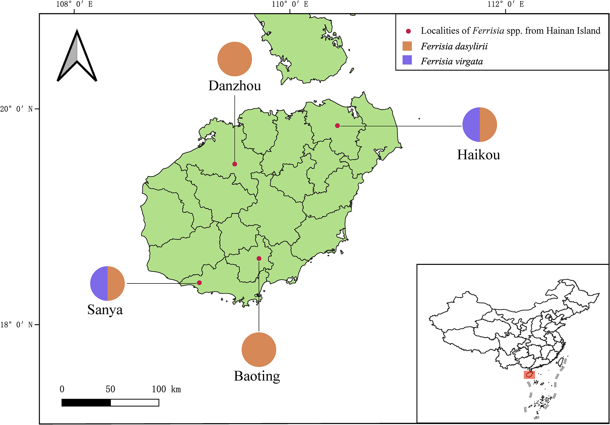
This study is based upon specimens obtained during field surveys on Hainan Island, which experiences a tropical monsoon marine climate characterised by year-round warm temperatures, abundant rainfall, and frequent tropical cyclones. Two field expeditions were conducted: the first from 4 to 7 August 2025 and the second from 13 to 16 September 2025. Mealybug samples were collected on various tropical fruit trees (fig. 1; Table S1), using visual inspection as the primary collection method. All collected specimens were preserved in 100% ethanol and transported to the laboratory for morphological examination and species identification.
Localities of sampling sites.

Figure 1 Long description
The map displays Hainan Island with marked localities of Ferrisia species using red dots. Pie charts represent the distribution of Ferrisia dasylirii (orange) and Ferrisia virgata (purple) in Danzhou, Haikou, Sanya and Baoting. An inset map shows Hainan Island's location within China. The scale bar indicates distances up to 100 km. The map includes latitude and longitude markers for precise location identification.
Species identification
For morphological identification, specimens were slide-mounted using the method described in Williams and Granara de Willink (Reference Williams and Granara de Willink1992). Specifically, slide-mounted specimens were stained in acid fuchsin and mounted in Canada balsam. Species were identified using the keys in Kaydan and Gullan (Reference Kaydan and Gullan2012). Specimen observation, measurement, and photography were conducted using a Leica DM750 microscope. The morphological terms used in our descriptions mainly follow Williams (Reference Williams2004) and Kaydan and Gullan (Reference Kaydan and Gullan2012). Measurements and counts were taken from all the specimens. Measurements are given in micrometres (μm) except for the length and width of the body, which are given in millimetres (mm).
To supplement morphological identification, we amplified the mitochondrial COI gene for molecular analyses. Genomic DNA was extracted from a female adult using a TIANamp Micro DNA Kit (Tiangen Biotech, Beijing, China), following a non-destructive DNA extraction protocol, as described in Taekul et al. (Reference Taekul, Valerio, Austin, Klompen and Johnson2014). According to Li et al. (Reference Li, Wang and Wu2020), there are two regions of the COI gene, the HCO–LCO region and region 2, commonly used in molecular phylogenetic analyses for mealybugs due to different primers used by different researchers. This is also the case for Ferrisia species. Thus, the two regions of the COI gene were also amplified in this study. The HCO–LCO region was amplified with LCO1490/HCO2198 (Folmer et al., Reference Folmer, Black, Hoeh, Lutz and Vrijenhoek1994), while region 2 was amplified with C1-J-2183/C1-N-2568 (Simon et al., Reference Simon, Frati, Beckenbach, Crespi, Liu and Flook1994; Brady et al., Reference Brady, Gadau, Ward, Austin and Dowton2000). Amplicons were directly sequenced in both directions using an Applied Biosystem (ABI) 3730XL from Sangon Biotech (Sanya, China). Chromatograms were assembled into contigs using SeqMan (DNASTAR Lasergene). The assembled sequences were translated to amino acids using the invertebrate mitochondrial code to check for stop codons and frameshifts and were blasted against the GenBank database to check for identification. All sequences generated in this study were deposited in the GenBank under accession numbers listed in Table S1. DNA-extracted specimens were slide-mounted and are deposited in the insect collection of the South China Botanical Garden (SCBG), Chinese Academy of Sciences, Guangzhou, China. Two datasets were used for phylogenetic analyses: the HCO–LCO region dataset and the region 2 dataset. The HCO–LCO region dataset (Table S2) contains published sequences of five Ferrisia species and the newly generated sequences in this study, with Pseudococcus longispinus (Ferris) (Accession no. KY687878 as an out-group. The region 2 dataset (Table S3) contains published sequences of eight Ferrisia species and the newly generated sequences in this study, with P. longispinus (Ferris) (Accession no. AY179439) as an out-group. Sequences were aligned using MUSCLE implemented in MEGA7 (Kumar et al., Reference Kumar, Stecher and Tamura2016). The aligned sequences were then analysed using RAxML as implemented in Geneious ver. 11.0.3 to generate a maximum likelihood (ML) tree. The model used was GTRGAMMA + I. Automatic bootstrapping criterion was selected as the appropriate number of bootstraps; 1000 replicates were run.
Imaging
Multifocal images of slide mounting specimens were produced using a microscopy imaging system of Leica DM750. Photographs of live insects were taken using a Canon 5D Mark IV (Tokyo, Japan) camera mounted with a 100-mm macro lens. Image plates were then post-processed with Adobe Photoshop CS6 Extended.
Results
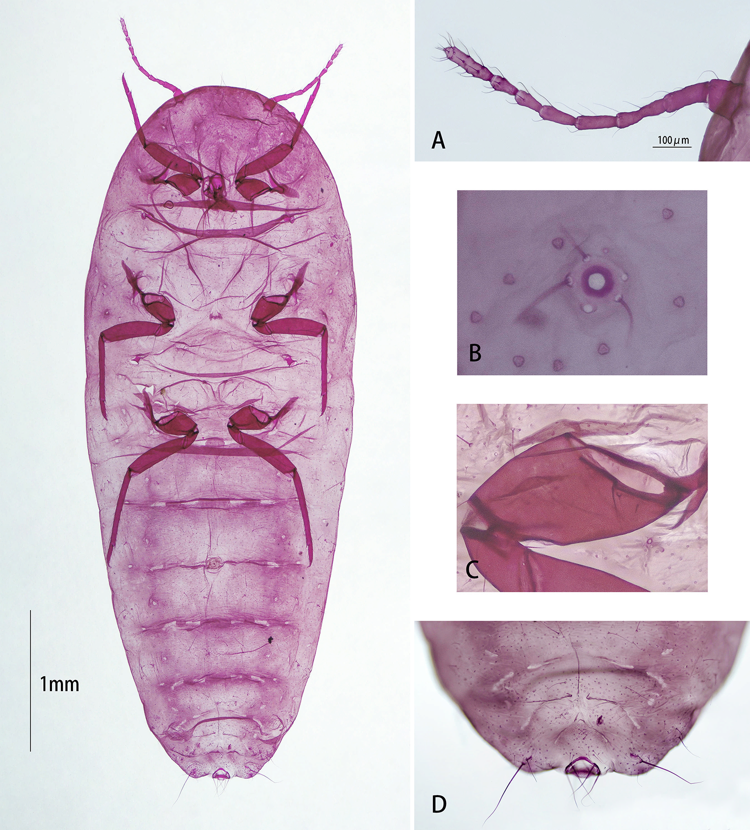
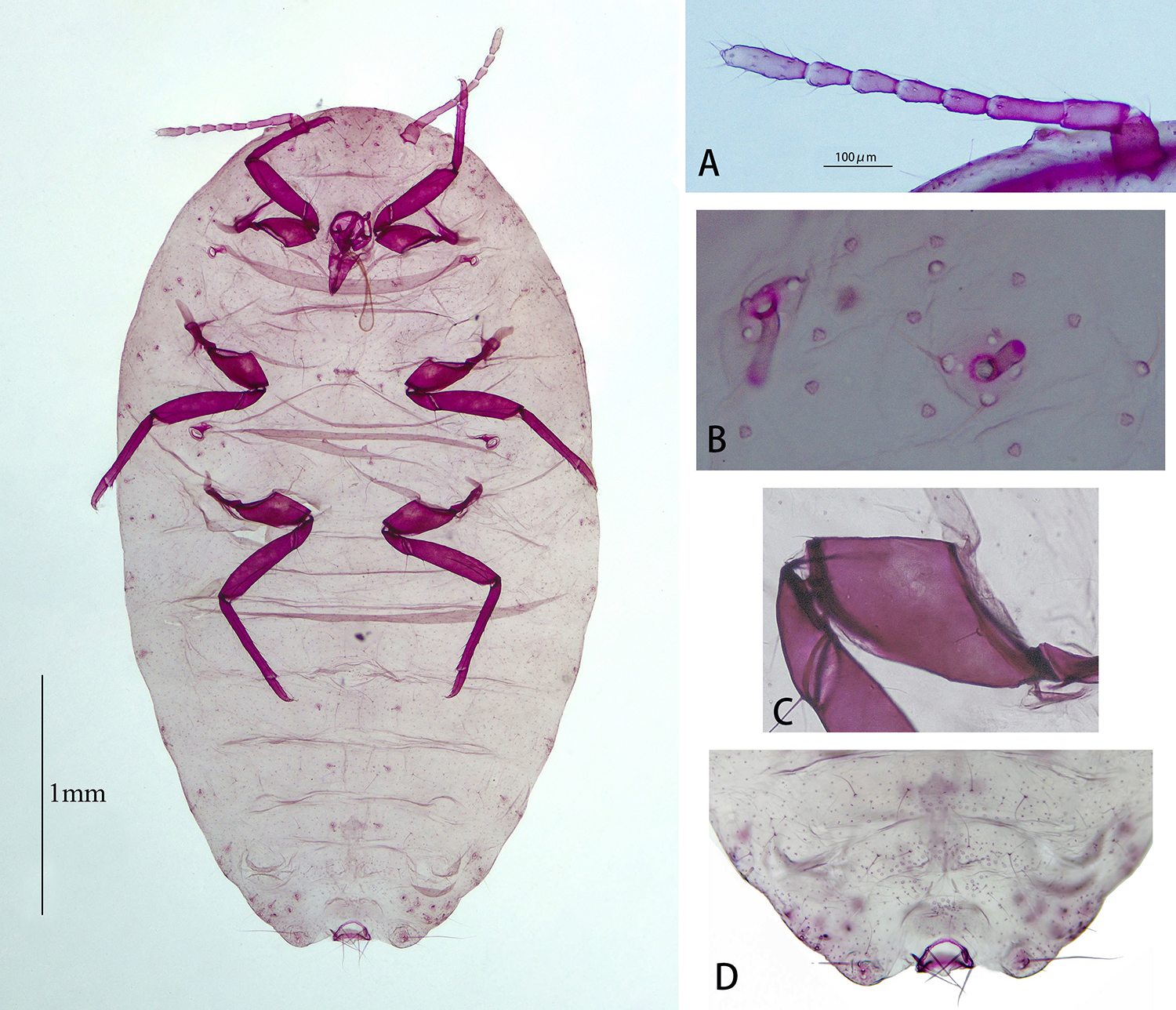
We collected 15 samples from 12 tropical fruit species at four locations in Hainan Province (Table S1). Each sample contained only one morphospecies. Morphological examination of slide-mounted specimens indicates that two species are present: F. dasylirii (fig. 2) and F. virgata (fig. 3). Ferrisia dasylirii represents a new record for China and a description of slide-mounted species is provided below.
Adult female of Ferrisia dasylirii (Cockerell). (A) Antenna. (B) Tubular duct with a minute discoidal pore touching the outer margin of the sclerotised area. (C) Hind coxa. (D) Multilocular disc pores in a row on abdominal segment VI–VIII.

Figure 2 Long description
The image A showing a segmented, curved structure with alternating darker and lighter purple sections on a pale background. A scale bar reads 100 micrometers. The image B showing a pale purple field with several small, round dots and one larger circular structure near the center with a darker ring. The image C showing a darker purple, elongated, folded structure with a smooth edge on a pale background. The image D showing a rounded, darker purple end region with several thin, hair-like strands extending outward. A large central specimen occupies the left side, stained purple on a white background. The body is elongated with a rounded end and multiple transverse segment-like bands. Several darker internal shapes are present near the upper half. A scale bar at the lower left reads 1 millimeter.
Adult female of Ferrisia virgata (Cockerell). (A) Antenna. (B) Tubular duct with a minute discoidal pore touching the inter margin of the sclerotised area. (C) Hind coxa. (D) Multilocular disc pores in a row on abdominal segment VI–VIII.

Figure 3 Long description
The image A showing a purple segmented structure on a pale background, with a scale bar labeled 400 µm. The image B showing several small purple rounded and ring-like structures scattered on a pale background. The image C showing a dark purple elongated structure with a broader central area and a thinner curved extension on a pale background. The image D showing a pale tissue area with faint purple staining and a darker purple curved structure near the lower edge.
Description of slide-mounted specimens of F. dasylirii (fig. 2)
Body elongate oval, 3.45–5.11 mm long (n = 4), 1.42–2.41 mm wide. Antenna 8 segmented, 715–842 μm long; segment I 69–94 μm, segment II 90–113 μm, segment III 128–142 μm, segment IV 81–108 μm, segment V 85–97 μm, segment VI 73–90 μm, segment VII 65–75 μm, apical segment 120–148 μm long. Legs well developed; lack translucent pores on the hind coxa; hind trochanter + femur 561–667 μm long, hind tibia + tarsus 607–711 μm long, hind claw 36–43 μm long. Ratio of lengths of hind tibia + tarsus to hind trochanter + femur 1.04–1.11. Tarsal digitules subequal, each 50–55 μm long. Translucent pores present on hind legs, but absent on coxae. Ostioles: both pairs present; each anterior ostiole poorly developed, with 20–26 trilocular pores and 3–5 setae; each posterior ostiole with 40–57 trilocular pores and 5–8 setae. Anal ring 133–154 μm wide, with 6 anal ring setae, each seta 175–204 μm long.
Dorsum. Anal lobe cerarii each with 2 conical setae, 30–41 μm long, with 50–70 trilocular pores and 4–6 auxiliary setae. Dorsal body setae slender, each 15–57 μm long. Trilocular pores each 4–5 μm in diameter. Enlarged tubular ducts totalling 96–122 on dorsum, each duct 21–36 μm long, 8.0–10 μm wide, rim of duct opening sclerotised, 12–14 μm wide, surrounded by a sclerotised circular area 18–28 μm wide, often enclosing 1 or 2 oval discoidal pores (touching outer margin of sclerotised area and sometimes projecting out from margin) and with 2–7 (generally 3) setae, each 16–29 μm long, usually either within rim or on edge of rim; ducts distributed marginally in clusters of 2–6 on head and thorax, on margins of all abdominal segments in groups of 1–4, but with 8–9 ducts on each side of abdominal segment VII, and also 7–10 medially to submarginally on head and thorax, 5–7 medially to submedially on abdominal segments.
Venter. Body setae slender, each 15–224 μm long, longest setae medially on head; apical setae of anal lobe 323 μm (n = 1). Multilocular disc pores present on posterior abdominal segments only: 10–21 pores on segment VI (usually less than 15; Kaydan and Gullan, Reference Kaydan and Gullan2012), 28–33 on segment VII, 20–28 on segments VIII + IX; each pore 7–8 μm in diameter. Trilocular pores each 4–5 μm in diameter. Minute discoidal pores each 3–4 μm in diameter. Oral-collar tubular ducts on most of venter each 8–10 μm long, 4 μm wide. Small oral-collar tubular ducts each 11–14 μm long, 3–4 μm wide.
Comments. According to the diagnostic characteristics described by Kaydan and Gullan (Reference Kaydan and Gullan2012), F. dasylirii can be distinguished from F. virgata by the following features, all of which are evident in our Hainan specimens: (i) discoidal pores are on the outer margin of the sclerotised area around the rim of the dorsal enlarged tubular ducts on the abdomen (2B) and often each pore and its surrounding sclerotisation projects out from the margin (in F. virgata discoidal pores associated with the sclerotised area around the rim of the dorsal enlarged tubular ducts on the abdomen usually not touching the outer margin of the sclerotised area and almost never projecting out from margin; 3B); (ii) antennae usually >600 μm long with apical segment 120–148 μm long (usually <600 μm long with apical segment 105–120 μm long in F. virgata); (iii) venter of abdominal segment VI usually with ≤15 multilocular disc pores typically forming a single, sometimes irregular, row (in F. virgata ≥15 pores, usually forming at least a partial double row medially); and (iv) translucent pores usually absent on hind coxa (in F. virgata present on dorsal surface of hind coxa, especially posterolaterally).
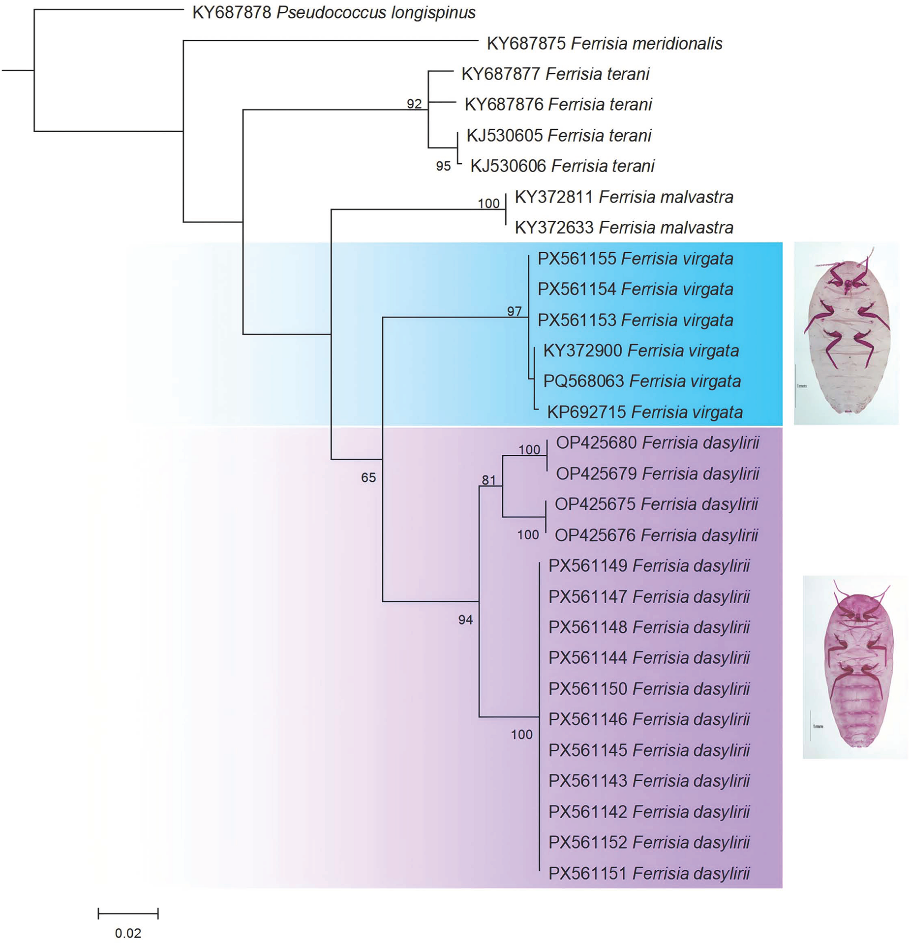
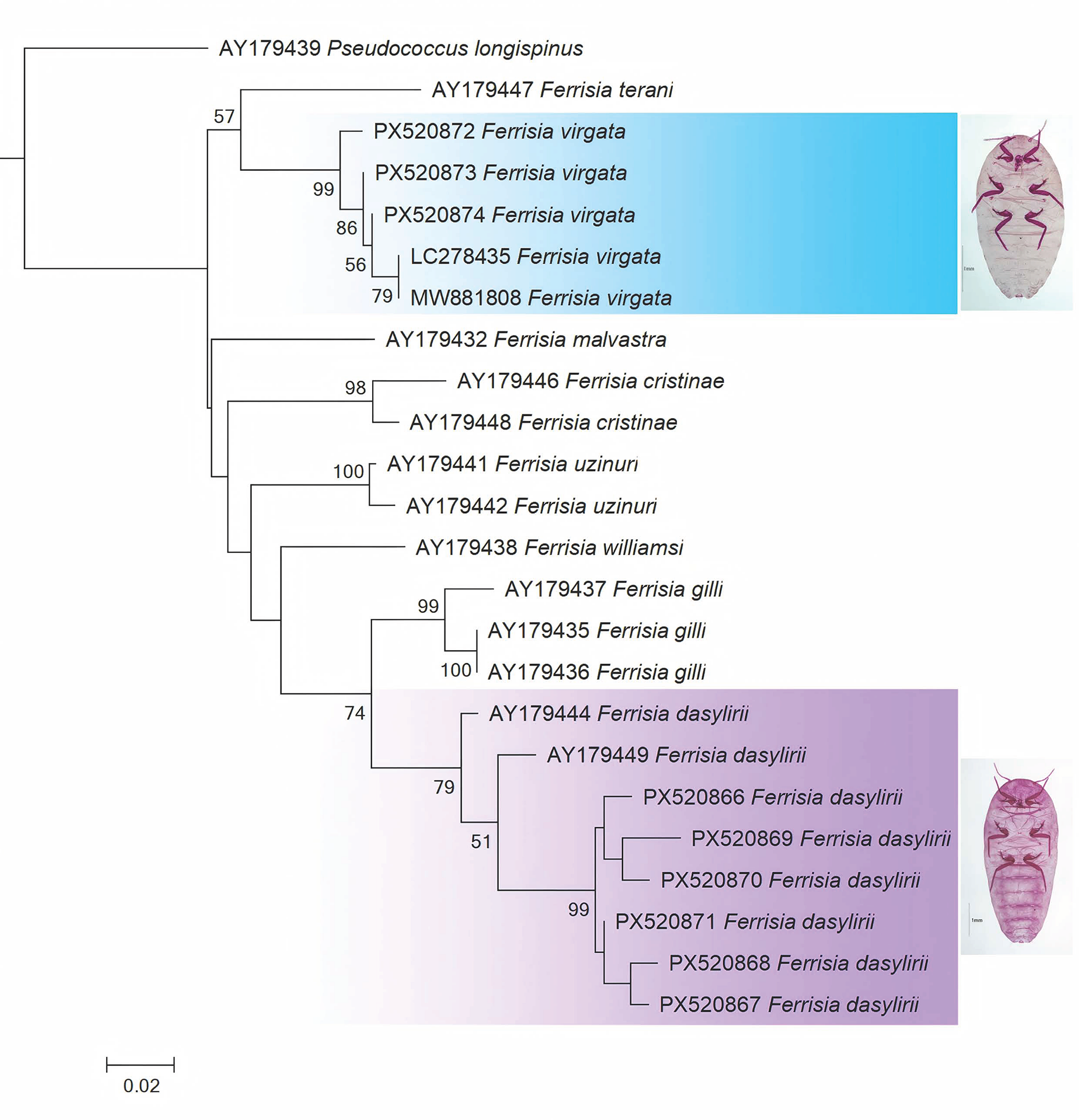
The morphological identification of the two Ferrisia species found in Hainan was also well supported by the molecular analyses of the HCO–LCO region and region 2 of the COI gene. We generated 11 sequences of F. dasylirii and 3 sequences of F. virgata for the HCO–LCO region. All the newly generated sequences for each species are identical, respectively. When blasted in the GenBank database, the HCO–LCO region sequences obtained in this study showed high consistency with morphological identification. Specifically, the sequences of F. dasylirii and F. virgata exhibited maximum similarity scores of 99.84% and 100%, respectively, with their corresponding referenced sequences. Phylogenetic analysis of the HCO–LCO region dataset showed that F. dasylirii and F. virgata, as well as another previously recorded Ferrisia species (Ferrisia malvastra) in China, were well separated as distinct species (fig. 4). For the COI region 2, six and three sequences were generated for F. dasylirii and F. virgata, respectively. Although not all identical, all the newly generated sequences for each species show more than 97.5% similarity among sequences. When blasted in the GenBank database, the region 2 sequences obtained in this study demonstrated ambiguous species discrimination: the F. dasylirii sequence achieved a 100% match with F. virgata (Accession no. PV050943), while the F. virgata sequence matched equally (100%) to both F. virgata (Accession no. PP275761) and F. dasylirii (Accession no. PV050944). These findings indicate a significant historical confusion between these two species in previous studies. Nevertheless, phylogenetic analysis of the region 2 dataset showed that F. dasylirii and F. virgata, as well as F. malvastra, were also well separated as distinct species (fig. 5). The morphological differences among F. dasylirii, F. virgata, and F. malvastra (Table S4) are highly consistent with the distinct clades identified in the COI phylogenies (figs. 4 and 5).
Maximum likelihood tree based on the COI HCO_LCO dataset, only values >50 for bootstrap are labelled. The number under the scale bar indicates the number of expected nucleotide substitutions per site.

Figure 4 Long description
The diagram is a phylogenetic tree illustrating the relationships among various Ferrisia species based on genetic data. The tree is oriented vertically, with branches extending from top to bottom. At the top, the outgroup KY687878 Pseudococcus longispinus is shown, followed by KY687875 Ferrisia meridionalis. Below, several branches represent Ferrisia terani, including KY687877, KY687876, KJ530605 and KJ530606, with bootstrap values of 52 and 92 indicating support for these groupings. Further down, KY372811 and KY372833 Ferrisia malvastra are grouped together with a bootstrap value of 101. The tree then splits into two main clades. The left clade, shaded in blue, includes PX561155, PX561153, PX561154, KY372809, PQ568283 and PQ568215 Ferrisia virgata, with a bootstrap value of 97. An image of Ferrisia virgata is shown adjacent to this clade. The right clade, shaded in purple, includes OP425680, OP425679, OP425678, OP425677, OP425676, PX561148, PX561147, PX561146, PX561145, PX561144, PX561143, PX561142, PX561141 and PX561151 Ferrisia dasylirii, with bootstrap values of 65 and 94. An image of Ferrisia dasylirii is shown next to this clade. The scale bar at the bottom indicates 0.02 expected nucleotide substitutions per site.
Maximum likelihood tree based on the COI region 2 dataset, only values >50 for bootstrap are labelled. The number under the scale bar indicates the number of expected nucleotide substitutions per site.

Figure 5 Long description
The diagram is a phylogenetic tree illustrating the relationships among various Ferrisia species based on the COI region 2 dataset. The tree is oriented vertically, with branches extending from top to bottom. At the top, AY179439 Pseudococcus longispinus serves as the outgroup. Below it, AY179447 Ferrisia terani branches off, followed by a cluster of Ferrisia virgata sequences labeled PX520287, PX520873, PX520874, LC278435 and MW881808, highlighted in blue. Bootstrap values above 50 are displayed at the nodes, indicating the confidence level of each branch. Moving down, AY179437 Ferrisia malvastra is shown branching separately. Further down, AY179446 and AY179448 Ferrisia cristinae, along with AY179441 Ferrisia uzinuri, form a distinct cluster. AY179438 Ferrisia williamsi branches off next, followed by AY179437 Ferrisia gilli, which is grouped with AY179435 and AY179436 Ferrisia gilli. The lower section of the tree, highlighted in purple, shows AY179444 Ferrisia dasylirii branching off, followed by AY179449 Ferrisia dasylirii and several sequences labeled PX520866, PX520865, PX520870, PX520871, PX520868 and PX520867 Ferrisia dasylirii. The scale bar at the bottom indicates the number of expected nucleotide substitutions per site, set at 0.02. Images of Ferrisia species are displayed on the right, corresponding to the blue and purple highlighted sections.
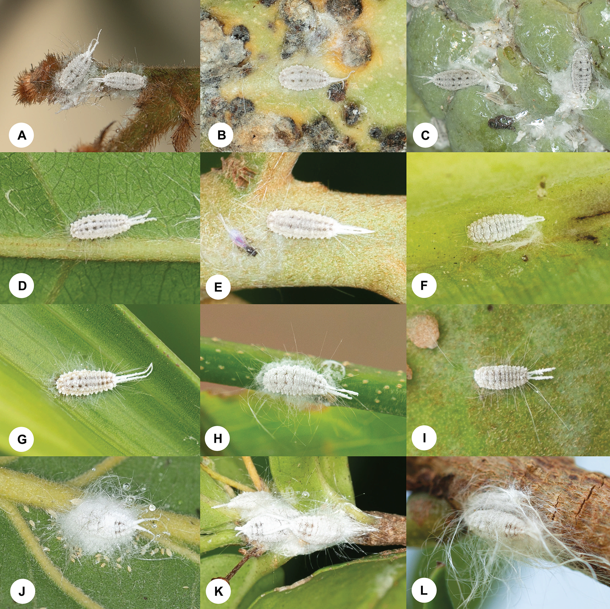
Of the 15 samples we collected, F. dasylirii was found feeding on 12 fruit species at all the four sampling locations, while F. virgata was found feeding on 3 fruit species at only two sampling locations (figs. 1 and 6; Table S1). Both species were found at Sanya and Haikou, but they fed on different fruit plants.
(A–I) Ferrisia dasylirii. (A) Adults on the flower of Davidsonia pruriens F. Muell (Cunoniaceae). (B) Adult on the fruit of Theobroma cacao Linn. (Malvaceae). (C) Adults and nymphs on the fruit of Annona squamosa L. (Annonaceae). (D) Adult on the leaf lower surface of A. squamosa L. (Annonaceae). (E) Adult on the stem of Pouteria campechiana (Kunth) Baehni (Sapotaceae). (F) Adult on the leaf lower surface of Musa nana Lour. (Musaceae). (G) Adult on the leaf upper surface of Areca catechu Linn. (Arecaceae). (H) Adult on the stem of Lecythis pisonis Cambess. (Lecythidaceae). (I) Adult on the fruit of Pouteria sapota (Jacq.) H.E.Moore & Stearn (Sapotaceae). (J–L) F. virgata. (J) Adult and nymphs on the leaf lower surface of Psidium guajava L. (Myrtaceae). (K) Adults on the stem of Malpighia emarginata Sesse & Noc.ex DC. (Malpighiaceae). (L) Adult on the stem of Phyllanthus emblica Linn. (Phyllanthaceae).

Figure 6 Long description
The image A showing a close-up view of a single white, wax-covered insect with long wax filaments at one end, positioned on a plant surface with hairlike plant structures nearby. The image B showing a close-up view of a single white, wax-covered insect on a mottled plant surface. The image C showing a close-up view of two white, wax-covered insects on a plant surface. The image D showing a close-up view of a single white, wax-covered insect on a leaf surface with a prominent vein running across the frame. The image E showing a close-up view of a single white, wax-covered insect on a plant surface with fine hairlike strands around it. The image F showing a close-up view of a single white, wax-covered insect on a smooth plant surface. The image G showing a close-up view of a single white, wax-covered insect on a leaf surface with parallel linear texture. The image H showing a close-up view of a single white, wax-covered insect on a cylindrical plant stem. The image I showing a close-up view of a single white, wax-covered insect on a leaf surface with scattered hairlike strands. The image J showing a close-up view of a white cottony mass on a leaf surface. The image K showing a close-up view of a white cottony mass on a plant stem junction. The image L showing a close-up view of a white cottony mass on a plant surface.
Discussion
This study provides the first confirmed record of F. dasylirii from China, based on combined morphological examination of slide-mounted adult females and mitochondrial COI sequence data. The detection of F. dasylirii on 12 different tropical fruit species across four localities in Hainan (Table S1; figs. 1 and 6) extends the known geographic and host range of this taxon and has immediate taxonomic and phytosanitary relevance for China. As the genus Ferrisia includes economically important, polyphagous pests and several species are morphologically similar (Kaydan and Gullan, Reference Kaydan and Gullan2012; Zhang and Wu, Reference Zhang and Wu2016), the co-occurrence of F. dasylirii and F. virgata in Hainan highlights the necessity of integrative approaches to avoid misidentification that could compromise pest management and quarantine decisions.
Ferrisia dasylirii was first described by Cockerell in 1896 but was later synonymised with F. virgata by Ferris (Reference Ferris1953), due to high morphological similarity and overlapping diagnostic characteristics. In the taxonomic revision by Kaydan and Gullan (Reference Kaydan and Gullan2012), morphological and molecular data clearly demonstrated that F. dasylirii represents a distinct species. Morphological characters observed in our slide-mounted specimens (notably the position of minute discoidal pores relative to the sclerotised rim of dorsal enlarged tubular ducts, typically longer antennae and apical antennal segment, the number/distribution of multilocular disc pores on venter abdominal segment VI, and absence of translucent pores on the hind coxa) match diagnostic differences used to separate F. dasylirii from F. virgata and agree with the distinctions emphasised by Kaydan and Gullan (Reference Kaydan and Gullan2012) (figs. 2 and 3). These characters are useful under careful slide-mounted examination, but morphological separation can be difficult in the field and subject to observer error. The molecular results further reinforce the identification while underscoring limitations of single marker reliance. The COI HCO–LCO region produced sequences that were identical within each species in our samples and yielded very high similarity scores in GenBank searches and clear resolution in the ML analysis (fig. 4). By contrast, the COI region 2 sequences showed more ambiguous BLAST matches (100% identity to sequences labelled as both F. virgata and F. dasylirii in GenBank) despite species specific clustering in the ML tree (fig. 5). This outcome corroborates the research by Li et al. (Reference Li, Wang and Wu2020), which demonstrated that the HCO–LCO region is more reliable than region 2 for species identification and phylogenetic analysis in Pseudococcidae, as it provides clearer genetic differentiation and higher accuracy in reflecting true evolutionary relationships. Taken together, our data demonstrate that the inclusion of the COI HCO–LCO region is valuable for routine barcoding of Ferrisia spp. and robust species delimitation should combine multiple loci (mitochondrial and nuclear markers), morphology, and where possible, broader geographic sampling and voucher-associated sequences.
The frequent occurrence of F. dasylirii across four sampled localities and 12 host species in Hainan suggests a recent but already well-established introduction with broad host associations, or a longer-term under-detected presence that was previously misidentified as F. virgata, or a combination of both. Our limited temporal and spatial sampling cannot discriminate among these scenarios, but the breadth of hosts recorded (Table S1) is consistent with the polyphagy reported for Ferrisia spp. elsewhere (Kaydan and Gullan, Reference Kaydan and Gullan2012; Rondelli et al., Reference Rondelli, Peronti, Dias, Fogaça, Dos Santos and Nery2018; Zarkani et al., Reference Zarkani, Apriyanto, Turanli and Kaydan2020, Reference Zarkani, Fauzi, Apriyanto and Kaydan2023), and indicates potential for rapid spread in Hainan’s tropical agroecosystems. By contrast, F. virgata in our survey was less widespread (three host species at two locations), but this does not necessarily reflect overall abundance differences outside the sampled sites. Although the precise pathway of introduction cannot be determined from our current data, the occurrence of F. dasylirii on a diversity of cultivated fruit species and at multiple localities suggests plant material movement (nursery stock, traded fruit, or ornamentals) as plausible pathways. Hainan’s active horticultural trade and plant introductions create multiple opportunities for mealybug translocations (Cai et al., Reference Cai, Ao, Meng, Xu, Wei and Yang2023; Xi et al., Reference Xi, Yan, Liu, Cai and Wu2024). Given the known capacity of mealybugs to be transported on plant material and packaging, and their potential to vector plant pathogens, the detection of F. dasylirii warrants rapid attention from quarantine and plant health authorities.
In conclusion, the combined morphological and molecular evidence presented here demonstrates that F. dasylirii occurs in Hainan Province, China, expanding the known distribution and host associations of this species. This record underscores the practical value of integrative taxonomy for accurate identification of cryptic, invasive mealybugs and the importance of voucher-based molecular references for surveillance and quarantine. Immediate follow-up actions such as expanded surveys, molecular confirmation of museum and recent collections, and engagement with plant health authorities, will be important to clarify the invasion history, assess the pest risk, and design appropriate management responses.
Supplementary material
The supplementary material for this article can be found at https://doi.org/10.1017/S000748532610100X.
Acknowledgements
We thank Zetao Chen (SCBG) and Xinyu Ding (Hainan Golden Palms EcoTechnology Development Co., Ltd) for their help in collecting samples.
Author contributions
Conceptualization: HJL, HYC; Methodology: HJL, SBH, ZXL, HYC; Software: HJL, ZXL, HYC; Data curation: HJL, SBH, ZXL, HYC; Investigation: HJL, ZXL, HJH, HYC; Validation: HYC; Formal analysis: HJL, SBH, ZXL, HYC; Visualization: HJL, SBH, ZXL, HYC; Project administration: HYC; Resources: HYC; Writing – original draft: HJL, SBH, HYC; Writing – review and editing: HJL, SBH, ZXL, HJH, HYC; Supervision: HYC; Funding acquisition: HJL, HYC. All authors have read and approved the final manuscript.
Financial support
This study was supported by the National Key R&D Program of China (2021YFC3100403), the Natural Science Foundation of Hainan Province (325MS100), and the Research Projects of the Hainan Academy of Agricultural Sciences (HAAS2024KJCX02).
Competing interests
There is no conflict of interest.
Data availability statement
All data are available in this paper.
Ethical approval
Not applicable.