INTRODUCTION
Infestation with the poultry red mite, Dermanyssus gallinae De Geer, costs the poultry industry an estimated €130 million per annum in the EU and has important animal welfare implications for laying hens as a result of anaemia, increased irritation and restlessness, feather-pecking and an increased incidence of cannibalism (Chauve, Reference Chauve1998; Van Emous, Reference Van Emous2005). Poultry mites have also been implicated as intermediate hosts for a number of important diseases (Valiente Moro et al. Reference Valiente Moro, De Luna, Tod, Guy, Sparagano and Zenner2009). Controlling mite populations is currently a major problem to the egg-producing industry, with most acaricides affording only a limited or short-lived reduction in the population of mites. Moreover, the withdrawal of current, effective acaricides along with the emergence of resistance to previously effective acaricides has exacerbated these problems with mite control (e.g. see Marangi et al. Reference Marangi, Cafiero, Capelli, Camarda, Sparagano and Giangaspero2009). As an alternative control strategy, vaccination offers advantages including prolonged efficacy, freedom from chemical residues/environmental pollution and reduced risk of resistance. It is now recognized that vaccines to blood-feeding ectoparasites can result in effective and sustainable control (de la Fuente and Kocan, Reference de la Fuente and Kocan2003; Willadsen, Reference Willadsen2004), including the commercial tick vaccine developed using the protective Bm86 immunogen (Willadsen, Reference Willadsen2004). Our previous work (Bartley et al. Reference Bartley, Nisbet, Offer, Sparks, Wright and Huntley2009; Wright et al. Reference Wright, Bartley, Nisbet, McDevitt, Sparks, Brocklehurst and Huntley2009) along with work published from other research groups (e.g. Harrington et al. Reference Harrington, Canales, de la Fuente, de Luna, Robinson, Guy and Sparagano2009a,Reference Harrington, Din, Guy, Robinson and Sparaganob) has clearly established that vaccination against the poultry red mite, using both native and recombinant antigens, is a feasible objective.
The identification of potential vaccine candidates by a ‘rational’ approach has been advocated in the field of ectoparasite vaccine for several years, with the 2 caveats that, to be practically useful, we must: (i) understand what molecules are truly essential to ectoparasite survival, and (ii) demonstrate that these molecules are accessible to the host immune system (Willadsen, Reference Willadsen2004). Thus far, for D. gallinae, candidate antigens have been identified through either a ‘pragmatic’ approach of fractionating native protein extracts of the mites and using these as vaccines (Wright et al. Reference Wright, Bartley, Nisbet, McDevitt, Sparks, Brocklehurst and Huntley2009) or a ‘rational-type’ approach of selecting suitable antigens based on homology with protective antigens from other species of ectoparasite (Bartley et al. Reference Bartley, Nisbet, Offer, Sparks, Wright and Huntley2009). A third approach, using antigens from ticks, without knowledge or evidence for the presence of the orthologous protein in D. gallinae, has also been attempted (Harrington et al. Reference Harrington, Canales, de la Fuente, de Luna, Robinson, Guy and Sparagano2009a).
We have previously demonstrated the presence of proteolytic enzymes, with activity against blood components, in D. gallinae extracts (Nisbet and Billingsley, Reference Nisbet and Billingsley2000) and shown that feeding inhibitors of these enzymes to the mites significantly increased their mortality (McDevitt et al. Reference McDevitt, Nisbet and Huntley2006). In addition we have demonstrated that immunoglobulins ingested during normal blood-feeding were present throughout the digestive system of the mites (Nisbet et al. Reference Nisbet, Huntley, MacKellar, Sparks and McDevitt2006). Taken together, these previous studies suggest that identification of vaccine candidates by a rational process of identifying feeding-induced molecules, in particular digestive enzymes, is a valid route to satisfying the criteria identified by Willadsen (Reference Willadsen2004). The aims of the work presented here were therefore to identify protease-encoding gene sequences of D. gallinae and to test the efficacy of antibodies raised against recombinant versions of these proteinases on the survival of mites in in vitro feeding assays.
MATERIALS AND METHODS
Mite collection and conditioning
Dermanyssus gallinae of mixed stage and gender were collected from a local commercial egg production unit. Mites were examined using light microscopy and deemed as ‘fed’ if more than 60% of mites appeared to contain fresh, bright red blood in their gut. Those for use as ‘fed’ mites were immediately snap-frozen in liquid nitrogen within 4 h of collection. ‘Starved’ mites were conditioned, without feeding, for 21 days as described previously (Wright et al. Reference Wright, Bartley, Nisbet, McDevitt, Sparks, Brocklehurst and Huntley2009). To extract RNA, mites were homogenized in liquid nitrogen using a pre-cooled mortar and pestle. TRIZOL® reagent (Invitrogen) was added, and the mites were ground to a fine powder after solidification. Total RNA was isolated according to the manufacturer's protocol. After DNaseI treatment and subsequent purification using RNeasy spin columns (Qiagen), mRNA was purified from total RNA using the Poly(A)Purist™ kit (Ambion).
Suppression subtractive hybridization (SSH) to enrich feeding-specific genes
SSH was performed according to the manufacturer's protocol (PCR-Select™ cDNA subtraction kit, Clontech) with minor modification (see Nisbet et al. Reference Nisbet, Redmond, Matthews, Watkins, Yaga, Jones, Nath and Knox2008). Briefly, this procedure involved the synthesis of cDNA, from mRNA extracted from fed or starved mites, by reverse transcription, digestion of the cDNA with endonuclease RsaI and ligation of adapters to the digested fragments to produce ‘tester’ cDNA for fed mites. The ‘fed tester cDNA’ was then hybridized with an excess of ‘starved driver cDNA’ to remove common cDNA species from the adapter-ligated, fed cDNA. Hybridized cDNAs were then subjected to PCR to amplify differentially expressed cDNAs. PCR-amplified, fed-specific cDNA, produced by SSH, was ligated into the pGEM®-T Easy vector (Promega) and transformed into competent Escherichia coli (strain JM109, Promega). Individual positive clones were picked (based on blue/white selection), plasmid was purified and sequenced using the M13 forward primer. Nucleotide sequences were compared with those in public databases, including the GenBank non-redundant database, using the Basic Local Alignment Search Tool (BLASTn and BLASTx) programme from the National Center for Biotechnology Information.
Amplification and sequencing of the D. gallinae cathepsin D and cathepsin L genes
Expressed sequence tag (EST) sequences, from the ‘fed-specific’ SSH library, with homology to cathepsin D and cathepsin L proteins from a variety of organisms, were selected for further analysis. Rapid amplification of cDNA ends (RACE) was performed to obtain the complete coding sequence of the D. gallinae cathepsin D and cathepsin L genes (Dg-CatD-1 and Dg-CatL-1). RNA from ‘fed’ mites was purified as detailed above and 5′ and 3′ RACE-ready cDNAs were generated from this material using the SMART™ RACE cDNA Amplification Kit (Clontech). RACE was performed with the Dg-CatD-1 and Dg-CatL-1 gene-specific oligonucleotide primers CatD-F1 (5′-GAGCTCACTCCCCTCGATTACGTCGTC-3′), CatD-R2 (5′-CTTGGAAGGCCAACACACAGAAGGTTTGGC-3′), CatL-F1 (5′-CCGCTTACAGACTTCGTTCAGAACAGGC-3′), and CatL-R1 (5′-GCCTGTTCTGAACGAAGTCTGTAAGCGG-3′), following the manufacturer's touchdown RACE protocol. RACE products were ligated into pGEM®-T Easy vector, transformed into competent JM109 E. coli cells (Promega) and, following plasmid cultivation and extraction, DNA sequences were obtained using automated sequencing with vector-specific primers (Eurofins MWG Operon). Consensus sequences were compiled using SeqMan software (DNAstar Inc.) and Dg-CatD-1 and Dg-CatL-1 DNA sequences were deposited in EMBL under Accession numbers HE565350 and HE565351. Further analyses were carried out with the SignalP algorithm (Emanuelsson et al. Reference Emanuelsson, Brunak, von Heijne and Nielsen2007) to predict the presence of signal peptides and Prosite to identify protein domains and active sites (de Castro et al. Reference de Castro, Sigrist, Gattiker, Bulliard, Langendijk-Genevaux, Gasteiger, Bairoch and Hulo2006; Sigrist et al. Reference Sigrist, Cerutti, de Castro, Langendijk-Genevaux, Bulliard, Bairoch and Hulo2010). Predictaprotein (Rost et al. Reference Rost, Yachdav and Liu2004), NetNGly (Gupta and Brunak, Reference Gupta and Brunak2002) and NetOGly (Julenius et al. Reference Julenius, Mølgaard, Gupta and Brunak2005) were employed to identify putative post-translational modification sites.
Semi-quantitative gene expression analysis of Dg-CatD-1 and Dg-CatL-1
RNA was isolated from ‘fed’ and ‘starved’ mites as described above. First-strand cDNA was synthesized from 4 μg RNA using 500 ng oligo(dT)12−18 primer and SuperScript™ III Reverse Transcriptase (Invitrogen) according to manufacturer's guidelines. Prior to gene-specific PCR amplification, residual RNA was removed using RNAse H treatment (Clontech). PCR reactions were performed with Platinum® Taq DNA polymerase (Invitrogen), 2 μl of first-strand fed or starved cDNA template, 0·2 mM dNTPs (Promega) and 0·2 mM oligonucleotide primers designed to generate short amplicons of Dg-CatD-1 (CatD-F 5′-GAGCTCACTCCCCTCGATTA-3′ and CatD-R 5′-ACATCATGCAACACGACGTT-3′, product size 205 bp), Dg-CatL-1 (CatL-F 5′-GAGCAGGACGTTGTGGAC-3′ and CatL-R 5′-TAGCGGGCCCTTAGAGAC-3′; product size 276 bp) and 2 house-keeping genes previously shown to be constitutively expressed in fed and starved mite populations (unpublished observation): M16 (M16-F 5′-GATTCGCTAACCCGTTCTCA-3′ and M16-R 5′-TCGTCGATATCATTGCCAAC-3′; product size 327 bp) and Fun14-like (Fun14-F 5′-TAACTGGCTCACCCGAACTC-3′ and Fun14-R 5′-CTTTCTCCACAGCCTTCCAG-3′; product size 208 bp). All reactions were simultaneously amplified for 1 cycle of 94°C for 30 sec and 23 cycles of 94°C for 30 sec, 54°C for 30 sec, 72°C for 30 sec. Equal volumes of each PCR product were examined by electrophoresis using a 0·8% (w/v) agarose gel containing GelRed™ (Biotium) and relative gene expression levels estimated by visual comparison of the band intensity of fed and starved mite PCR products. The constitutively expressed genes M16 and Fun14-like were included as controls to confirm equal addition of template to the RT-PCR reactions and equal product loading of the gel. The rRNA subunit fragments present in total RNA from fed and starved mites were also visualized to demonstrate template integrity.
Expression of recombinant Dg-CatD-1 and Dg-CatL-1 proteins
The coding regions (CDS) of Dg-CatD-1 and Dg-CatL-1, minus the predicted signal peptides, were amplified by PCR with the oligonucleotide primers CatDpet22-F3 (5′-TCGTAGAATTCCGATCTCATCAGGGTGCCTCTG-3′), CatDpet22-R3 (5′-TCGGCTCGAGAGCGGCATCGGCAAAGCCAACG-3′), CatLpet22-F2 (5′-AATTCGGATCCGTCTCCGGAGGCGGCGGAAAG-3′) and CatLpet22-R1 (5′-CCGCAAGCTTCTCGACGAAAAAGACACCAAAC-3′), which incorporated EcoRI, XhoI, BamHI and HindIII restriction site (italicized) to facilitate in-frame and directional ligation into the cloning site of the pET22b(+) expression plasmid (Novagen). PCR was performed using the Advantage® 2 PCR Kit (Clontech) according to manufacturer's instructions with the cycling conditions: 25 cycles of 94°C for 30 sec, 64°C (Dg-CatD-1) or 57°C (Dg-CatL-1) for 30 sec, 72°C for 1 min, followed by one cycle of 72°C for 7 min. The PCR products were ligated into pGEM T-Easy vector and plasmid DNA purified and DNA sequenced (as described previously). Plasmid clone DNA was digested with EcoRI and XhoI (Dg-CatD-1) and HindIII and BamHI (Dg-CatL-1) (Roche) and the excised coding region ligated into pET22b(+) vector using the Rapid Ligation Kit (Promega).
Expression of the recombinant proteins (rDg-CatD-1 and rDg-CatL-1) was carried out in BL21-CodonPlus®(DE3)-RIPL E. coli cells (Stratagene) and the protein purified from the insoluble inclusion body by binding to His-Trap™ HP columns under 8 M urea reducing conditions as described previously (Bartley et al. Reference Bartley, Nisbet, Offer, Sparks, Wright and Huntley2009). Purified rDg-CatD-1 and rDg-CatL-1 proteins were eluted from His-Trap™ HP columns with imidazole concentrations ranging from 350 to 500 mM.
Generation of immunoglobulins specific for Dg-CatD-1 and Dg-CatL-1
Two female Lohmann Brown hens were injected intra-muscularly with 25 μg rDg-CatD-1 and 2 hens with 25 μg rDg-CatL-1. The recombinant proteins were diluted in PBS with 200 μg QuilA adjuvant in a final volume of 500 μl. Two further injections were administered 2 and 4 weeks later. IgY was purified from the yolks of eggs laid prior to injection (negative control IgY) and 2 weeks after the third injection (positive IgY) using the Eggstract kit (Promega) according to the manufacturer's instructions. Purified IgY was reconstituted in phosphate-buffered saline (PBS) and stored at −20°C. Care of animals and experimental procedures were carried out with institute ethics committee approval in accordance with United Kingdom Animals (Scientific Procedures) Act, 1986.
PAGE and Western blotting
Soluble, membrane-associated, integral-membrane and insoluble D. gallinae proteins were prepared from fed mites using sequential fractionation in PBS, Tween-20 (Sigma), Triton-X100 (Sigma) and 8 M urea following a method described previously (Wright et al. Reference Wright, Bartley, Nisbet, McDevitt, Sparks, Brocklehurst and Huntley2009). Native and recombinant proteins were separated by electrophoresis on 4–12% NuPAGE® Novex Bis-Tris Gels minigels (Invitrogen) under reducing conditions using the NuPAGE® reducing agent and 2-(N-Morpholino)ethanesulphonic acid (MES)-SDS running buffer according to the manufacturer's guidelines. Protein bands were visualized by staining with SimplyBlue™ SafeStain (Invitrogen) and, where necessary, were subjected to MALDI-TOF-MS (Moredun Proteomics Unit) to confirm identity. Western blotting was performed as described previously (Bartley et al. Reference Bartley, Nisbet, Offer, Sparks, Wright and Huntley2009). Immuno-blots were visualized with SIGMAFAST™ 3,3′-Diaminobenzidine (DAB; Sigma) or ECL™ plus Western blotting detection reagent (Amersham) and exposure to X-ray film (Fuji).
In vitro feeding assay
The in vitro feeding device described by McDevitt et al. (Reference McDevitt, Nisbet and Huntley2006), as modified by Wright et al. (Reference Wright, Bartley, Nisbet, McDevitt, Sparks, Brocklehurst and Huntley2009), was employed to test the effect of ingested antibodies on mite survival. Briefly, starved, conditioned mites were fed with fresh, heparinized chicken blood (36 USP-units of heparin sodium salt (Sigma) ml−1 blood) enriched with purified polyclonal chicken IgY (2 mg ml−1) generated against rDg-CatD-1 and rDg-CatL-1 or with IgY from a negative control hen (no immunization with recombinant proteins) which was housed in a mite-free environment. Ten in vitro feeding devices were set up for each treatment group. After a 24-h feeding period, mites that had recently fed were pooled according to treatment group and transferred into a 96-well plate (1 mite per well) for observation. Isolated fed mites were immediately scored as dead/alive and scored thereafter every 24 h. The experiment was repeated on 3 separate occasions.
Statistical analysis
Data were analysed using 2 different statistical approaches. A survival analysis was conducted based on Cox's proportional hazards model with frailty function that ignored interval censoring. Mites that survived after 120 h were treated as censored data. The frailty function incorporated experiment as a random effect, with the estimate of random effect obtained using the restricted maximum likelihood method, assuming a Gaussian distribution.
In the second approach, the cumulative proportions of dead mites in each treatment group at the end of the 120 h monitoring period were analysed with a generalised linear mixed model. The model incorporated treatment group as a fixed effect and experiment as a random effect. The generalised linear mixed model was fitted using a Binomial distribution and logit link function. Daily mortality rates (number of dead mites/number of mites at risk) in each treatment group at 24 and 48 h were also analysed separately with a generalised linear mixed model in order to examine differences in mean mortality between groups at different time-points.
For both approaches, treatment groups were compared and the estimated P-values were adjusted (P F value) using the False Discovery Rate (FDR) approach (Benjamini and Hochberg, Reference Benjamini and Hochberg1995) to allow for multiple comparisons of treatments. All statistical analyses were carried out using the R software version 2.13.1 (R Development Core Team, 2011).
RESULTS
Suppression subtractive hybridization (SSH) to enrich feeding-specific genes
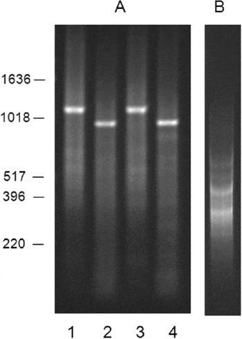
The process of SSH effectively removed cDNA molecules representing transcripts expressed in both ‘fed’ and ‘starved’ D. gallinae (Fig. 1). In particular, one highly abundant cDNA species of ca. 1050 bp (lanes 1 and 3, Fig. 1A (ca. 950 bp following RsaI digestion during SSH, lanes 2 and 4)) was absent from the ‘feeding-specific’ cDNA pool after the SSH procedure (Fig. 1B). It is thought that this cDNA represents mitochondrial 16S ribosomal RNA which forms a large percentage of the molecules present in D. gallinae cDNA, irrespective of the method of synthesis (Bartley et al. unpublished). In total, 770 individual sequences were generated from the feeding-specific SSH library and were assembled into 310 contigs, 133 of which had significant homology to known proteins. The largest proportion of these sequences (9·2%) had homology to the egg precursor protein vitellogenin and the second highest proportion (2·9%) represented homologues of a vitellogenin proteolytic product, GP80 (Tellam et al. Reference Tellam, Kemp, Riding, Briscoe, Smith, Sharp, Irving and Willadsen2002). Proteinases, peptidases and other metabolic enzymes were highly represented with 7·1% of the total number of individual sequences. From this group of proteolytic enzymes, we selected ESTs representing an aspartyl proteinase (cathepsin D) and a cysteine proteinase (cathepsin L) to generate full-length sequence and use for further study.
Enrichment of feeding-induced gene expression in Dermanyssus gallinae by suppression subtractive hybridization. (A) cDNA synthesized from fed D. gallinae (lane 1) and starved D. gallinae (lane 3). Lanes 2 (fed) and 4 (starved) show the same material after RsaI digestion prior to cDNA subtraction by SSH. (B) cDNA from fed D. gallinae after removal of common transcripts by cDNA subtraction using cDNA from starved mites as the driver.

Sequence analysis of D. gallinae cathepsin D1 and cathepsin L1 (Dg-CatD-1 and Dg-CatL-1)
The CDS of Dg-CatD-1 (Accession number HE565350) was 1152 bp in length, encoding a 383 amino acid (aa) protein (Dg-CatD-1) that exhibited up to 53% identity (4.3E-101) to cathepsin D-like lysosomal aspartyl proteases (peptidase A1 family) from a variety of diverse organisms. The consensus signature sequence of the active site of eukaryotic and viral aspartyl proteases [LIVMFGAC]-[LIVMTADN]-[LIVFSA]-D-[ST]-G-[STAV]-[STAPDENQ]-{GQ}-[LIVMFSTNC]-{EGK}-[LIVMFGTA] was present at 80–89 amino acids (VIFDTGSSDLWV), with the second active site present at 267–278 amino acids (GIADTGTSLFVG) with the active aspartic acid residue (underlined) conserved in both sites. SignalP analysis of Dg-CatD-1 predicted a putative 16 amino-acid N-terminal signal peptide.
The CDS of Dg-CatL-1 (Accession number HE565351) encoded a 341 aa protein (Dg-CatL-1) that exhibited the greatest identity (37%, 2.5E-48) to cathepsin L proteases (C1 peptidase family) derived from the haematophagous arthropods Aedes aegypti (Q1PA55) and Haemaphysalis longicornis (C6L6E2). Prosite motif scanning identified 2 different signature sequences of eukaryotic thiol (cysteine) protease active sites. The sequence located at 133 to 141 amino acid (QLYCGSCYAFAV) site matched with the protease consensus signature sequence Q-{V}-x-{DE}-[GE]-{F}-C-[YW]-{DN}-x-[STAGC]-[STAGCV] which has a cysteine active site (underlined). A second protease signature sequence [FYCH]-[WI]-[LIVT]-x-[KRQAG]-N-[ST]-W-x(3)-[FYW]-G-x(2)-G-[LFYW]-[LIVMFYG]-x-[LIVMF] with an asparagine active site (underlined) matched with amino acids 304 to 323 (YWLIKNSWGTEWGVGGYGKI). The residues Gln133 and His289 which are predicted to be important for C1 peptidase function, were also conserved in Dg-CatL1. A 20 amino acid N-terminal signal peptide was identified by Signal P analysis. In addition, cathepsin L proteins characteristically undergo a self-cleavage event resulting in the formation of mature proteins. The ‘ERFNIN’ motif of non-cathepsin B papain propeptides (Karrer et al. Reference Karrer, Peiffer and DiTomas1993) is mostly conserved in the predicted Dg-CatL-1 proprotein. The cleavage site of Dg-CatL-1 proprotein was predicted by alignment with several characterized cathepsin L proteins, to occur between the Ser114 and Met115. The mature Dg-CatL-1 protein was predicted to be 227 amino acids with a proline in the second amino acid position, which is a conserved feature in most C1 peptidases and is thought to contribute to resistance to proteolysis (Rawlings and Barrett, Reference Rawlings and Barrett1993).
Both Dg-CatD-1 and Dg-CatL-1 are predicted to be glycosylated. A predicted N-linked glycosylation site is present in the pro-region of Dg–CatL-1 at Asn100 (potential=0·7491). Dg-CatD-1 is predicted to be glycosylated at 2 sites: N-linked at position Asn120 (potential=0·729) and O-linked at Thr84 (G=00515).
Differential gene expression of Dg-CatD-1 and Dg-CatL-1 in fed and starved mite populations
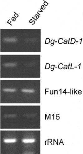
In order to determine whether expression of Dg-CatD-1 and Dg-CatL-1 is increased after feeding, semi-quantitative RT-PCR was performed on total RNA purified from fed and starved mites (Fig. 2). The band intensities for the Dg-CatD-1 and Dg-CatL-1 RT-PCR products are clearly greater in fed mites when compared to starved mites, demonstrating upregulation of Dg-CatD-1 and Dg-CatL-1 post-feeding. In contrast, the comparable band intensity observed in fed and starved mite RT-PCR products for the 2 control genes M16 and Fun14-like supports the previous observation (unpublished) that these genes are constitutively expressed and demonstrates the equal addition of template to all of the RT-PCR reactions and equivalent loading of the RT-PCR products on the gel.
Semi-quantitative RT-PCR demonstrating differential expression of Dg-CatD-1 and CatL-1 genes in fed and starved mites. Equal volumes of RT-PCR products generated from equivalent amounts total RNA purified from fed and starved mites were separated by electrophoresis on a 0·8% (w/v) agarose/TAE gel. The constitutively expressed genes M16 and Fun14-like were included as controls to confirm equal addition of template to the RT-PCR reactions. Total RNA from fed and starved mites was also visualized to demonstrate template integrity and equal loading of the gel.

Expression and characterization of rDg-CatD-1 and rDg-CatL-1 and comparison to native cathepsins
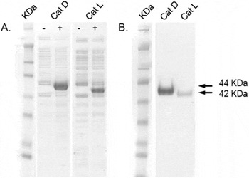
rDg-CatD-1 was produced in large quantities in the bacterial expression system, whilst rDg-CatL-1 was less well expressed. Despite the addition of N-terminal pelB leader sequences and expression at low temperatures; both expressed cathepsin proteins were directed to inclusion bodies from which protein was subsequently purified using nickel affinity columns under denaturing conditions (Fig. 3). The molecular masses of rDg-CatD-1 and rDg-CatL-1, including the pelB leader and C-terminal His fusion, were estimated, from PAGE gel analysis, to be 44 and 42 KDa respectively which correlated well with the calculated predicted masses of 44·2 and 41·1 KDa. The identities of the insoluble recombinant proteins were verified by MALDI-TOF-MS (59% and 51% sequence coverage for rDg-CatD-1 and rDg-CatL-1 respectively). Despite adjustment of expression conditions and the use of different prokaryotic vectors (not shown), soluble forms of the two cathepsins could not be obtained nor could the yield of rDg-CatL-1 be improved.
Expression (A) and purification (B) of Dermanyssus gallinae cathepsin D1 and cathepsin L1 recombinant proteins (rDg-CatD-1 and rDg-CatL-1). Expression of rDg-CatD-1 (Cat D) and rDg-CatL-1 (Cat L) was carried out in the presence (+) and absence (−) of 1 mM isopropyl β-D-1-thiogalactopyranoside (IPTG) Insoluble cell lysate fractions (panel A) and purified rDg-CatD-1 and rDg-CatL-1 (panel B) were separated by electrophoresis on 4–12% (w/v) Bis-Tris Novex gel (Invitrogen) under reducing conditions. Molecular weights were estimated by comparison with pre-stained SeeBlue plus2 markers (Invitrogen).

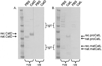
Anti-Dg-CatD-1 IgY, raised in hens using rDg-CatD-1 as an immunogen, showed strong immunoreactivity with rDg-CatD-1 (Fig. 4). A protein corresponding in molecular mass to the native Dg-CatD-1 (39·9 KDa) was detected in the PBS-soluble fraction of mites, but not the Tween-20, Triton-X100 or 8 M urea fractions; suggesting that the natural state of native Dg-CatD-1 is as a soluble protein.
Western blot analysis of recombinant and native Dermanyssus gallinae cathepsin D1 (CatD, blot A) and L1 (CatL, blot B). Samples of 100 ng per well of recombinant CatD-1 (rDg-CatD-1), 200 ng of recombinant CatL-1 (rDg-CatL-1) and 25 μg soluble mite extract per lane were denatured and separated by electrophoresis on a 4–12% BisTris Novex gel (Invitrogen) under reducing conditions prior to transfer to nitrocellulose. Blots were incubated in 40 μg/ml yolk-derived antibodies generated against recombinant cathepsins (+ve) or with negative yolk antibodies (−ve). Specifically bound yolk antibodies were detected with anti-IgY-horseradish peroxidase (HRP)-conjugated antibody (Sigma) and visualized with SIGMAFAST™ 3,3′-diaminobenzidine. Molecular weights of proteins were estimated by comparison with SeeBlue plus2 protein standards. Putative recombinant and native forms of the cathepsins detected by +ve IgYs are indicated by the arrows. Background detection of contaminating heavy and light IgY fragments present in the soluble mite extract (originating from the gut contents of mites) occurred with +ve and –ve antibodies (A and B) and anti-IgYHRP conjugate (not shown) and is indicted by the brackets.

Anti-Dg-CatL-1 antibodies clearly detected the rDg-CatL-1 pro-protein (41·1 KDa) and several smaller proteins (Fig. 4). One of the smaller proteins detected was estimated to be 28 KDa and corresponded to the calculated mass of the C-terminal His-labelled mature rDg-CatL-1 (27·2 KDa). Detection of the 28 KDa protein may indicate that a minority of rDg-CatL-1 pro-protein underwent self-cleavage or intra-molecular cleavage with bacterial proteinases prior to purification. Confirmation of the identity of the 28 KDa protein by MALDI-TOF-MS was not possible due to low abundance. Two further small proteins with an estimated mass of 16 and 17 KDa were recognized by the Anti-Dg-CatL-1 and the identities of these proteins are unknown.
Anti-Dg-CatL-1 antibodies did not visibly detect any bands in the PBS-soluble fraction of mites corresponding in weight to the native pro-protein (calculated MW=38·6 KDa), but did react with a band of approximately 25 KDa corresponding to the calculated MW of the native mature Dg-CatL-1 (25·7 KDa). Detection of the native mature Dg-CatL-1 is visible as a band amongst a background smear of co-localizing light chains of IgY derived from circulating IgY which is also present in the PBS-soluble mite fraction and also bound by the anti-IgY-conjugated antibody.
Immunoreactivity of naturally infested hens to rDg-CatD-1 and rDg-CatL-1
Western blots of rDg-CatD-1 and rDg-CatL-1 were probed with yolk-derived IgY obtained from hens housed for 20 weeks in a mite-infested commercial egg production system to determine whether the hens generated a specific antibody response to these antigens during prolonged natural exposure to mites.
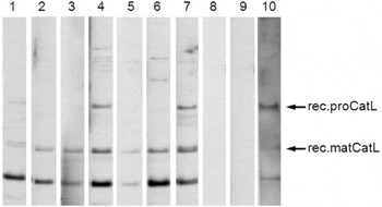
IgY in the eggs of all hens clearly bound to a 25 KDa protein predicted to be the mature rDg-CatL-1 (Fig. 5). An identical profile of detection was observed with the positive control IgY (lane 10, experimental hen injected with rDg-CatL-1). The positive control and commercial hen IgYs also detected 2 additional bands both approximately 15 and 17 KDa in size and unknown identity.
Immunoreactivity of naturally infested hens with recombinant Dermanysus gallinae CatL-1 (rDg-CatL-1). The rDg-CatL-1 (200 ng per well) was separated by electrophoresis on a 4–12% BisTris Novex gel (Invitrogen) and immobilized onto nitrocellulose. The Western blot was probed with 75 μg/ml yolk-derived IgY obtained from hens housed in a mite-infested commercial poultry unit (lanes 1 to 7), a mite-naïve hen (lane 9, negative control) and a hen inoculated with rDg-CatL-1 (lane 10, positive control). Specific binding of IgY to rDg-CatL-1 was detected with anti-IgY-horseradish peroxidase (HRP)-conjugated antibody (Sigma) and chemiluminescence. Lane 8 served as a control for non-specific binding of anti-IgY-horseradish peroxidase (HRP)-conjugated antibody.

The negative control IgY (lane 8, mite-naive hen) and the anti-IgY-horseradish peroxidase (HRP)-conjugated antibody control (lane 9) did not react with any protein present in purified rDg-CatL-1, suggesting that the reactivity observed with the battery hen and positive control IgYs is specific to rDg-CatL-1.
There was no detection of rDg-CatD-1 with the battery hen IgY samples suggesting that none of the hens were exposed to this antigen and that they did not develop specific immunoglobulin to Dg-CatD-1 during natural infestation (results not shown).
Survival of mites following ingestion of anti-cathepsin IgY
The mortality of mites fed with chicken blood enriched with IgY specific for rDg-CatD-1 or rDg-CatL-1 were compared to mites fed on blood enriched with IgY obtained from a mite-naive hen and the effect monitored over a 5-day period. The experiment was repeated on 3 separate occasions with different batches of chicken skin and conditioned mites to control for any variable effects from these parameters. Only fed mites were recovered from the feeding devices, pooled according to group and scored for mortality. The total numbers of fed mites recovered was variable between the groups and between the individual experiments. The group sizes for the 3 experiments are as follows: rDg-CatD-1: n=99, 26, 65; rDg-CatL-1: n=68, 42, 48 and control: n=32, 19, 44.
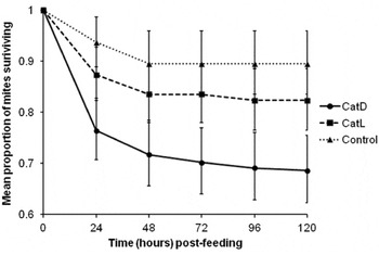
The mean proportion of mites that survived in each treatment group at different time-points (24-h interval) is presented graphically in Fig. 6. The highest mean survival proportion at 24 h was seen in the control group [0·94; (95% confidence interval (CI): 0·89, 0·99)] followed by the rDg-CatL-1 (0·87; CI: 0·82, 0·93) and rDg-CatD-1 (0·76; CI: 0·71, 0·83) groups. A similar pattern was observed at 120 h where the mean survival proportions in descending order were estimated in the control (0·89; CI: 0·84, 0·96), rDg-CatL-1 (0·82; CI: 0·77, 0·89) and rDg-CatD-1 (0·69; CI: 0·62, 0·76) groups. Under Cox's proportional hazards model, the risk that mites in the rDg-CatD-1 group would die at anytime during the experiment was 4·42 times higher than the control group (P F<0·001) and 2·08 times higher than the rDg-CatL-1 group (P F=0·002). Mites in the rDg-CatL-1 group also had a statistically significant 2·13 times higher risk of dying (P F=0·042) than mites in the control group.
The mean survival proportion of Dermanyssus gallinae mites following ingestion of anti-cathespsin IgY. Mites were fed with fresh heparinized chicken blood enriched with 2 mg/ml polyclonal IgY generated against recombinant D. gallinae cathepsin D1 (CatD), cathepsin L1 (CatL) or with control IgY from a mite-naïve hen. Fed mites were recovered from the feeding devices and scored daily for mortality for 5 consecutive days. The experiment was repeated on 3 separate occasions. The total number of fed mites monitored per treatment group for each of the 3 experiments are as follows: CatD: n=99, 26, 65; CatL: n=68, 42, 48; Control: n=32, 19, 44. The mean survival proportion (expressed as a proportion of number of mites survived and number of mites at risk during the specified interval of time) is plotted against time post-feeding. The error bars represent the 95% upper and lower confidence intervals.

Mite mortality after 120 h was analysed in a generalised linear mixed model. The results showed that mites fed on antibodies raised against rDg-CatD-1 had the highest overall mean mortality rate (0·234; CI: 0·153, 0·340) followed by rDg-CatL-1 (0·143; CI: 0·086, 0·230) and control (0·081; CI: 0·040, 0·159) groups. The mean mortality rate in the rDg-CatD-1 group was significantly higher in comparison to the mean mortality rate of rDg-CatL-1 (P F=0·003) and control (P F<0·001) groups. Likewise mites in the rDg-CatL-1 had a higher mean mortality than the control group but this difference was not statistically significant (P F=0·062) at the 120 h time-point.
Individual analysis of the early time-points revealed a statistically significant difference in mean mortality rates between the control group compared to the rDg-CatD-1 and rDg-CatL-1 groups (P F<0·001 and 0·026 respectively) at 24 h. At 48 h; the difference in mean mortality between the control and the rDg-CatD-1 and rDg-CatL-1 groups was not statistically significant (P F=0·929 for both group comparisons).
DISCUSSION
Here we have described the rational selection and in vitro testing of 2 proteolytic enzymes, an aspartyl proteinase and a cysteine proteinase, as vaccine candidate molecules for the control of D. gallinae, based on their inferred role(s) in a critical physiological process, food digestion. In previous studies we have demonstrated the presence of, and have partially characterized, these enzymes in D. gallinae extracts (Nisbet and Billingsley, Reference Nisbet and Billingsley2000) and shown that feeding inhibitors of these enzymes to the mites significantly increased their mortality (McDevitt et al. Reference McDevitt, Nisbet and Huntley2006). Both aspartyl proteinases and cysteine proteinases have shown promise as vaccine candidate molecules in other species of haematophagous parasites: Na-APR-1, an aspartyl proteinase which digests haemoglobin in the gut of Necator americanus is one of the two lead vaccine candidates in the development of a vaccine against human hookworm infection (reviewed by Bethony et al. Reference Bethony, Cole, Guo, Kamhawi, Lightowlers, Loukas, Petri, Reed, Valenzuela and Hotez2011); and the aspartyl proteinases HcPEP-1 and HcPEP-2 are integral components of the galactose-containing glycoprotein complex ‘H-gal-GP’, a highly immunoprotective complex extracted from the gut of Haemonchus contortus (Longbottom et al. Reference Longbottom, Redmond, Russell, Liddell, Smith and Knox1997; Smith et al. Reference Smith, Skuce, Newlands, Smith and Pettit2003). Protection against H. contortus has also been induced in sheep after vaccination with a native extract enriched for cysteine proteinases and the levels of protection were associated with the proteolytic activity of the extract (Knox et al. Reference Knox, Smith, Redmond and Smith2005). Cathepsin D and L proteases have been identified in a variety of tick and mite tissues (Mulenga et al. Reference Mulenga, Sugimoto, Ingram, Ohashi and Onuma1999; Renard et al. Reference Renard, Garcia, Cardoso, Richter, Sakanari, Ozaki, Termignoni and Masuda2000, Reference Renard, Lara, de Cardoso, Miguens, Dansa-Petretski, Termignoni and Masuda2002; Seixas et al. Reference Seixas, Estrela, Ceolato, Pontes, Lara, Gondim and Termignoni2010) and cleave several proteins including haemoglobin, albumin, gelatin and vitellin (Renard et al. Reference Renard, Garcia, Cardoso, Richter, Sakanari, Ozaki, Termignoni and Masuda2000; Seixas et al. Reference Seixas, Dos Santos, Velloso, da Silva Vas, Masuda, Horn and Termignoni2003; Yamaji et al. Reference Yamaji, Tsuji, Miyoshi, Islam, Hatta, Alim, Anisuzzaman, Takenaka and Fujisaki2009; Estrela et al. Reference Estrela, Seixas, Teixeira, Pinto and Termignoni2010). Experimental vaccination with tick cathepsins involved in yolk processing have yielded encouraging results (da Silva Vaz Jr et al. Reference da Silva Vas, Logullo, Sorgine, Velloso, Rosa de Lima, Gonzales, Masuda, Oliveira and Masuda1998; Leal et al. Reference Leal, Seixas, Pohl, Ferreira, Logullo, Oliveira, Farias, Termignoni, da Silva Vas and Masuda2006; Seixas et al. Reference Seixas, Leal, Nascimento-Silva, Masuda, Termignoni and da Silva Vas2008). Proteolytic enzymes therefore represent a promising group of molecules to target for vaccine candidates in both ecto- and endoparasites (reviewed by Nisbet and Huntley, (Reference Nisbet and Huntley2006) and Pearson et al. (Reference Pearson, Ranjit and Loukas2010) respectively).
Production of rDg-CatD-1 and rDg-CatL-1 was successful using a prokaryotic system and the recombinant antigens were used to generate antibody responses in chickens. Both rDg-CatD-1 and rDg-CatL-1 antigens generated measurable IgY responses in chickens as determined by Western blotting. The response against rDg-CatD-1 appeared to be much stronger than against rDg-CatL-1. The difference in response was observed despite injecting hens with equivalent amounts of rDg-CatD-1 and rDg-CatL-1, using twice as much rDg-CatL-1 protein than rDg-CatD-1 on the Western blot and screening with purified IgY of the same concentration. These data indicate that rDg-CatL-1 is less immunogenic than rDg-CatD-1. Similarly, variation in the response to several different Fasiola hepatica recombinant cathepsin L proteins has been observed in rats (Jayaraj et al. Reference Jayaraj, Piedrafita, Dynon, Grams, Spithill and Smooker2009). There may be several reasons for the poor immunogenicity of rDg-CatL-1: rDg-CatL-1 was expressed and purified in the pro-protein form. Incorrectly folded papain-like proteins have been shown to be susceptible to the action of other proteases (Vernet et al. Reference Vernet, Berti, de, Musil, Tessier, Menard, Magny, Storer and Thomas1995), and susceptibility of non-native conformation antigens to proteolysis in the harsh in vivo environment may, in effect, reduce the dose of vaccine and result in a poor response (Jayaraj et al. Reference Jayaraj, Piedrafita, Dynon, Grams, Spithill and Smooker2009). Post-translational processing may also impact on correct conformation and/or immunogenicity. Dg-CatL-1 has one potential N-linked glycosylation site located in the pro-region at Asn−100, which is conserved with the parasitic nematode cathepsin L proteins (Britton and Murray, 2002; Murray et al. Reference Murray, Geldhof, Clark, Knox and Britton2007). The importance of glycosylation of the pro-region of cathepsin L in terms of correct folding and immunogenicity has yet to be determined.
In our analyses we demonstrated that naturally infested hens produced an antibody response against Dg-CatL-1, indicating natural exposure to this antigen. IgY from naturally infested hens detected the rDg-CatL-1 pro-protein and several other smaller proteins present in the purified fraction. These low molecular weight proteins are likely to be Dg-CatL-1–derived because they were specifically detected with both anti-rDg-CatL-1 IgY and IgY from naturally infested hens but not with negative control IgY; nor were these bands seen when the same natural infestation IgY samples were used to screen other recombinant D. gallinae proteins produced using identical expression and purification systems (rDg-CatD-1 (this paper) and rDg-Histamine release factor (Bartley et al. Reference Bartley, Nisbet, Offer, Sparks, Wright and Huntley2009)). Furthermore, 1 protein corresponded with the predicted molecular mass (27·2 KDa) of the mature rDg-CatL-1 protein and raises the question: is rDg-CatL-1 pro-protein being partially processed into its mature form during prokaryotic expression? Alternatively expression of rDg-CatL-1 in a prokaryotic system may result in a low level production of truncated proteins.
Interestingly, detection of the putative mature Dg-CatL-1 protein was stronger with IgY from naturally infested hens than with generated anti-rDg-CatL-1 IgY suggesting that not only is the mature form of Dg-CatL-1 exposed to the host during natural infestation, but it is a highly immunogenic protein; and elicits a vigorous IgY response. Cathepsin L-like molecules are abundant in the excretory/secretory (E/S) products of other parasites, for example adult and juvenile liver flukes (Dalton et al. Reference Dalton, Neill, Stack, Collins, Walshe, Sekiya, Doyle, Mulcahy, Hoyle, Khaznadji, Moire, Brennan, Mousley, Kreshchenko, Maule and Donnelly2003) and it appears that D. gallinae must similarly produce these enzymes extracellularly, potentially in salivary exudate and/or feces.
In contrast to Dg-CatL-1, infested hens did not possess naturally-occurring antibodies against Dg-CatD-1. This latter candidate generated a strong antibody response in hens and induced statistically significantly higher mortality than control and Dg-CatL-1 antibodies in the in vitro feeding assay, suggesting that it is both a ‘hidden’ antigen and a potential vaccine candidate molecule. The potential for use of hidden or ‘concealed’ antigens in ectoparasitic arthropods has, of course, been recognized since the groundbreaking work on Bm86, and has more recently been extended into other species of tick (reviewed by de la Fuente and Kocan, Reference de la Fuente and Kocan2006).
Ingestion of anti-rDg-CatL-1 IgY exhibited a modest but statistically significant (P F=0·042) decrease in survival compared to the control mites when survivability at each time-point was taken into account in the Cox's proportional hazards model. Analysis of overall mean mortality rate at 120h post-feeding in a generalised mixed linear model showed that the difference in mean mortality rates of mites of anti-rDg-CatL-1 and control group was close to statistical significance at the 5% level (P F=0·062). The daily mean mite mortality in the rDg-CatL-1 group was statistically significant at 24 h (P F=0·026) when compared the control group, but not statistically significant at later time-points. These data indicate that the miticidal effect of ingested antibodies occurs rapidly, in the first 24 h following ingestion. The rapid effect of ingested antibodies was also observed in mites fed with anti-rDg-CatD-1 IgY (P F<0·001) and similarly with other anti-mite antibodies (Bartley et al. Reference Bartley, Nisbet, Offer, Sparks, Wright and Huntley2009; Wright et al. Reference Wright, Bartley, Nisbet, McDevitt, Sparks, Brocklehurst and Huntley2009).
There may be several reasons for the weak performance of anti-rDg-CatL-1 IgY in the feeding assay. The rDg-CatL-1 pro-protein appeared to be a poor immunogen and may have failed to induce an IgY titre sufficiently high enough to kill the mites. In addition, IgY generated against rDg-CatL-1 pro-protein demonstrated only limited detection of the putative mature protein when compared with IgY from naturally infested hens. It may be important for the vaccine Dg-CatL-1 antigen to be correctly folded into the active mature protein form and glycoslated in order to induce a vigorous and appropriate immune response capable of disrupting the Dg-CatL-1 function when ingested by mites. The use of native proteins or alternative expression systems (e.g. insect cell/yeast expression) may prove as vital with Dg-CatL-1 antigen production as they have with nematode protease based vaccines where correct conformation and glycosylation are generally required in order to induce good protection (reviewed by Knox et al. Reference Knox, Redmond, Newlands, Skuce, Pettit and Smith2003). If Dg-CatL-1 is secreted as the evidence suggests; the effect of antibodies may not be immediately apparent and may not directly kill the mites. For example, if Dg-CatL-1 is salivary digestive enzyme, disruption of function may lead to long-term effects such as reduced digestion efficiency, fat storage, fecundity and long-term survival. A limitation of the feeding device is that long-term effects are not measurable. Secreted cathepsin L has other defined functions in parasites, for example host immune-modulation in Fasiola hepatica infection (Dowling et al. Reference Dowling, Hamilton, Donnelly, La Course, Brophy, Dalton and O'Neill2010), if Dg-CatL-1 has a similar function it would likewise not be measurable in the feeding assay.
Harrington et al. (Reference Harrington, Robinson, Guy and Sparagano2010) suggested that commercial laying hens are exposed to a range of mite proteins but fail to mount a sufficiently effective immune response to control mite populations. The natural response to Dg-CatL-1 we detected in commercial layer hens supports this observation. Despite repeatedly ingesting blood containing anti-Dg-CatL-1 IgY, mite populations continue to rise throughout the laying life of the hens in commercial poultry houses, unless additional control measures are taken.
Ingested immunoglobulins are able to cross the tick/mite gut barrier intact (reviewed by Jeffers and Roe, Reference Jeffers and Roe2008). It has been hypothesized that induced immunity to ticks and mites may function by ingested immunoglobulins binding directly to gut antigens and inducing damage to the gut barrier via activation of the complement cascade (Kemp et al. Reference Kemp, Pearson, Gough and Willadsen1989; Pettit et al. Reference Pettit, Smith, Richardson and Munn2000). Additionally immunogloblins that have crossed the gut barrier retain their antigen-binding capability (Jasinskas et al. Reference Jasinskas, Jaworski and Barbour2000). Experimental vaccination with tick aspartic and cysteine yolk-processing proteases results in a significant reduction in tick fecundity (da Silva Vaz et al. Reference da Silva Vas, Logullo, Sorgine, Velloso, Rosa de Lima, Gonzales, Masuda, Oliveira and Masuda1998; Leal et al. Reference Leal, Seixas, Pohl, Ferreira, Logullo, Oliveira, Farias, Termignoni, da Silva Vas and Masuda2006; Seixas et al. Reference Seixas, Leal, Nascimento-Silva, Masuda, Termignoni and da Silva Vas2008), indicating that ingested immunogloblins can access hidden antigens located beyond the gut barrier and interfere with function. In the case of Dg-CatD-1, a modest level of mortality was observed within 24 h of feeding; suggesting that ingestion of anti-Dg-CatD-1 IgY causes immediate damage. Given the immediate effect on mite survival and the extensive involvement of cathepsin D protease in bloodmeal digestion, it is likely that Dg-CatD-1 will be a hidden gut protein.
Using the in vitro feeding device we have demonstrated an immediate rise in mite mortality in response to a single bloodmeal enriched with anti-rDg-CatD-1/L-1 IgY, thus indicating the potential of cathepsins as D. gallinae vaccine antigens, probably as part of a multi-component vaccine. It is possible that vaccine efficacy could be further increased by use of the mature glycosylated forms of the cathepsin proteins. In addition to the immediate mortality, investigating long-term effects such as fecundity and the effect of successive bloodmeals will be important in determining the overall efficacy of any potential D. gallinae vaccine.
ACKNOWLEDGEMENTS
This work was funded by The British Egg Marketing Board Trust and by Defra (Grant number AW 00186).