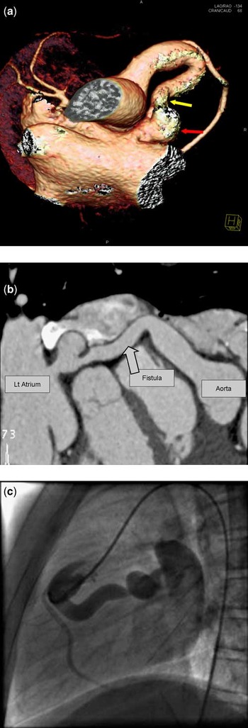
A 13-year-old girl presented with a continuous murmur and was found to have a tortuous right coronary artery to the left atrial fistula on transthoracic echocardiography. Multi-detector computed tomography coronary angiography showed that the fistula had a long isthmus and a saccular aneurysm that arose distally before it emptied into the roof of the left atrium (Fig 1a and b). The mid and distal right coronary artery and left coronary artery were normal.
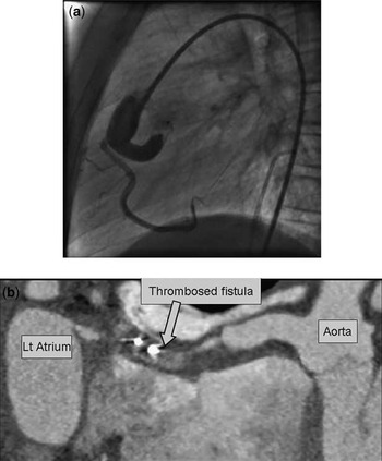
The computed tomography images were helpful in planning an optimum position for the vascular occluder. A 10 mm × 2 mm Amplatzer™ vascular plug was delivered via a retrograde approach proximal to the saccular aneurysm, with complete occlusion of the fistula and normal antegrade flow through the right coronary system (Figs 1c and 2a). There were no complications and repeat computed tomography 1 month later showed extensive thrombosis of the fistula (Fig 2b).
(a) and (b) Three-dimensional rendered images from a computed tomography coronary angiogram showing a right coronary artery to the left atrial fistula. Featured are the distal isthmus (yellow arrow) and saccular aneurysm (red arrow). The distal right coronary artery is of normal calibre. (c) Selective right coronary angiography showing the large fistula.

(a) Right coronary angiogram showing complete occlusion of the fistula with normal flow into the right coronary artery. (b) Dual-source computed tomography showing extensive thrombosis of the fistula 1 month later.

Coronary artery fistulas are rare malformations between a coronary artery and a cardiac vessel or chamber.Reference Qureshi1 Reported complications of non-occluded lesions include cardiac failure, bacterial endocarditis, myocardial ischaemia, dissection, and rupture. Right coronary artery fistulae have communications to the left atrium or ventricle in less than 10% of cases. Transcatheter device closure is an effective and safe alternative to surgery with good short-to-medium-term outcomes. We found that multi-detector computed tomography was useful to evaluate the course of the fistula and to aid in planning the optimal position for the occluding device.