Introduction
The prevalence of thyroid disorders increases with advancing age.Reference Finucane and Anderson1 Making a clinical diagnosis of both hypothyroidism and hyperthyroidism can be difficult because of the atypical presentation and attribution of symptoms to the normal ageing process, or to other co-existing diseases.Reference Finucane and Anderson1 Many signs and symptoms of hypothyroidism can be present in the euthyroid elderly. On the other hand, typical features of hyperthyroidism are mostly absent in older people. A good understanding of age-related changes in thyroid structure and physiology, as well as awareness of the atypical presentation of the disease in older individuals, is very important for accurate diagnosis and management. A high degree of suspicion is the key to the diagnosis of thyroid problems in older people.
The ageing thyroid
Anatomical and histological changes
In old age, the overall mass of the thyroid gland declines from the normal range of 15–20 grams.Reference Mochizuki, Mowafy and Pasternack2 Its weight may increase with the development of a nodular goitre which becomes more common.Reference Mariotti, Franceschi, Cossarizza and Pinchera3 However, one study using ultrasound found little change in the size of the thyroid in healthy ageing subjects.Reference Hegedus, Perrild, Poulsen, Andersen, Holm, Schnohr, Jensen and Hansen4
Microscopic changes are characterized by fibrosis, lymphocytic infiltration, reduction in follicle size and flattening of follicular epithelium.Reference Mariotti, Franceschi, Cossarizza and Pinchera3 The gland becomes increasingly nodular with age.Reference Gupta83 The presence of thyroid nodules carries a risk of autonomous function and hyperthyroidism. However, anatomical and histological changes do not always correlate with abnormalities of thyroid function.Reference Oiknine, Mooradian, Pathy, Sinclair and Morley5
Thyroid axis regulation and thyroid hormone metabolism
There are age-related physiological changes in the hypothalamic–pituitary–thyroid axis and thyroid hormone metabolism. These can be summarized as follows:Reference Oiknine, Mooradian, Pathy, Sinclair and Morley5–Reference Mariotti, Chiovato, Franceschi and Pinchera12
1. Release of thyroid-stimulating hormone (TSH) from the pituitary in response to thyrotrophic-releasing hormone (TRH) is sluggish in old age.
2. The 24-hour secretion of TSH has been reported to be decreased, but the pituitary and serum concentrations of TSH are unaltered because of reduced clearance, except for a mild decrease in octogenarians.
3. There is a significant reduction in pituitary sensitivity to thyroxine (T4) and TSH rise is blunted. Elevations in TSH levels in older people should be considered indicative of a failing thyroid.
4. There is smaller circadian fluctuation in TSH secretion in comparison with younger adults. It may be indicative of lesser biological activity of TSH.
5. Thyroid hormone release in response to TSH is not altered.
6. There is reduced uptake of iodine by the thyroid gland.
7. The T4 production is decreased from 80 to 60 micrograms per day and triiodothyronine (T3) production decreases from 30 to 20 micrograms a day.
8. There is decreased peripheral conversion of T4 to T3.
9. Thyroid hormone clearance is reduced, leading to unaltered serum concentrations of T3 and T4 with age. The half-life of T4 increases from 6.7 days in the third decade to 9.1 days in the seventh decade. By the age of 90 years, the T4 degradation rate is approximately half that of younger subjects. This has been attributed to the reduction in activity of 5’-monodeiodinase enzyme in peripheral tissues. This explains the lower replacement levothyroxine dose required by elderly hypothyroid subjects.
10. Circulating levels of free T3 and free T4 remain constant over the age span and a decrease in concentration should be considered as secondary to illness rather than to ageing.
11. The generation of reverse triiodothyronine (rT3) is unaffected by ageing. An increase in levels should be considered to be secondary to illness or drug-induced alteration in T4 degradation.
12. There are changes in transport of T3 across plasma membranes and alterations in post-receptor gene expression resulting in relative resistance to thyroid hormone action. This may be a possible cause of presence of features of hypothyroidism in older people despite normal plasma levels of thyroid hormones. Alternatively this may explain paucity of clinical features in patients with apathetic hyperthyroidism.
13. A few studies have shown an elevation of thyroid autoantibodies, but this seems secondary to age-associated disease rather than the ageing process itself.
Clinical assessment
Localization of the thyroid gland can be difficult in elderly subjects. The gland can shrink secondary to atrophy or fibrosis. In the presence of kyphosis, lower poles or most of the gland is located below the suprasternal notch. However, visual examination under side-lighting of the pretracheal area, with the patient's neck slightly in extension, can display the anatomy of a slightly enlarged gland satisfactorily in the elderly patient with symmetrical loss of strap-muscle mass.Reference Jameson, Weetman, Fauci, Braunwald and Kesper13 The incidence of carotid bruit is high in older people and it may be misinterpreted as thyroid bruit.
In contrast to younger patients, goitre is less likely in the setting of hyperthyroidism or Hashimoto's thyroiditis. A large retrosternal goitre can lead to venous dilatation over the neck and breathing difficulty, especially after raising the arms (Pemborton's sign).Reference Jameson, Weetman, Fauci, Braunwald and Kesper13 Lymphadenopathy in the cervical and supraclavicular regions should be carefully investigated because of the higher risk of malignant lesions with increasing age.
Laboratory assessment of thyroid function
Hormonal assays. Third generation laboratory radioimmunoassay methods are very sensitive and cost effective. They have facilitated the diagnosis of thyroid dysfunction. Measurement of blood TSH, total circulating T4 and T3 concentrations and free T4 (FT4) and free T3 (FT3) concentrations through use of radioimmunoassay or immunometric assays is useful to screen the secretory status of thyroid glands.Reference Hay and Klee14
TSH assay may be the best initial screening test of thyroid dysfunction.Reference Robuschi, Safran, Braverman, Gnudi and Roti15 Free T4 is inferior to TSH for screening because it can be normal in subclinical hypothyroidism. TSH is suppressed in hyperthyroidism and elevated in hypothyroidism. Measurement of circulating thyroid hormones should follow the finding of an abnormal TSH.
T4 and T3 are highly protein bound, 99.8 and 99.7%, respectively. Multiple factors including non-thyroid-related illness, medications and genetic factors can influence protein binding. Total thyroid hormone levels become elevated when thyroid binding globulin (TBG) is increased due to oestrogens (hormone supplements, tamoxifen) and decrease when TBG is reduced (androgens, nephrotic syndrome). Drugs such as phenytoin, carbamazepine and salicylates can also interfere with thyroid hormone binding. Hence it is useful to measure free, or unbound, hormone levels.Reference Jameson, Weetman, Fauci, Braunwald and Kesper13
TSH can be misleading in a few clinical conditions. Rare causes of elevated TSH include TSH-secreting pituitary tumour and thyroid hormone resistance. In iatrogenic hypothyroidism (e.g. post-radioiodine ablation of thyrotoxicosis), TSH can be suppressed for a prolonged period and unbound T4 may be a better indicator of hypothyroid state. Acute non-thyroidal illnesses can give rise to erroneous TSH estimates. Similarly, low levels of TSH can be due to other causes such as secondary hypothyroidism, high-dose glucocorticoids and dopamine.
Antibody assays. Autoimmune thyroid diseases are characterized by the presence of antibodies against thyroperoxidase (TPO) and thyroglobulin (Tg). Tg antibodies are less specific because moderate to high titres may be present with non-thyroid autoimmune disorders.Reference Hay and Klee14
TPO antibodies are more specific to thyroid dysfunction and commonly used. Almost all patients with Hashimoto's thyroiditis causing hypothyroidism, and 80% of patients with Graves’ disease, have TPO antibodies.
The prevalence of thyroid antibodies in serum progressively increases with increasing age. Above 50 years of age, 20–25% of women and 5–10% of men have been reported to have thyroid antibodies,Reference Robuschi, Safran, Braverman, Gnudi and Roti15, Reference Blumenthal and Perlstein16 although in a study comparing healthy elderly adults with healthy younger people, antibody prevalence was low and did not differ between the two groups.Reference Mariotti, Franceschi, Cossarizza and Pinchera3 This finding suggests that high prevalence of antibody in the older population is indicative of disease and not a result of the normal ageing process.
It is debatable whether the presence of thyroid antibody in the serum is predictive of future development of hypothyroidism. In a prospective study, women with both raised TSH and thyroid antibodies developed overt hypothyroidism at a rate of 5% per year.Reference Tunbridge, Brewis and French17 Other studies did not support a predictive value but showed strong association with the presence of antibodies and increased TSH and clinical hypothyroidism.Reference Sawin, Bigos, Land and Bacharach18
Thyroid-stimulating immunoglobulins (TSI) stimulate the TSH receptors and are found in over 85% of patients with untreated Graves’ disease and 100% with severe Graves’ ophthalmopathy.Reference Jameson, Weetman, Fauci, Braunwald and Kesper13 TSI is rarely present in toxic nodular goitre.
Serum thyroglobulin. Serum thyroglobulin levels are increased in all types of thyrotoxicosis except thyrotoxicosis factitia, thyroiditis and thyroid cancer.Reference Jameson, Weetman, Fauci, Braunwald and Kesper13 The clinical role is in the follow-up of cancer patients as they become undetectable after total thyroidectomy and radioablation. During follow-up, measurable quantity indicates either incomplete ablation or recurrence.Reference Jameson, Weetman, Fauci, Braunwald and Kesper13
Radioiodine uptake thyroid scan
The thyroid gland selectively transports radioisotopes of iodine (123I, 125I, 131I) and 99mTc pertechnetate, allowing nuclear imaging of thyroid and estimation of the amount of radioactive tracer uptake.Reference Jameson, Weetman, Fauci, Braunwald and Kesper13 As with iodine uptake, 10–25% of radioactive iodine tracer is taken up by the normal thyroid gland and the rest is excreted in urine.
Graves’ disease is characterized by diffusely homogenous increased tracer uptake of as much as 70–90%. Toxic adenoma shows focal areas of increased uptake and suppressed uptake in the rest of the gland. Multinodular toxic goitre is characterized by multiple areas of relatively high or low uptake. Very low uptake and low uptake are suggestive of acute thyroiditis and thyrotoxicosis factitia, respectively.
In the evaluation of solitary nodules, uptake also has some prognostic significance. About 5–10% of cold nodules showing decreased uptake are likely to be malignant, whereas malignancy is very unlikely in hot nodules. It is also useful in the follow-up of thyroid cancer after thyroidectomy and radioiodine ablation, allowing the detection of recurrence/metastatic disease.
The amount of tracer uptake is helpful to determine the required dose of radioactive iodine for thyroablation in hyperthyroidism.
Thyroid ultrasound and fine needle aspiration (FNA) biopsy
Ultrasound is easy to perform, widely available, does not involve ionizing radiation and, when combined with FNA cytology, gives better tissue yield. It is therefore an ideal investigation of choice for evaluating thyroid nodules.Reference Wong and Ahuja19
It is commonly used to investigate nodular thyroid disease and allows detection of nodules and cysts larger than 3 mm in diameter. It is very useful in the surveillance of thyroid cancer.
MRI and CT scans
Their main clinical role is to assess the extent of the disease in cases of thyroid cancer.
Hypothyroidism
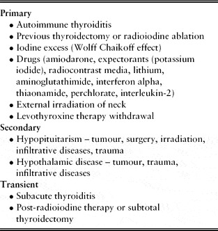
Most commonly, hypothyroidism is the result of decreased thyroid hormone production from the thyroid gland (primary hypothyroidism). Less commonly, it can be due to inadequate TSH release from the pituitary (secondary hypothyroidism). Causes are listed in Table 1.
Causes of hypothyroidism

Demography
Primary hypothyroidism is predominantly a disease of older females. The reported prevalence of hypothyroidism in older people ranges from 1 to 17.5%.Reference Levy20–Reference Tunbridge, Evered, Hall, Appleton, Brewis, Clark, Evans, Young, Bird and Smith24 The disparity between these estimates is because of geographical variations, differences in the definition of elderly, sex distribution within the samples, sample size, iodine intake and laboratory tests used. In the UK-based Wickham survey, the incidence of increased TSH increased from 4.0 to 5.7% of women under the age of 45, and to 17.4% of women over the age of 75 years. In men, elevated serum TSH incidence increased from 1.6 to 3.5% of those under the age of 65 years, and to 3.5–6.9% over the age of 65 years. The prevalence was ten times greater in females than in males.Reference Tunbridge, Evered, Hall, Appleton, Brewis, Clark, Evans, Young, Bird and Smith24 However, a study from Israel showed a prevalence of hypothyroidism of 9.7% in males and 18.2% in females in the 65–92 year age group.Reference Flatau, Trougouboff, Kaufman, Reichman and Luboshitzky23 The prevalence in whites was double that in blacks in a study from the USA.Reference Bagchi, Brown and Parish25 In healthy out-patient subjects followed in the Framingham Heart study, the prevalence of clinically unrecognized hypothyroidism was found to be about 2.5% in subjects of 60 years or older.Reference Sawin, Castelli, Hershman, McNamara and Bacharach26
Aetiology (Table 1)
Autoimmune hypothyroidism (Hashimoto's thyroiditis) is the commonest cause of thyroid failure in the elderly.Reference Hurley27, Reference Diez28 In a study of 655 patients older than 55 years (599 women, 96 men), the causes of hypothyroidism were autoimmune in 308 (47%), post-operative hypothyroidism in 175 (26.7%), therapy for previous thyrotoxicosis in 63 (9.6%), thyrotropin deficiency in 15 (2.3%), iodine excess in 6 (0.9%), subacute thyroiditis in 2 (0.3%) and unknown aetiology in 86 (13.1%). Most of the autoimmune thyroiditis patients in the study did not have a goitre.Reference Diez28
Hashimoto's thyroiditis is characterized by replacement of normal thyroid tissue by lymphocytes and fibrous tissue leading to reduced thyroid mass and thyroid hormone production. Initially the pituitary tries to compensate for this deficiency by producing more TSH and thereby normalizing thyroid hormone production (subclinical hypothyroidism) but, in the later phase of disease, the thyroid fails to produce hormones despite high TSH levels. As with most other autoimmune diseases, increased susceptibility has been linked to environmental and genetic factors, the elaboration of which is beyond the scope of this review. Iodine and iodine-containing drugs can lead to reduced thyroid hormone synthesis (Wolff-Chaikoff effect). This effect is reversible, with the thyroid resuming normal hormonogenesis upon removal of the cause. The most common culprits are amiodarone, radiographic iodine containing contrast media, expectorants (potassium iodide) and topical antiseptics (betadine).
Secondary hypothyroidism occurs because of impaired production or release of TSH from the pituitary. It is uncommon. Possible causes are pituitary surgery, radiotherapy, or infiltrative diseases such as amyloidosis, sarcoidosis or tuberculosis.
Iodine deficiency remains a cause of hypothyroidism in a few parts of the world. This problem has largely been solved by the fortification of salt and bread in iodine-deficient areas.
Clinical presentation
Making a clinical diagnosis of hypothyroidism on the basis of presenting clinical findings is very challenging. The disease evolution is insidious and involves multiple systems. Many of the manifestations may mimic or be attributed to old age and/or to other diseases. In one study, only 10% with a laboratory diagnosis of hypothyroidism were recognized clinically as having a hypothyroid state.Reference Lloyd and Goldberg29
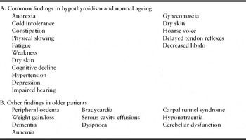
As the classic signs and symptoms such as lassitude, cold intolerance, constipation, dry skin, hair loss, delayed relaxation phase of deep tendon jerks, psychomotor retardation, fatigue and anorexia occur in both hypothyroid and euthyroid elderly patients, they can be mistakenly attributed to old age.Reference Mintzer30–Reference Case, Mooradian, Morley, Armbrecht, Coe and Vellas32Table 2 summarizes the signs and symptoms found both in hypothyroidism and normal ageing.Reference Case, Mooradian, Morley, Armbrecht, Coe and Vellas32, Reference Mohandas and Gupta33
Signs and symptoms of hypothyroidism
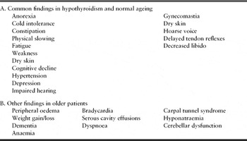
Doucet and colleagues compared clinical features of hypothyroidism in 67 elderly patients with a younger hypothyroid population. Older patients had fewer signs or symptoms. Some of the classic signs (e.g. cold intolerance and weight gain) were mostly absent.Reference Doucet, Trivalle, Chassagne, Perol, Vuillermet, Manchon, Menard and Bercoff34 In a similar study, mean number of clinical signs per patient were significantly fewer in the elderly, and all four classical signs (i.e. cold intolerance, parasthesia, weight gain and cramps) occurred significantly less frequently in older patients.Reference Trivalle, Doucet, Chassagne, Landrin, Kadri, Menard and Bercoff35 The most common clinical features were fatigue and weakness.Reference Trivalle, Doucet, Chassagne, Landrin, Kadri, Menard and Bercoff35 Paradoxically, there may be weight loss in older individuals secondary to anorexia. Any change in behaviour and slow engagement during rehabilitation should lead to clinical suspicion.
Hypothyroidism can affect almost all systems of the body and give rise to a myriad of symptoms and signs. Many of the signs are due to accumulation of glycosaminoglycans (myxoedematous infiltration).
Hypothyroidism is associated with increased cardiovascular risk as evidenced by augmented central aortic pressures and central arterial stiffness, and reversal of these abnormalities in response to T4 replacement.Reference Obuobie, Smith, Evans, John, Davies and Lazarus36 Hypertension, particularly diastolic, can be secondary to increased vascular resistance. Reduced myocardial contractility, decreased stroke volume and bradycardia aggravate pre-existing heart failure. Pericardial effusion is present in up to 30% of patients but rarely large enough to cause clinical compromise.Reference Jameson, Weetman, Fauci, Braunwald and Kesper13 Myxoedematous infiltration of myocardium results in cardiac enlargement and symptoms of ischaemic heart disease.Reference Vanhaelst, Neve, Chailly and Bastenie37 Patients may present with pleural effusion. Dyspnoea and hypercapnia can be a feature because of decreased chemoreceptor sensitivity and weak respiratory musculature.Reference Jameson, Weetman, Fauci, Braunwald and Kesper13, Reference Miller, Tallis and Fillit38
ECG changes include bradycardia, low-voltage complexes, flattening and/or inversion of T-waves.Reference Miller, Tallis and Fillit38 Many older people are already on beta blockers or other rate-limiting medications, hence bradycardia may be unhelpful as a clinical sign. However, if associated with decreased core body temperature, hypothyroidism should be considered. Extremities are cool, secondary to low core body temperature and decreased cardiac output. The face may be puffy with thickened pale skin (myxoedematous facies). Despite the presence of myxoedema (classically non-pitting oedema), pitting oedema is present in about 40% of elderly patients,Reference Davis, Katz, Evans, Williams and Beattie39 even in the absence of heart failure. Reduced cardiac output leads to reduced glomerular filtration rate and consequent fluid and water retention.
Proximal myopathy, cereballar dysfunction, motor retardation, carpal tunnel syndrome and neuropathy have been described.Reference Davis, Katz, Evans, Williams and Beattie39–Reference Rehman, Cope, Senseney and Brzezinski41 Myopathy is evidenced by elevated serum creatinine phosphokinase (CPK). Cerebellar signs include locomotor and truncal ataxia, a finding more common in older than younger patients. Motor retardation can be demonstrated by poor performance of tasks such as buttoning. Motor retardation is improved with replacement of thyroid hormone, but ataxia can be persistent.
Delayed contraction and relaxation phases of the deep tendon reflexes are common findings in all age groups. Coarsening of voice and slurred speech is due to infiltration of vocal cords. Similarly, fluid deposition in the middle ear can cause hearing impairment.
Cognitive dysfunction is one of the commoner features in the older population. Hypothyroidism may cause decreased memory, and slowed speech and thinking. Dementia has been found to be associated with hypothyroidism in many studies. A recently published long-term follow-up study comprising 1864 original cohort patients from the Framingham study concluded that low and high thyrotropin levels were associated with an increased risk of incident Alzheimer's disease in women but not in men.Reference Tan, Bieser, Vasan, Au, Auerbach, Kiel, Wolf and Seshadri43
A popularly held classical view recognizes hypothyroidism as a cause of reversible dementia. This seems debatable. In an extensive literature review, this view was only partially supported. Successful treatment of clinically evident hypothyroidism may be associated with only partial and a typically inconsistent pattern of improvement in overall neurocognitive function.Reference Dugbartey42
Both hypothyroidism and hyperthyroidism can cause depressive symptoms.Reference Green and Robinson44, Reference Tapp45
Myxoedema madness with psychotic features is infrequent.
Normocytic or macrocytic anaemia can be found in up to one-third of patients.Reference Bahemuka and Hodkinson46 Possible pernicious anaemia should be investigated because of autoimmune association. Hyponatraemia is a common association, the picture being that of the syndrome of inappropriate antidiuretic hormone secretion. The postulated mechanisms of dilutional hyponatraemia are increased release of antidiuretic hormone and increased tubular resorption of sodium and water due to altered renal flow. Serum cholesterol and triglyceride levels and LDH can be elevated.Reference Khaira and Franklin47, Reference Aronow48
Management
A high degree of clinical suspicion is the key for diagnosis, which is confirmed by laboratory assays of hormones. TSH is used for initial screening. A normal TSH excludes primary hypothyroidism. If TSH is high, free T4 should be measured. Diagnosis of primary hypothyroidism is confirmed by low FT4 and high TSH.
Free T3 is not measured commonly as it can be normal in 25% of patients because of adaptive responses, i.e. increased peripheral conversion of T4 to T3. Thyroid antibodies are helpful in establishing the cause. In cases of normal clinical examination and exclusion of substernal goitre through chest radiograph, no further investigation is required. The possibility of sick euthyroid syndrome (described below) should be kept in mind.
If the gland is asymmetric or hard, FNA biopsy of the gland is useful.
In pituitary disease, TSH can be low or normal, with low FT4 (secondary hypothyroidism). Referral to an endocrinologist should be made. Occasionally measuring response to thyrotropin-releasing hormone in the TRH test is valuable as patients with secondary hypothyroidism show a blunted or absent rise in TSH.
The treatment of hypothyroidism is replacement of levothyroxine (T4). It can be complex in elderly patients because of the high incidence of coronary artery disease in this population and the chance of iatrogenic unmasking of ischaemic symptoms and cardiac instability resulting in angina, infarction or arrhythmias. ‘Start low and go slow’ is the best approach.Reference Mohandas and Gupta33 The usual starting dose of T4 is 25 μg, but if there is concern regarding cardiac status, an initial dose of 12.5 μg is recommended.Reference Davis and Davis31, Reference Ellyin, Fuh, Singh and Kumar49 The dose should be increased by 12.5–25 μg every 2–3 months.Reference Jameson, Weetman, Fauci, Braunwald and Kesper13 Pulse rate should be monitored for tachycardia.Reference Ladenson50 The mean dose of T4 decreases with age, because of slower clearance and increased half-life.Reference Case, Mooradian, Morley, Armbrecht, Coe and Vellas32
The physiological dose in older patients is about 75 μg.Reference Synder and Utiger7 TSH is used for monitoring51 but, in the initial phase, it may lag behind in down-regulation. It may be necessary to measure FT4 as the patient may be euthyroid in the presence of a still slightly elevated TSH. The TSH returns to normal in a few months without dose alteration in these cases. If after 1 year of therapy TSH remains elevated, then a dose increase may be warranted. Once a steady state is reached, TSH can be monitored every 6–12 months. In cases of cardiac instability, the dose can be withheld for a few days, pending further cardiac evaluation, without risk because of the sufficiently long half-life of T4 in older patients.Reference Rehman, Cope, Senseney and Brzezinski41 In known ischaemic heart disease patients, antianginals should be maximized.Reference Levine52
Most patients need lifelong therapy. One study reported the possibility to reduce or withdraw hormone therapy in frail elderly patients with multiple medical problems.Reference Coll and Abourizk53 Ideally only one standard preparation should be used to ensure predictable drug levels because gastrointestinal absorption varies between different preparations.
Adjustment of therapy to ensure adequate replacement can be tricky in older patients. Because of the risk of cardiac complications, over-treatment is not advisable. In addition, over-replacement leads to suppressed TSH and increased risk of osteoporosis and fractures due to increased bone resorption.Reference Stall, Harris, Sokoll and Dawson54, Reference Solomon, Wartofsky and Burman55 In patients with secondary hypothyroidism, thyroid hormone replacement should not be commenced unless adrenal sufficiency has been confirmed, or glucocorticoid therapy started in insufficiency.
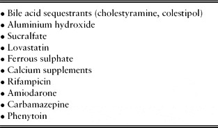
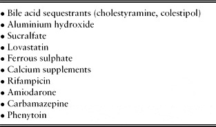
Many drugs can decrease absorption or interfere with thyroxine clearance rendering patients hypothyroid again, or requiring increased replacement therapy, an important element to be considered in older people because of polypharmacy (Table 3).Reference Jameson, Weetman, Fauci, Braunwald and Kesper13, Reference Stall, Harris, Sokoll and Dawson54 Malabsorption syndromes, like small bowel surgery or coeliac disease, have similar effects.
Drugs decreasing levothyroxine absorption or interfering with clearance
Sick euthyroid syndrome (low T3 syndrome)
Thyroid function tests should ideally not be performed during any acute illness and, if done, must be interpreted with caution. The hypothalamic–pituitary–thyroid axis is affected by non-thyroidal illness.Reference Solomon, Wartofsky and Burman55 Almost any illness, starvation, recent surgery or stress can lead to disarray in thyroid function test results. The syndrome is reversible. It mimics hypothyroid state with low T3, and either normal or low T4 and TSH.
The factors implicated are high cortisol and release of cytokines such as interleukins. The activity of 5’-monodeiodinase, the enzyme responsible for peripheral conversion of T4 to T3 by outer ring deiodination, gets inhibited. The other physiological component of T4 degradation involves deiodination from the inner ring of T4, resulting in formation of physiologically inactive reverse T3 (rT3). Rise in rT3 parallels decreased T3 formation in sick euthyroid state, an effect useful in differentiating this condition from primary hypothyroidism. As serum rT3 is not affected by normal ageing, an increase must be regarded as a consequence of illness or drug-induced alteration of T4 degradation.Reference Nishikawa, Inada, Naito, Ishii, Tanaka, Mashio and Imura56 The drugs implicated are propranolol, amiodarone and iodinated contrast materials. Treatment is not required because the condition is reversible.
Subclinical hypothyroidism
Normal thyroid hormone levels in the presence of elevated TSH levels is termed subclinical hypothyroidism (SCH). The prevalence may be as high as 20% in women over 60 years.Reference Canaris, Manowitz, Mayor and Ridgway57 It is considered to be an indicator of a failing thyroid and most individuals are asymptomatic.
In one long-term follow-up study, women with TSH values greater than 6 mIU/l and positive thyroid antibodies developed overt hypothyroidism at a rate of 5% per year. No case developed in patients with borderline elevation of TSH only (6–10 mIU/l) and only one patient developed overt hypothyroidism who had normal TSH with positive anti-thyroid antibodies.Reference Tunbridge, Brewis, French, Appleton, Bird, Clark, Evered, Evans, Hall, Smith, Stephenson and Young58 In another recent study from Spain, 107 subjects older than 55 years were followed for 6–72 months. Twenty-eight (26.8%) developed overt hypothyroidism and 40 (37.4%) showed normalization of their TSH value. Higher TSH concentration was the most powerful predictor of overt hypothyroidism in this study.Reference Hak, Pols, Visser, Drexhage, Hofman and Witteman59
A cross-sectional study of 295 patients with subclinical thyroid dysfunction reported that, after the confounding effects of comorbid conditions and use of medications were controlled for, subclinical thyroid dysfunction was not associated with depression, anxiety or cognitive dysfunction.Reference Roberts, Pattison, Roalfe, Franklyn, Wilson, Hobbs and Parle88
There are observational data suggesting SCH as a risk factor for accelerated atherosclerosis and ischaemic heart disease (IHD) independent of cholesterol levels.Reference Hak, Pols, Visser, Drexhage, Hofman and Witteman59, Reference Kahaly60 A recent meta-analysis concluded that SCH is associated with increased IHD and cardiovascular mortality only in subjects from a younger population, not in those of 65 years or older.Reference Razvi, Shakoor, Vanderpump, Weaver and Pearce89
The treatment is controversial in asymptomatic older individuals and should be individualized because of dangers of accelerated bone loss, atrial fibrillation and worsening of heart failure. A reasonable approach seems to be to treat symptomatic patients (e.g. those with memory loss or elevated lipid levels) and patients with high TSH and TPO antibodies who are more likely to progress into overt hypothyroidism.Reference Mohandas and Gupta33, Reference Jameson, Weetman, Fauci, Braunwald and Kesper13
Myxoedema coma/stupor (profound hypothyroidism)
This is a medical emergency almost exclusively affecting older patients and has a high mortality rate despite intensive treatment. It is precipitated by various factors causing physiological stress such as sepsis, myocardial infarction, gastrointestinal bleeding, alcohol intoxication and drugs causing respiratory compromise (e.g. sedatives, antidepressants and anaesthetic medications).Reference Jameson, Weetman, Fauci, Braunwald and Kesper13 Hypoventilation causing hypoxia and hypercapnia plays a major role in pathogenesis.
Patients may present with reduced level of consciousness, severe mental deterioration, disorientation or psychosis, profound hypothermia and bradycardia, besides having other usual features of hypothyroidism.
Patients should be managed in an intensive care unit. The treatment of choice is intravenous liothyronine sodium (T3); the usual dose is 5–20 μg every 12 h.61 Alternatively an intravenous loading dose of 250–500 μg of levothyroxine is given to replenish the body stores.Reference Jameson, Weetman, Fauci, Braunwald and Kesper13, Reference Rehman, Cope, Senseney and Brzezinski41 If an intravenous preparation is not available, the same dosage can be given through a nasogastric tube, though absorption may be impaired. Supportive therapy for underlying precipitant illness and treatment for low cortisol levels if present is necessary before replenishing thyroid hormone.
Hyperthyroidism
The data for the demography of hyperthyroidism are sparse but it is no longer considered as rare in old age. The estimated prevalence rate in the population over the age of 60 is 2%, and women predominate.Reference Bagchi, Brown and Parish25 The proportion of hyperthyroid patients who are over 60 years of age is estimated as 15–20%.Reference Davis and Davis62, Reference Ronnov-Jessen and Kirkegaard63
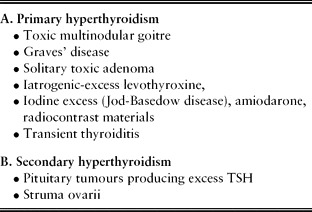
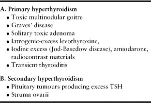
Aetiology (Table 4)
The commonest cause in older people is multinodular toxic goitre.Reference Davis and Davis62 The incidence of Graves’ disease, the commonest cause of hyperthyroid state in younger population, is lower. In a large cross-sectional study, the different aetiologies of hyperthyroidism in older people were as follows: toxic multinodular goitre 43.1%; Graves’ disease 21.4%; iatrogenic thyrotoxicosis 16.0%; toxic adenoma 11.8%; iodine-induced thyrotoxicosis (Jod-Basedow disease) 1.2%; subacute thyroiditis 1.0%; painless thyroiditis 0.3%; factitious thyrotoxicosis 0.3%; TSH-secreting pituitary adenoma 0.6%, and unknown etiology 3.8%.Reference Diez64
Aetiology of hyperthyroidism
Clinical presentation
Hyperthyroidism in older people is difficult to recognize because of a paucity of clinical signs and symptoms. Frequently, patients present with only one symptom, referred to sometimes as monosymptomatic hyperthyroidism.Reference Federman65 The paucity of symptoms in old age is postulated to be secondary to hormone resistance, but this needs to be confirmed.
A prospective cohort studyReference Trivalle, Doucet, Chassagne, Landrin, Kadri, Menard and Bercoff35 compared classical signs and symptoms between older and younger populations. The mean number of clinical signs found in the older subjects was significantly smaller than the number found in younger patients. Only three signs were found in more than 50% of older patients: tachycardia, fatigue and weight loss. Seven signs were found significantly less frequently in older patients: hyperactive reflexes, increased sweating, heat intolerance, tremor, nervousness, polydipsia, and increased appetite. Only anorexia (32 vs. 4%) and atrial fibrillation (35 vs. 2%) were more frequent in older people (P < 0.001). Goitre was present in 94% of the younger and in 50% of the older patients (P < 0.001). Comparison with older controls showed three signs that were highly associated with thyrotoxicosis in older people: apathy (odds ratio (OR): 14.8), tachycardia (OR: 11.2), and weight loss (OR: 8.7).Reference Trivalle, Doucet, Chassagne, Landrin, Kadri, Menard and Bercoff35 Neuromuscular symptoms such as myopathy, fatigue, weakness, lethargy, agitation and dementia are common.Reference Waldenstorm66, Reference Seed and Lindsay67
A prospective studyReference Nordyke, Gilbert and Harada68 examining 880 hyperthyroid patients reported that findings were fewer after the fifth decade, and older people with Graves’ disease may not have goitre, exopthalmos or other ophthalmopathy. Atrial fibrillation and weight loss were commoner with increasing age in this study. Osteoporosis in the axial skeleton may be present in all age groups.Reference Seeman, Wahner, Offord, Kumar, Johnson and Riggs69
Not uncommonly, patients may present with an apathetic form of hyperthyroidism, a term described by Lahey in 1937, with stuporous depression and placid apathetic facies.Reference Felicetta40 They may simulate severely demented patients.Reference Lahey70, Reference Hall, Beresford and Cavenar71
T3 toxicosis
This condition is almost exclusive to older people with isolated finding of excessive T3.Reference Gambert72 The most common cause is a toxic solitary nodule producing excessive T3.Reference Marsden, Facer, Acosta and McKerron73 The serum T4 may be normal or suppressed and TSH remains suppressed.Reference Federman65 If suspected, FT3 must be checked to avoid misinterpretation as a hypothyroid state.
Subclinical hyperthyroidism
This condition is characterized by low or suppressed TSH with normal serum T4 and/or T3 and is not uncommon in elderly people. In a study of 1210 elderly patients in Oxford, low TSH was found in 6.3% of women and 5.5% of men but, on repeat measurement a year later, the majority of patients showed return to the normal range.Reference Parle, Franklyn, Cross, Jones and Sheppard74 Similar findings have been observed in other studies.Reference Sawin, Geller, Kaplan, Bacharach, Wilson and Hershman75, Reference Diez64
These patients generally do not present with features of hyperthyroidism, but are reported to have increased risk of atrial fibrillation, atrial ectopy, increased left ventricular mass index and decreased ventricular filling.Reference Foldes, Tarj, Szathmari, Varga, Krasznai and Horvath76, Reference Biondi, Palmieri, Fazio, Cosco, Nocera, Sacca, Filetti, Lombardi and Perticone77
Because of variability in the natural history of disease and high chances of return to normality over time, current evidence favours an observant approach if patients are asymptomatic. The US Preventive Task Force recommends treatment if TSH is <0.1 mIU/l, in the presence of Graves’ disease or nodular thyroid disease.Reference Weetman78
Periodically these patients should be followed by hormonal assays as a few of them might develop overt hyperthyroidism.
Management
Diagnosis of primary hyperthyroidism is established by elevated FT4 and suppressed TSH. If FT4 is normal, serum T3/FT3 should be checked to rule out T3 toxicosis. Presence of thyroid antibodies suggests Graves’ disease. Graves’ disease is the likely diagnosis if the thyroid gland is diffusely enlarged.Reference Federman65 Further investigations are not required in these cases. In patients with nodular disease, nuclear scan or ultrasound is useful.
Associated laboratory findings in hyperthyroidism may be microcytic anaemia, thrombocytopenia, deranged liver function tests and decreased ferritin.
Treatment methods are similar in all age groups and are described below briefly.
Medications. A beta blocker (propranolol) is used for the rapid relief of the symptoms of tachycardia, tremor and anxiety. Thionamides (propylthiouracil and carbimazole) are available to treat hyperthyroidism in the UK and act by reducing thyroid hormone production. In addition, propylthiouracil blocks the action of 5’-deiodinase at higher dosage, reducing peripheral conversion of T4 to T3, an effect useful in thyrotoxic crisis. Between 30 and 40% of patients treated with anti-thyroid drugs go into a remission lasting 10 years or longer.Reference Weetman78
Dosage should be titrated and the lowest possible dose should be used in elderly patients. The side-effects, such as arthralgia, rashes, hepatotoxicity and agranulocytosis, should be looked for. Agranulocytosis is estimated to occur in 1 in 400 patients.Reference Sheng, Hung, Chen, Fang, Hsieh, Chang and Hsieh79 Patients should be educated to report any signs of infection immediately to the treating physician.
Another approach is to use a fixed dose to block thyroid hormone production and replace levothyroxine, called the block and replacement regime. Using this method, Weetman et al. reported an insignificant difference in remission rate one year after cessation of thionamide therapy, whether given for 6 or 12 months (59 vs. 65%).Reference Weetman, Pickerill, Watson, Chatterjee and Edwards80 Another prospective, randomized trial of the titration method indicated that there was no increase in the rate of remission when treatment was continued beyond 18 months.Reference Massart, Guilhem, Lorcy, Lescouarch, Herry and Allannic81
Radioactive iodine (RAI). Ablation of overactive thyroid tissue with 131I is the preferred treatment in older patients. It is safe, efficacious and time tested.Reference Gross, Shapiro and Sisson82 Potential risks of surgery can be avoided. Patients should preferably be rendered euthyroid by pharmacotherapy before undergoing radioablation, to avoid possible thyroid storm because of excess hormonal release secondary to thyroid tissue destruction. A lag of one to two months usually occurs before a clinical improvement is seen, during which time patients can be treated with medications. Repeated treatments may be necessary to achieve remission. Subjects may become hypothyroid over time and should be monitored. With all dosing regimens, by 12 months post-therapy, 40–50% of patients were hypothyroid and hypothyroidism continued to occur thereafter at the rate of 2–3% per year.Reference Holm90
Surgery. Because of multiple coexisting morbidities, surgery is not the preferred treatment in older patients. The indications are suspected malignancy, failed RAI, or large goitre producing pressure symptoms such as dysphagia or respiratory compromise.
Thyroid storm
Acute hyperthyroidism can be precipitated by stressful conditions such as surgery, anaesthesia, sepsis or after RAI therapy in patients who did not receive anti-thyroid therapy prior to ablation. Patients may present with fever, tachycardia, acute heart failure, dehydration, diarrhoea and abnormal liver function. It requires intensive management. Treatment options available are iodide (inhibits hormone release), beta blockers, glucocorticoids (blunts peripheral action) and ipanoic acid (reduces peripheral conversion of T4).Reference Miller, Tallis and Fillit38, Reference Rehman, Cope, Senseney and Brzezinski41
Nodular thyroid disease and cancer
The prevalence of thyroid nodules increases with age and 6–10% of the elderly population may have solitary nodules.Reference Gupta83 The vast majority of these are benign. The differential diagnosis includes cysts, benign adenoma, or inflammation. Only few solitary adenomas produce excess T3 causing T3 toxicosis and can be ablated with RAI. Malignancy is rare, estimated to occur in less than 10% of cases.Reference Oiknine, Mooradian, Pathy, Sinclair and Morley5 Most neoplasms are euthyroid.
TSH is the initial investigation in nodular thyroid disease. If TSH is suppressed, then a radioactive iodine uptake scan should be organized. If it shows increased uptake, no further work-up for cancer is necessary. A cold nodule on a nuclear scan is malignant in 5–10% and requires further work-up/FNA biopsy. FNA biopsy may be done with normal thyroid function and suspicion of malignancy. External radiation to the neck in the past is associated with increased cancer risk.
Papillary cell carcinoma is the commonest type of cancer, followed by follicular type. Papillary carcinoma is locally invasive and primarily metastasizes through lymphatics to bone and lungs. Follicular cell carcinoma is commoner in iodine-deficient areas. The peak incidence is in the sixth decade of life. It metastasizes through the haematogenous route and is more aggressive.
In a retrospective analysis of 204 cancer patients aged 60 years or older, 142 cases (70%) were well differentiated (68% papillary, 30% follicular and 2% Hurthle cell), 3 (2%) medullary cell carcinoma, and 59 (29%) of the cancers were poorly differentiated (67% anaplastic, 15% metastatic, 4% lymphoma, 7% squamous cell and 7% inconclusive).Reference Lin, Chao, Chen, Weng and Lin84
Medullary carcinoma is rare in elderly people. Sometimes lymphoma can be present. Old age is associated with bad prognosis and mortality irrespective of cancer type.Reference Hundahl, Fleming, Fremgen and Menck85
Anaplastic cancer comprises 1–2% of all thyroid cancer and is almost exclusive to the elderly population. The peak incidence is in the seventh decade. It mostly presents with a rapidly growing mass with pressure symptoms. Not uncommonly it can be a rapidly progressive growth on the background of well-differentiated cancer or pre-existing goitre. It is the most aggressive thyroid cancer. In a review of 84 patients, the mean survival time was 6.2 months from tissue diagnosis, despite aggressive management.Reference Aldinger, Samaan, Ibanez and Hill86
Treatment
The approach to treatment of thyroid cancer is similar at all ages. Total or near-total thyroidectomy is done. The American Thyroid Association recommends RAI ablation of remnants in all papillary or follicular carcinoma >1.5 cm and follow-up measurements of thyroglobulins at 4–6 month intervals to detect any cancer recurrence.Reference Cooper, Doherty, Haugen, Kloos, Lee, Mandel, Mazzaferri, McIver, Sherman and Tuttle RM87 Most of the cancer patients are treated with levothyroxine suppression therapy to reduce TSH-stimulated thyroid growth.
Conclusion
The atypical presentation of thyroid disorders leads to difficulty in diagnosis despite the high prevalence of thyroid disease in older people. Symptoms are subtle and often attributed to normal ageing or considered secondary to co-existing illnesses. High suspicion is the key to the diagnosis and it is not unreasonable to screen older people if there is any doubt.
Interpretation of thyroid function tests should be done cautiously because of the high possibility of impaired assays secondary to co-existing illnesses, malnutrition or medications. Subclinical thyroid disorders should be monitored periodically and preferably not treated except in high-risk cases, to avoid complications related to treatment.
Elderly hypothyroid patients should be given low-dose thyroxine and the dose should be titrated up very slowly to avoid ischaemic cardiac complications. Radioiodine ablation is an effective and well-tolerated therapy for older hyperthyroid patients.
Conflicts of interest
The authors declare no conflicts of interest.