Introduction
Lithos is Latin for stone. Stone formation is a common pathological entity which may occur in many sites and especially within tubular organs.Reference Walter and Israel1 Although the literature describes the term ‘lithos’ as a synonym for stone and cites cases of rhinolith,Reference Vink, Hasselt and Warmald2 tonsillolith,Reference Benjamin, Bingham, Hawke, Stammberger and Hawke3 sialolithReference Work, Hecht, Paparella and Shumrick4 and even broncholith,Reference Schwartz, Paparella and Shumrick5 the term ‘laryngolith’ has rarely been mentioned.
The only case of laryngolith previously reported in the literature occurred in a New York patient in 1972. In this case, the laryngolith was a sequestration of ossified laryngeal cartilages presenting 50 years after neck irradiation for hyperthyroidism. The sequestered cartilage projected inside the laryngeal lumen, with further accumulation of inspissated mucus and additional calcification.Reference Baker and Karlan6
In the present case, there was no history of neck irradiation, and histopathological examination revealed a true phosphate stone with no evidence of any chronic, specific inflammatory process. This is a completely different pathology to that described in the previously reported case.
Case report
In June 2002, a 44-year-old woman was referred to our ENT clinic by a chest physician due to increasing laryngeal stridor and incapacitating dyspnoea unresponsive to medical treatment. On questioning, the patient revealed that, because of a right-sided Erb's palsy, she had undergone some sort of orthopaedic surgery at the age of 16 years. Shortly after that procedure, she had suffered coughing, noisy breathing and dyspnoea on exertion. The condition had been diagnosed and managed as a chest allergy for the past 28 years.
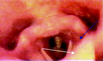
On examination, the patient's ears and nose were unremarkable. Her neck showed a mild, multinodular goitre, while endoscopy of the larynx revealed two lesions (Figure 1): (1) a yellowish-white, spiky lesion projecting from the posterior cricoid lamina, and (2) an anterior subglottic, soft tissue mass. Both vocal folds were freely mobile.
Endoscopic view of the subglottic lesions: the posterior, spiky lesion (blue arrow) and the anterior, soft tissue lesion (white arrow).

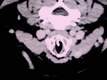
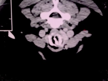
Computed tomography of the larynx confirmed the presence of a hyperdense, ossified lesion projecting from the posterior cricoid lamina below the level of the vocal folds and almost dividing the subglottis postero-anteriorly into right and left spaces (Figures 2 and 3).
Axial computed tomography scan of the uppermost level of the subglottis.

Axial computed tomography scan of the lower level of the subglottis, showing complete division of the subglottic area into right and left spaces.

The patient's hormonal and biochemical profiles were all within normal limits.
The patient underwent laryngeal litholapaxy (i.e. stone evacuation) in a two-step procedure (within the one surgical session) due to the presence of a large mass in the subglottis. In step one, after establishing general anaesthesia intravenously without intubation, direct laryngoscopy was performed and a posterior, tough, spiky lesion was evacuated with cupped laryngeal forceps. In step two, a small endotracheal tube was introduced, a detailed examination of the whole larynx conducted using an operating microscope, and excision of the anterior subglottic soft tissue lesion accomplished. The operative specimens were sent for histopathological examination.
After decalcification, the posterior, hard lesion was found to be negative for tuberculosis, specific granulomas, fungi and neoplasia. The specimen was sent for biochemical examination; the report described a single, irregular, 0.5 × 0.3 mm, rough, hard, brownish, calcular mass which on chemical analysis was found to be composed mainly of phosphate salts. The anterior, soft lesion was found to represent a chronic inflammatory process with evidence of acanthosis and keratosis.
The patient recovered extremely well from the procedure and was subsequently able to cease all her previous medication.
Discussion
Theoretically speaking, the pathogenesis of stone formation in the respiratory tract is the same in all its possible locations (i.e. nose, larynx, trachea and bronchus). The larynx provides the least favourable environment for stone formation, due to its continuous motion (apart from the subglottis) and the fact that it is a wide airway connecting the upper and lower air passages and has a high velocity airflow. Furthermore, there is no lithogenic medium present.
Three basic aetiologies have been suggested for stone formation in the respiratory tract: (1) calcium deposition around a foreign body nucleus within the lumen; (2) ossification of elastic cartilage with subsequent sequestration; and (3) protrusion of calcified hilar or para-tracheal lymph nodes into the respiratory tract.Reference Bridges and Proctor7 Inspissation of mucus, with further fibrin formation and calcification, may be a possible fourth aetiology.Reference Moersch and Schmidt8
Many cases of laryngeal perichondritis and necrosis have been reported, but not a laryngolith.Reference Clerf9–Reference Kapur13 Bearing in mind that the larynx is derived embryologically from the laryngotracheal groove, which eventually evolves into a tube, and despite the fact that the cricoid has two ossification centres,Reference Spector and Ballenger14 it is not possible that this would be the cause of stone formation.
In the present patient, the most likely pathogenesis of a laryngolith in this particular site would seem to be trauma from an endotracheal tube, followed by some sort of granuloma formation (similar to intubation granuloma) which subsequently ran an indolent course over many years and calcified (as it was located in the main path of the mucus stream through the larynx).