Introduction
Time-lapse (TL) technologies provide an opportunity to perform continuous monitoring of the in vitro development of embryos. Beyond being able to note cleavage time, constant dynamic monitoring also enables accurate morphokinetic evaluation of embryos from initial stages to blastocyst formation (Wong et al., Reference Wong, Loewke, Bossert, Behr, Jonge, Baer and Reijo Pera2010; Meseguer et al., Reference Meseguer, Herrero, Tejera, Hilligsøe, Ramsing and Remoh2011; Cruz et al., Reference Cruz, Garrido, Herrero, Pérez-Cano, Muñoz and Meseguer2012; Hashimoto et al., Reference Hashimoto, Kato, Saeki and Morimoto2012; Milewski et al., Reference Milewski, Ku, Kuczyska, Stankiewicz, Ukaszuk and Kuczyski2015). It is therefore possible to detect alterations in expected normal development that cannot be identified through classical morphological evaluation (Rubio et al., Reference Rubio, Kuhlmann, Agerholm, Kirk, Herrero, Escribá, Bellver and Meseguer2012; Athayde Wirka et al., Reference Athayde Wirka, Chen, Conaghan, Ivani, Gvakharia, Behr, Suraj, Tan and Shen2014; Liu et al., Reference Liu, Chapple, Roberts and Matson2014b; Carrasco et al., Reference Carrasco, Arroyo, Gil, Gómez, Rodríguez, Barri and Veiga2017).
TL systems have been used to study the fertilization process (Payne et al., Reference Payne, Flaherty, Barry and Matthews1997; Montag et al., Reference Montag, Liebenthron and Köster2011; Azzarello et al., Reference Azzarello, Hoest and Mikkelsen2012; Aguilar et al., Reference Aguilar, Motato, Escribá, Ojeda, Muñoz and Meseguer2014; Liu et al., Reference Liu, Chapple, Roberts, Ali and Matson2014a; Iwata and Mio, Reference Iwata and Mio2016), and are also used to detect and evaluate embryos derived from zygotes that show pronuclear patterns other than two pronuclei (no pronuclei, one pronucleus, three or more pronuclei).
The evaluation of embryos originating from tripronucleated (3PN) zygotes using TL systems has shown differences in morphokinetic behaviour when compared with 2PN-derived embryos (Joergensen et al., Reference Joergensen, Agerholm, Hindkjaer, Bolund, Sunde, Ingerslev and Kirkegaard2014; Grau et al., Reference Grau, Escrich, Galiana, Meseguer, García-Herrero, Remohí and Escribá2015).
The presence of a single pronucleus is another abnormal pronuclear pattern. Although it only represents 2–5% of intracytoplasmic sperm injection (ICSI) zygotes (Staessen and Van Steirteghem, Reference Staessen and Van Steirteghem1997; Itoi et al., Reference Itoi, Asano, Shimizu, Honnma and Murata2015; Bradley et al., Reference Bradley, Traversa, Hobson, Gee and McArthur2017; Mateo et al., Reference Mateo, Vidal, Parriego, Rodríguez, Montalvo, Veiga and Boada2017), it is still interesting to study this pattern. Preliminary TL studies have assessed monopronucleated zygotes (1PN), generally focusing on the fertilization process and early development (Boada et al., Reference Boada, Gil, Mateo, Jorda, Barri and Veiga2012; Yamaguchi et al., Reference Yamaguchi, Takayanagi, Minamoto, Suzuki and Ando2013; Igashira et al., Reference Igashira, Otsuki, Furuhashi, Katada, Sumimoto, Kishi, Matsuura, Mukai, Sumi and Tsuji2016; Tanaka et al., Reference Tanaka, Yumoto, Iwata, Mizoguchi, Tsuneto and Mio2016; Araki et al., Reference Araki, Itoi, Honnma, Asano, Oguri and Nishikawa2018). Using this technology, some authors have been able to detect a novel mechanism of 1PN formation involving the complete extrusion of female genetic material in a third polar body-like cell (Mio et al., Reference Mio, Iwata, Yumoto and Maeda2014; Iwata and Mio, Reference Iwata and Mio2016). There is little knowledge about the developmental morphokinetics of 1PN 2PB-derived embryos, and no studies to date have employed TL technology up to the blastocyst stage.
Considering that a unique pronucleus is not a normal pronuclear pattern, embryos derived from monopronucleated zygotes may show a different morphokinetic behaviour to 2PN-derived embryos, as was previously observed for 3PN zygotes (Joergensen et al., Reference Joergensen, Agerholm, Hindkjaer, Bolund, Sunde, Ingerslev and Kirkegaard2014).
The aim of this study was to increase our understanding of 1PN ICSI zygotes. To achieve this objective, we investigated whether all 1PN-derived embryos showed a similar morphokinetic pattern and if the morphokinetic behaviour of 1PN-derived embryos was comparable to that of 2PN-derived embryos.
Materials and methods
Study and control groups
This retrospective observational study was carried out at the assisted reproductive technology (ART) laboratory at Women’s Health Dexeus in Barcelona, Spain. Zygotes from both study and control groups were obtained under the same conditions. The study was approved by the institutional review board of the centre.
The study group included 149 monopronucleated zygotes. Only zygotes displaying a single pronucleus and two polar bodies were included in the study. Zygotes temporarily showing 1PN due to asynchronous formation or fusion of 2PN or zygotes showing one pronucleus and micronuclei were not included in the study group. Furthermore, zygotes in which 1PN did not disappear or those with only one polar body were also excluded from the study. The control group included 195 zygotes with two pronuclei and two polar bodies.
Ovarian stimulation and oocyte retrieval
Patients underwent ovarian stimulation with the use of gonadotrophins (FSH or hMG) and GnRH analogues (Martinez et al., Reference Martinez, Rodríguez, Devesa, Buxaderas, Gómez and Coroleu2016). Monitoring of the response was performed by transvaginal ultrasound scan, and the levels of estradiol and progesterone were determined according to our protocols. Patients received hCG when the diameter of the leading follicle was >18 mm. The oocyte retrieval procedure was carried out 36 h after hCG administration.
In vitro fertilization and embryo culture
Oocytes were microinjected 40 ± 2 h after hCG administration. After ICSI, oocytes from both groups were individually placed on a pre-equilibrated EmbryoSlide (Vitrolife®). Zygotes were cultured in Vitrolife® culture medium in an Embryoscope™ (Vitrolife®) at 37°C, 5% O2 and 6% CO2 up to blastocyst formation or until their arrest.
Embryo development
TL images of each zygote and embryo were acquired at five equidistant focal planes (20 µm) every 15 min.
The developmental capacity was analyzed from day 1 to day 7, at which point the maximum stage of cleavage achieved for each embryo was recorded. The blastocyst rate at a maximum of 164 h post-insemination (hpi) was also calculated and recorded.
The occurrence of abnormal cleavage was evaluated, differentiating irregular division (ID; cellular fusion/reverse cleavage and failed cytokinesis) from direct cleavage (DC; division of a single blastomere directly into three daughter cells at any stage of development) (Ciray et al., Reference Ciray, Campbell, Agerholm, Aguilar, Chamayou, Esbert and Sayed2014; Liu et al., Reference Liu, Chapple, Roberts and Matson2014b).
Morulae were classified as ‘fully compacted’ when all the cells of the embryo were included in the morulae and ‘partially compacted’ when at least one cell was excluded.
Blastocyst morphology was assessed when a full blastocyst was observed. The characteristics of the inner cell mass and trophectoderm cells were evaluated, and blastocysts were further classified into A, B, C or D categories according to the ASEBIR score categorization (ASEBIR, 2015), in which A was the best and D was the worst.
Morphometric analysis
The maximum diameter and area of pronuclei (PN) were acquired at the mid-time of visible pronucleus (mtvPN).
For the control group, the average diameter and area of the two pronuclei were calculated and used for comparison with the corresponding measures from the 1PN ICSI zygotes.
Kinetic analysis
Embryo kinetics was evaluated retrospectively using the images acquired periodically (every 15 min) from the time of ICSI until a maximum of 7 days of development. The reference starting point (t0) was the mid-time between the first and last microinjected oocyte.
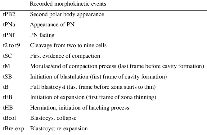
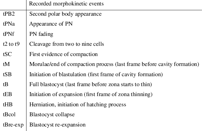
The times recorded for each morphokinetic variable were adapted from those proposed by Ciray et al. (Reference Ciray, Campbell, Agerholm, Aguilar, Chamayou, Esbert and Sayed2014) (Fig. 1). The time of PN appearance for the 2PN group was recorded when the first pronuclei was visible for the first time. The time of PN fading was the first frame when neither of the two pronuclei were visible. The morphokinetic variables for cleavage, morula, blastocyst and other events observed during embryo development are shown in Fig. 1.
Events recorded during the kinetic analysis of 1PN and 2PN ICSI zygotes (adapted from Ciray et al, Reference Ciray, Campbell, Agerholm, Aguilar, Chamayou, Esbert and Sayed2014).
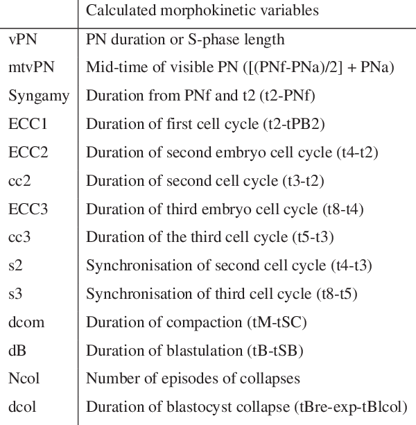
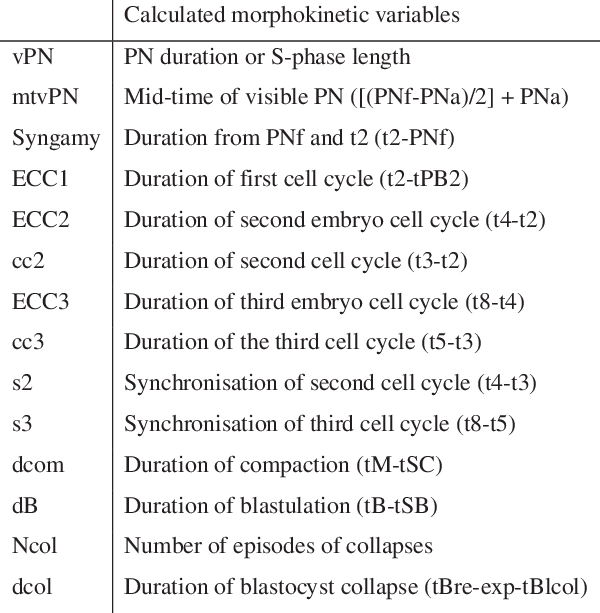
The associated calculated variables were adapted from previous publications (Meseguer et al., Reference Meseguer, Herrero, Tejera, Hilligsøe, Ramsing and Remoh2011; Aguilar et al., Reference Aguilar, Motato, Escribá, Ojeda, Muñoz and Meseguer2014; Athayde Wirka et al., Reference Athayde Wirka, Chen, Conaghan, Ivani, Gvakharia, Behr, Suraj, Tan and Shen2014; Ciray et al., Reference Ciray, Campbell, Agerholm, Aguilar, Chamayou, Esbert and Sayed2014) and are summarized in Fig. 2. The times at which each event occurred were recorded as the number of hours post-insemination. If blastocyst collapses were observed, the number and timing of collapses were recorded, as well as the time between the first collapse and the following re-expansion.
Calculated morphokinetic variables during the kinetic analysis of 1PN and 2PN ICSI zygotes (adapted from Meseguer et al., Reference Meseguer, Herrero, Tejera, Hilligsøe, Ramsing and Remoh2011; Aguilar et al., Reference Aguilar, Motato, Escribá, Ojeda, Muñoz and Meseguer2014; Athayde Wirka et al., 2014; Ciray et al., Reference Ciray, Campbell, Agerholm, Aguilar, Chamayou, Esbert and Sayed2014).
Statistical analysis
The data obtained in development, morphometric and kinetic analyses were compared between the 1PN group and 2PN group. Furthermore, comparisons were performed between: (i) 1PN zygotes that reached the blastocyst stage (1PN-derived blastocysts) and 1PN zygotes that arrested their development before the blastocyst stage; and (ii) 1PN-derived blastocysts and 2PN-derived blastocysts.
Continuous variables are described using median, ranges (minimum–maximum) and the mean and standard deviation (±SD). Nominal variables are presented in frequency tables.
To compare the distribution of the continuous variables, the non-parametric Wilcoxon–Mann–Whitney test or T-test was used in when appropriate, according to the assumptions. Associations between nominal variables were evaluated using the Pearson chi-squared test. All the tests were bilateral with a significance level of 5%. All statistical analyses were performed using IBM SPSS Statistics 22 software.
Results
Patients in the 1PN and 2PN groups showed similar maternal ages (36.6 ± 4.01 vs. 36.21 ± 3.15 years; P = 0.432) and ART indications (P = 0.191). No differences were observed in semen characteristics (volume, sperm concentration, percentage and concentration of progressive mobility; P = 0.691, P = 0.757, P = 0.83 and P = 0.737, respectively) or in seminal diagnosis (normozoozpermia, astenozoospermia and oligozoospermia; P = 0.44). The two groups were homogeneous in terms of cycle characteristics, with no differences in the gonadotrophin dose administrated, number of retrieved oocytes and number of fertilized oocytes (P = 0.403, P = 0.296 and P = 0.713, respectively). The ICSI time was 40.33 h post-hCG in the 1PN group and 39.75 h post-hCG in the 2PN group (P < 0.001).
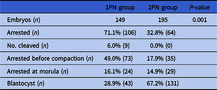
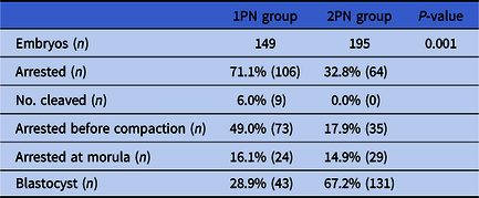
Monopronucleated zygotes showed impaired development compared with 2PN zygotes, evidencing higher rates of embryo arrest at early stages (prior to compaction; 71.1% vs. 32.8%) and a lower blastocyst rate (28.9% vs. 67.2%; Table 1).
Embryo development from 1PN and 2PN ICSI zygotes
The morphometric analysis showed differences between 1PN and 2PN zygotes in PN diameter (26.74 ± 2.91 vs. 24.09 ±1.51 µm; P < 0.001) and area (571.84 ± 106.38 vs. 478.43 ±54.34 µm2; P < 0.001). The 1PN zygotes that reached the blastocyst stage were those with a larger PN diameter and area (27.51 ± 3.42 µm and 609.07 ± 127.48 µm2, respectively; Supporting Information Table S1).
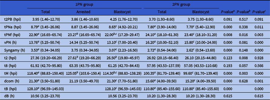
Differences in PN appearance and fading were observed between the 1PN and 2PN groups (Table 2). In 1PN zygotes, the pronucleus appeared later and faded earlier than pronuclei in 2PN zygotes, which resulted in a shorter time of visible pronuclei in the 1PN group (13.76 vs. 16.20 h; P < 0.001). Syngamy, defined as the duration between pronuclear fading and the first mitotic cleavage (Fig. 2), was longer in 1PN-derived embryos than in 2PN-derived embryos.
Results of the kinetic analysis of selected parameters of 1PN and 2PN ICSI zygotes
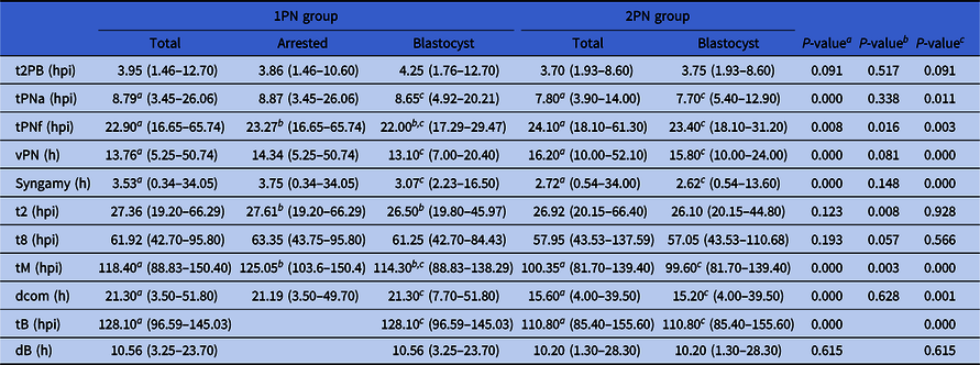
a1PN vs. 2PN groups. bArrested 1PN vs. 1PN blastocyst groups. c1PN blastocyst vs. 2PN blastocyst groups.
All values are presented as the median and range (in parentheses).
Different morphokinetic behaviour was observed among 1PN ICSI zygotes and their derived embryos. Comparison of 1PN zygotes that reached the blastocyst stage to 1PN zygotes whose development was arrested prior to this stage revealed similar behaviour in PN appearance. Earlier PN fading was recorded in the 1PN-derived blastocysts (Table 2). The duration of syngamy did not differ between 1PN zygotes that reached the blastocyst stage and those that were arrested beforehand (P = 0.148).
Cleavages in the 1PN group showed a longer second embryo cell cycle (ECC2) and more asynchronous second (s2) and third (s3) cell cycles than in 2PN-derived embryos. Abnormal cleavages were observed more frequently in the 1PN group than in the 2PN group, with similar rates of direct cleavage but higher rates of ID (33.6% vs. 10.3%; P < 0.001; Table 3). No differences in abnormal cleavages (neither ID nor DC) were observed in the two groups of 1PN-derived embryos (P = 0.454; Table 3). Different kinetics were observed in t2, t4, t5 and t7, showing delayed cleavage in those 1PN zygotes that did not reach the blastocyst stage.
Results from the analysis of abnormal cleavages observed in 1PN and 2PN ICSI zygotes

ID, irregular division; DC, direct cleavage.
a1PN vs. 2PN total groups. bArrested 1PN vs. 1PN blastocyst groups. c1PN blastocyst vs. 2PN blastocyst groups.
The first evidence of compaction (tSC) and time of morula (tM) were delayed in 1PN zygotes that did not reach the blastocyst stage compared with 1PN-derived blastocysts. The morulae characteristics were different between 1PN- and 2PN-derived embryos that reached the blastocyst stage, with a higher percentage of morulae that excluded some cells during compaction observed in 1PN-derived blastocysts (29/43, 67.4%) compared with 2PN-derived blastocysts (61/131, 46.6%; P = 0.017).
Monopronucleated zygotes achieved the blastocyst stage on day 5 (48.8%) or day 6 (51.2%), while most of the 2PN zygotes formed a blastocyst on day 5 (92.4%). The morphology of blastocysts from 1PN zygotes was significantly poorer than those blastocysts from 2PN zygotes. Good quality blastocysts (grades A and B) were observed in 32.5% of the 1PN-derived blastocysts compared with 61.9% in the 2PN group (P = 0.001).
Kinetics analysis of 1PN- and 2PN-derived blastocysts showed that the time taken for the PN to become visible was shorter in 1PN-derived blastocysts due to later appearance and earlier disappearance of the pronucleus (Table 2). The kinetics of 1PN-derived blastocysts and 2PN-derived blastocysts were similar from t2 to t9. The first evidence of delayed development in 1PN-derived blastocysts started at compaction. Blastocyst collapse occurred more frequently in 1PN-derived blastocysts than in 2PN-derived blastocysts (2 ± 1.05 vs. 1.17 ± 0.51; P = 0.001). The time of first collapse and re-expansion occurred later in 1PN-derived blastocysts than 2PN-derived blastocysts, but the duration of the collapse was similar.
Detailed data for all kinetic analyses are included in Table S2.
Discussion
Time-lapse systems are not commonly used to analyse 1PN ICSI zygotes, and the few published studies that have employed this technique focus mainly on analyzing pronuclear formation. Studies that evaluated the potential of 1PN-derived embryos to reach the blastocyst stage are performed in traditional incubators or do not evaluate embryo kinetics (Yamaguchi et al., Reference Yamaguchi, Takayanagi, Minamoto, Suzuki and Ando2013; Igashira et al., Reference Igashira, Otsuki, Furuhashi, Katada, Sumimoto, Kishi, Matsuura, Mukai, Sumi and Tsuji2016; Mateo et al., Reference Mateo, Vidal, Parriego, Rodríguez, Montalvo, Veiga and Boada2017; Araki et al., Reference Araki, Itoi, Honnma, Asano, Oguri and Nishikawa2018). The novelty of our study lies in the kinetics of embryo development of 1PN ICSI zygotes, which were comprehensively analyzed from fertilization to blastocyst stage, and the identification of different morphokinetic behaviours that can be associated with the abnormal pronuclear pattern of 1PN-derived embryos. The data obtained not only increased our knowledge about 1PN-derived embryos, but also revealed clear differences between those that reached the blastocyst stage and those that did not. We acknowledge that the high number of statistical comparisons may increase the risk of type 1 errors, and some spurious associations may have been found. Even so, as this was an observational retrospective study, their inclusion was considered relevant to estimate the effect of the specific morphokinetic characteristics observed during embryo development, rather than a simple P-value estimation.
We observed a longer time until injection in 1PN ICSI zygotes compared with those with 2PN, this is in accordance with a recently published study by Fabozzi et al. (Reference Fabozzi, Rega, Starita, Amendola, Colicchia, Giannini and Piscitelli2019), and this finding has been associated with in vitro oocyte ageing. Despite the slight delay in ICSI, we did not find any effect on oocyte activation. The timing of the second polar body appearance was equivalent in both groups, and similar to that previously published (Azzarello et al., Reference Azzarello, Hoest and Mikkelsen2012; Aguilar et al., Reference Aguilar, Motato, Escribá, Ojeda, Muñoz and Meseguer2014). Relationships between the appearance of monopronucleated zygotes and some patient characteristics (younger female age, more oocytes retrieved and astenozospermic male patients) noted in a previous study by Fabozzi et al. (Reference Fabozzi, Rega, Starita, Amendola, Colicchia, Giannini and Piscitelli2019) were not observed in the current study.
Differences in pronuclear kinetics were observed between the 1PN and 2PN groups. The delayed appearance of the pronucleus in 1PN ICSI zygotes could be due to impaired oocyte factors or anomalies that occur during fertilization that promote the formation of a unique pronucleus. The earlier fading could result from the absence of juxtaposition of the pronuclei, as it usually occurs in 2PN zygotes (Coticchio et al., Reference Coticchio, Mignini Renzini, Novara, Lain, Ponti, Turchi, Fadini and Dal Canto2018). The presence of a unique pronucleus in the zygote would probably hinder the metaphase plate organization, and this could explain the elongated time for syngamy observed in all 1PN ICSI zygotes. Based on the observed timings, it is important to highlight that some 1PN ICSI zygotes would never be identified if dynamic monitoring was not routinely performed.
Regarding early in vitro development, TL methodology made it possible to identify that 1PN-derived embryos were more likely to undergo ID than 2PN-derived embryos. This abnormal cleavage behaviour could be due to the formation of abnormal mitotic spindles that would lead to an imbalance in chromosomal distribution. This mechanism was previously described by Chatzimeletiou et al. (Reference Chatzimeletiou, Morrison, Prapas, Prapas and Handyside2005) and supported by the results of a previous study by our group, in which we observed 70.4% of chaotic embryos derived from 1PN ICSI zygotes (Mateo et al., Reference Mateo, Parriego, Boada, Vidal, Coroleu and Veiga2013).
Two populations with different morphokinetic behaviours were identified in the 1PN zygote group: those with early arrested development and a slow cleavage rate, and those that reached the blastocyst stage with faster cleavages and similar kinetics to 2PN-derived blastocysts. It was observed that those 1PN-derived embryos that arrested their development usually did so before compaction, which could be related to major alterations in chromosomal content or genome activation (Sultan et al., Reference Sultan, Munné, Palermo, Alikani and Cohen1995; Staessen and Van Steirteghem, Reference Staessen and Van Steirteghem1997; Lim et al., Reference Lim, Goh, Su and Yu2000; Liao et al., Reference Liao, Zhang, Cheng, Ouyang, Lin, Gu, Lu, Gong and Lu2009; Van Der Heijden et al., Reference Van Der Heijden, Van Den, Baart, Derijck, Martini and Boer2009; Mateo et al., Reference Mateo, Parriego, Boada, Vidal, Coroleu and Veiga2013, Reference Mateo, Vidal, Parriego, Rodríguez, Montalvo, Veiga and Boada2017; Jukam et al., Reference Jukam, Shariati and Skotheim2017; Ottolini et al., Reference Ottolini, Kitchen, Xanthopoulou, Gordon, Summers and Handyside2017).
All 1PN-derived embryos that continued their development presented a particular behaviour during compaction. A delayed and longer compaction was observed in 1PN-derived blastocysts, this could be related to an abnormal chromosomal content as this behaviour is similar to that described in embryos carrying aneuploidies (Campbell et al., Reference Campbell, Fishel, Bowman, Duffy, Sedler and Hickman2013). Furthermore, a partial compaction pattern was more frequently observed in 1PN-derived morulae that could be interpreted as an attempt at self-correction excluding aneuploid cells (Lagalla et al., Reference Lagalla, Tarozzi, Sciajno, Wells, Santo, Nadalini, Distratis and Borini2017).
Related to the capability to reach the blastocyst stage, our results are in accordance with other studies that reported a lower blastocyst rate in the 1PN group (3.4–14.8%) compared with the 2PN group (24.3–56.6%) (Mateo et al., Reference Mateo, Parriego, Boada, Vidal, Coroleu and Veiga2013, Reference Mateo, Vidal, Parriego, Rodríguez, Montalvo, Veiga and Boada2017; Itoi et al., Reference Itoi, Asano, Shimizu, Honnma and Murata2015; Bradley et al., Reference Bradley, Traversa, Hobson, Gee and McArthur2017; Capalbo et al., Reference Capalbo, Treff, Cimadomo, Tao, Ferrero, Vaiarelli, Colamaria, Maggiulli, Orlando, Scarica, Scott, Ubaldi and Rienzi2017). The blastocyst rate for 1PN ICSI zygotes observed in this study (28.9%) was higher than that previously reported by our group (3.4–14.8%) (Mateo et al., Reference Mateo, Parriego, Boada, Vidal, Coroleu and Veiga2013, Reference Mateo, Vidal, Parriego, Rodríguez, Montalvo, Veiga and Boada2017). The lower blastocyst rate observed previously could be attributed to the non-continuous culture conditions used in our earlier studies instead of the uninterrupted culture performed in the present work. Furthermore, with regard to the lowest blastocyst rate obtained (3.4%; Mateo et al., Reference Mateo, Vidal, Parriego, Rodríguez, Montalvo, Veiga and Boada2017), it should be highlighted that this study was undertaken in patients enrolled for preimplantation genetic testing for aneuploidies (PGT-A) on day 3. Therefore, embryo biopsy and patient characteristics could have had a detrimental effect on the blastocyst rate.
In summary, this work evidences for the first time that not all 1PN ICSI zygotes show the same pattern of development. The 1PN zygotes that achieved the blastocyst stage showed similar kinetics to 2PN-derived blastocysts. Considering that the 1PN zygotes that reached the blastocyst stage were those with the largest PN diameter, it could be interpreted that formation of the pronucleus in 1PN-derived blastocysts is the result of having a diploid genome englobed in a single pronucleus (Levron et al., Reference Levron, Munné, Willadsen, Rosenwaks and Cohen1995; Otsu et al., Reference Otsu, Sato, Nagaki, Araki and Utsunomiya2004; Van Der Heijden et al., Reference Van Der Heijden, Van Den, Baart, Derijck, Martini and Boer2009; Kai et al., Reference Kai, Iwata, Iba and Mio2015). This would favour development until the blastocyst stage, and would explain the similarity in morphokinetics observed among 1PN- and 2PN-derived blastocysts.
Recent studies have demonstrated that some 1PN-derived embryos might be euploid with a biparental origin, and can therefore be considered for transfer (Bradley et al., Reference Bradley, Traversa, Hobson, Gee and McArthur2017; Capalbo et al., Reference Capalbo, Treff, Cimadomo, Tao, Ferrero, Vaiarelli, Colamaria, Maggiulli, Orlando, Scarica, Scott, Ubaldi and Rienzi2017; Destouni et al., Reference Destouni, Dimitriadou, Masset, Debrock, Melotte, Van Den, Zamani Esteki, Ding, Voet, Denayer, de Ravel, Legius, Meuleman, Peeraer and Vermeesch2018; Xie et al., Reference Xie, Tang, Hu, Ouyang, Gu, Gong, Leng, Zhang, Xiong, Lu and Lin2018). The interest in rescuing embryos derived from 1PN zygotes has emerged as an option for some couples that have no other viable embryos. From our study, it can be concluded that TL data alone cannot be used to come to a decision about the fate of these embryos. Culture until the blastocyst stage should be mandatory if reproductive use of an 1PN-derived embryo is under consideration. If blastocysts from 1PN zygotes are considered for embryo transfer or cryopreservation, complementary genetic testing, including assessment of genome parental origin and aneuploidy screening, together with appropriate counselling, should be mandatory.
Acknowledgements
The authors thank all members of the Reproduction Medicine Service of Women’s Health Dexeus for their support.
Financial support
This work was funded by ‘Cátedra de Investigación en Obstetricia y Ginecología’ of the Department of Obstetrics, Gynaecology and Reproduction, Dexeus Women’s Health (Barcelona).
Conflicts of interest
The authors declare that they have no conflict of interest.
Ethical standards
This retrospective observational study was carried out at the ART laboratory at Women’s Health Dexeus in Barcelona. The study was approved by the institutional review board of the centre.
Supplementary material
To view supplementary material for this article, please visit https://doi.org/10.1017/S0967199420000027