Carbapenemase-producing Enterobacteriaceae (CPE) pose a serious public health threat, causing significant morbidity and mortality worldwide.1, Reference Nordmann, Cuzon and Naas2 In the United States, the predominant carbapenemase is Klebsiella pneumoniae carbapenemase (KPC),3 and hospitals remain a focal point for transmission of KPC-producing Enterobacteriaceae.Reference Logan and Weinstein4 To prevent transmission of these organisms in the hospital setting, multiple infection control interventions are implemented, including transmission-based isolation precautions, enhanced environmental cleaning, and care of KPC-positive patients using dedicated staff and equipment.5 Despite a very low number of CPE cases (<5 annually) at baseline, our hospital experienced an unusually high number of KPC-producing isolates among solid-organ transplant recipients between 2016 and 2017. We describe the epidemiologic and genetic characteristics of the outbreak and the intervention that allowed us to successfully stop horizontal transmission of KPC-producing organisms within our institution.
Methods
Our facility is a 566-bed academic hospital in Milwaukee, Wisconsin, with ∼150,000 admissions per year. The unit initially involved in this outbreak was the solid-organ transplant intensive care unit (TICU), with subsequent cases detected in the transplant-step-down-unit (3SW) and the inpatient rehabilitation unit. In 2016 and 2017, the TICU consisted of 10 private rooms, each with a sink (Supplemental Fig. 1A online). Rooms 1 and 10 had private patient bathrooms, and rooms 2–9 had contiguous macerators to grind human waste to a slurry, which was pumped into the sewer system. Five additional sinks were located outside patient rooms. The cohorting unit (Supplemental Figure 1B online) was an 8-bed hospital ward initially designed for containment of Ebola hemorrhagic fever cases and was used for cohorting KPC cases during this outbreak. The transplant step-down unit had 25 single-occupancy rooms and was located on a different floor (Supplemental Fig. 1C online). The inpatient rehabilitation unit consisted of 28 patient rooms and a large therapy gym (Supplemental Fig. 1D online). For this report, a clinical case was defined as a patient with a KPC-producing organism detected in cultures ordered by the managing team. A surveillance case was defined as a patient detected through surveillance cultures ordered by the infection control department.
Timeline of Klebsiella pneumoniae carbapenemase (KPC)–producing organism cases admitted to the hospital from January 2016 through April 2017. Note. *Time of first KPC detection. Rehab, inpatient rehabilitation unit; 3SW, transplant step-down unit. A black bar reflects patient expired.

Infection control methods
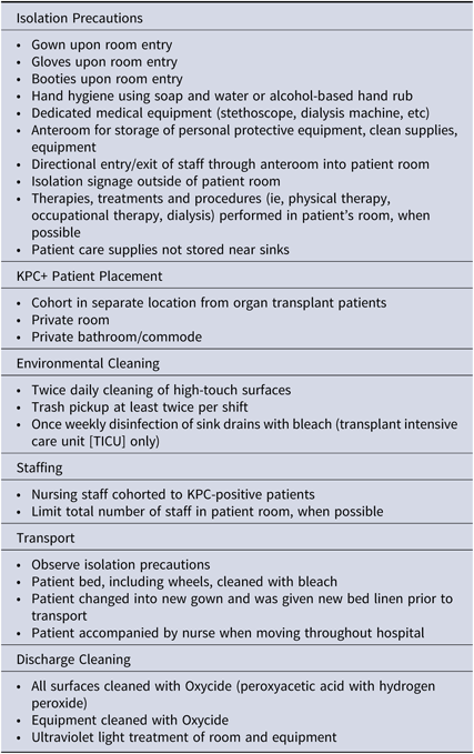
The infection control bundle had been implemented since September 2016 starting in TICU and included isolation precautions, dedicated nursing personnel, and equipment (Table 1). Physical therapy was performed either in the patient’s room or in the rehabilitation gym at the end of the day. Any equipment used by the patient during physical therapy sessions was supposed to be cleaned with a hospital-approved quaternary ammonium disinfectant.
Components of the Infection Control Bundle

Cohorting of patients and providers
KPC cases were initially cohorted in TICU rooms 1–3 (Supplemental Fig. 1A online). Once the cohorting-unit was implemented, KPC patients had dedicated nurses and nursing aids and were placed in single rooms with an adjoining room, which served as the anteroom for donning and doffing personal protecting equipment and storing clean supplies, equipment, and linen. In the inpatient rehabilitation unit, dedicated staff, equipment, and contact precautions were used, although in a regular patient room.
Stool/rectal surveillance culture specimens
After the first KPC patient was identified, surveillances (stool samples or rectal swabs on admission and weekly thereafter) were used to determine asymptomatic colonization and were performed on all patients housed in the TICU and transplant step-down unit beginning in September 2016. In February 2017, surveillance cultures were also performed weekly in the inpatient rehabilitation unit.
Environmental sampling
In September 2016, horizontal surfaces in the TICU were sampled using premoistened swabs. Sink drains and overflows were then sampled in the TICU and the transplant step-down unit in February 2017. Sampling was performed by introducing a culture swab through the drains as far down into the drain as possible (to swab the inner walls) or inside the overflow space, respectively.
Environmental cleaning and disinfection
All high-touch surfaces in occupied patient rooms were cleaned daily and after the patient was discharged with the standard hospital disinfectant (OxyCide; Ecolab, Saint Paul, MN). Floor tiles were cleaned daily with Stride Neutral Cleaner (Diversey, Charlotte, NC). Automated ultraviolet disinfection machines (Surfacide Helios System, Waukesha, WI) were used for discharge cleaning throughout all the phases of the outbreak. To evaluate compliance with daily and discharge cleaning, a fluorescent marker gel (Dazo, Ecolab, Saint Paul, MN) was applied to high-touch surfaces in patient rooms, including bed rails, intravenous pumps, tray tables, and computers. These gel marks were evaluated 48 hours after they were made.
Microbiology methods
Human samples and environmental swabs. Environmental surfaces, sink drains, and patient rectums were swabbed using sterile swabs that were transported to the hospital laboratory (Wisconsin Diagnostic Laboratories, Milwaukee, WI) in Amies transport gel. Swabs were suspended in 5 mL tryptic soy broth (TSB) containing a 10-µg ertapenem disk and incubated overnight at 35°C. Turbid broth cultures were subcultured to MacConkey agar plates and incubated overnight at 35°C. All unique colonies were identified to the species using MALDI-TOF MS (Bruker Daltonik, Billerica, MA). Colonies consistent with Enterobacteriaceae were subsequently suspended in saline to make a 0.5 McFarland standard dilution. The suspension was then streaked onto Mueller-Hilton agar plates and overlaid with Etest meropenem and ertapenem test strips (bioMérieux, Marcy-l’Étoile, France). Any isolates that were nonsusceptible to ertapenem or meropenem (>0.5 μg/mL or >1 μg/mL, respectively) underwent real-time polymerase chain reaction (RT-PCR) for detection of bla KPC and bla NDM.6
Disassembled sink drains. Portions of sink drain pipes were removed on February 27, 2017, and were cultured at our facility as described in the previous section. The removed drainpipes (both P-trap water and biofilm) were cultured for carbapenemase-producing isolates. Biofilm sampling was performed by swabbing an inner pipe at the levels of the drain, tail piece, P-trap, and J-arm (distal to the P-trap) with BBL CultureSwab EZ swabs (Becton Dickinson, Franklin Lakes, NJ). Swabs and sink trap water were processed as previously described.Reference Mathers, Vegesana and German Mesner7
Generation of transformants/transconjugants. Total plasmids were extracted by phenol/chloroform extraction and were used to transform E. coli TOP10 by electroporation, using ampicillin 50 mg/L as the selective agent. When transformants were obtained, carriage of bla KPC was confirmed by PCR. For strains for which transformants could not be obtained, broth mating was performed between the strains and E. coli J53AziR, and transconjugants were selected using ampicillin 50 mg/L and sodium azide 100 mg/L. As with the transformants, carriage of bla KPC was confirmed by PCR.Reference Kim, Qureshi, Adams-Haduch, Park, Shutt and Doi8
S1 nuclease PFGE. The sizes of bla KPC-carrying plasmids in the transformants/transconjugants were estimated by S1 nuclease PFGE, as described previously.Reference Bueno, Francisco, O’Hara, De Oliveira Garcia and Doi9
Whole-genome sequencing and analysis. Genomic DNA was extracted using a Qiagen Blood and Tissue kit (Qiagen, Hilden, Germany). Next-generation sequencing was performed using the paired-end method on a NextSeq500 instrument (Illumina, San Diego, CA). De novo contigs were generated using the CLC Genomics Workbench. When applicable, sequence types (STs) were determined by in silico multilocus sequence typing (MLST). Resistance-gene contents and plasmid replicons were queried through the ResFinder and PlasmidFinder, respectively (https://cge.cbs.dtu.dk/services). For E. cloacae and K. pneumoniae, the core single-nucleotide polymorphism (SNP)–based phylogeny was obtained using snippy (https://github.com/tseemann/snippy) with assembled genomes of representative study isolates as internal references. Sequencing data are accessible in the NCBI Genome database under BioProject PRJNA482007.
Results
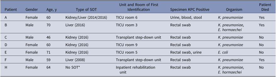
From April 2016 to April 2017, 8 patients were identified as either clinical (n = 3) or surveillance (n = 5) KPC cases. One of these patients was positive for KPC-producing Citrobacter spp upon admission to an unrelated unit and was removed from further analysis. Of the remaining 7 patients, 6 (86%) patients were aged 60 or older (median age, 60 years), 4 (57%) were females, and 6 (86%) had received solid-organ transplantation (kidney, liver, or both) (Table 2). The mean number of days from admission to KPC identification was 52.7 days. The KPC-producing species identified during this outbreak included Klebsiella pneumoniae, Enterobacter cloacae complex (E. hormaechei), and Escherichia coli (Table 2; Fig. 1). KPC-producing K. pneumoniae were detected from multiple sources, including urine (n = 2), blood (n = 1), and stool or rectal swabs (n = 7). Of these 7 patients, 3 (43%) died during hospitalization: 1 due to sepsis and 2 due to noninfectious causes.
KPC Outbreak Patients

Note. SOT, solid-organ transplant; KPC, Klebsiella pneumoniae carbapenemase.
* Patient had a solid malignancy. Transplant step-down unit was 3SW.
Phase 1. Infection control bundle (July 2016–October 31, 2016)
The index case (patient A) was admitted to TICU room 2 in April 2016 and was identified as a clinical case in July 2016 with KPC-producing K. pneumoniae in urine (Fig. 1). Surveillances began in September 2016. Patient B was admitted to TICU room 1 in June 2016 and KPC-producing K. pneumoniae was identified on surveillance cultures in September 2016 (TICU room 3). Both cases were cohorted in TICU (rooms 1 and 2; Supplemental Fig. 1A online). In September 2016, after patient B was identified as a KPC carrier, the full infection control bundle was implemented for all TICU patients (Table 1).
In October 2016, surveillance cultures detected 2 additional KPC patients (patients C and D; Fig. 1). Patient C was located in the transplant step-down unit (3SW), and patient D was located in TICU room 9 (Supplemental Figure 1A and 1C). After these 2 new cases, we decided to cohort patients in the cohorting unit.
Phase 2. Cohorting unit (November 1, 2016–January 7, 2017)
Patient C was discharged, and the remaining KPC cases, patients A, B, and D, were admitted to cohorting unit rooms 1, 5, and 6 (Supplemental Fig. 1B online). Access to the unit was limited to staff needing to perform patient care; all other foot traffic was restricted by locked entrances. In November 2016, patient D was discharged from the hospital, and in December 2016, patient A died of sepsis. In January 2017, only patient B remained in contact isolation within a single room in the cohorting unit. No additional cases were identified by surveillance.
Phase 3. Infection control bundle (January 8, 2017–February 23, 2017)
In January 2017, patient B was transferred back to TICU (room 4), and 2 weeks later was moved to the transplant step-down unit (3SW-13; Fig. 1), where he became positive for KPC-producing E. hormaechei in addition to KPC-producing K. pneumoniae. In January 2017, patient F was identified by surveillance as a carrier of KPC-producing K. pneumoniae in the transplant step-down unit. In February 2017, patient E was identified as KPC positive while in TICU room 5 (Fig. 1). Patient B was transferred to the inpatient rehabilitation unit (Fig. 1; Supplemental Fig. 1). Approximately 1 week after patient B was admitted to the rehabilitation unit, a new KPC patient was identified by surveillance culture in the rehabilitation unit (patient H), with both KPC-producing K. pneumoniae and E. hormaechei, just as patient B.
Phase 4. Cohorting unit (February 24, 2017–April 25, 2017)
Due to the identification of patients E, F, and H, the cohorting unit was reopened. Patient F died and patient H was discharged prior to the transfer, so the only patient transferred to the cohorting unit was patient E.
Hand hygiene
Hand hygiene observations obtained using the “secret shopper” method showed an overall hospital hand hygiene compliance of 82% during the outbreak. In the TICU, the transplant step-down unit, and the rehabilitation unit, hand hygiene compliance rates were 75%, 64%, and 77%, respectively.
Environmental cleaning compliance
Despite the instruction to staff to perform more frequent cleaning and disinfection in rooms occupied by KPC cases, infection control audits reported a cleaning compliance of 75.5% for 118 surfaces marked, and environmental services staff reported 71% compliance for 396 surfaces marked.
Environmental cultures
The results of environmental cultures are shown in Supplemental Table 1 (online). Of 14 sink drains in the TICU, 64% were bla KPC positive by PCR and 29% grew KPC-producing isolates by culture (Supplemental Fig. 2 online). Of 3 overflow drains in TICU, 2 (66%) were bla KPC-positive by PCR; however, none grew KPC-producing isolates by culture. All disassembled drains and sink trap water cultures remained negative.
Surveillance cultures
Surveillance cultures were conducted on a weekly basis for all patients in affected units from September 2016 to April 2017. Of 2,067 specimens, 117 (5.6%) had Enterobacteriaceae phenotypically nonsusceptible to ertapenem and/or meropenem. Direct testing of these isolates using targeted real-time PCR identified 21 of 117 isolates (17.9%) to be positive for bla KPC. The 21 KPC-positive isolates were obtained from 10 unique patients.
Strain and plasmid typing
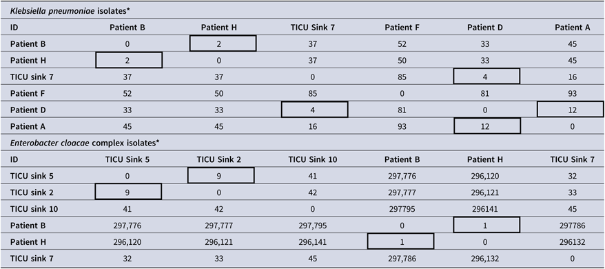
A total of 6 K. pneumoniae isolates and 6 E. cloacae complex strains were subjected to whole-genome sequencing (WGS) to determine genetic relatedness (Table 3 and Supplemental Table 2 online). All 6 K. pneumoniae isolates were within 2–93 SNPs of each other. Furthermore, 5 genomes were ST258 and carried bla KPC-3 (n = 4) or bla KPC-31 (n = 1) on a ∼60-kb IncN plasmid based on S1 PFGE. One isolate (from patient A) belonged to ST1084, a single-locus variant from ST258 differing in rpoB. In addition, the bla KPC-3-bearing plasmid in this isolate was much smaller (Supplemental Fig. 3 online). Isolates from patient B and patient H, first identified in TICU and the rehabilitation unit, respectively, were within 2 SNPs of each other, though they carried bla KPC-31 and bla KPC-3, respectively, which differ from each other by one amino acid. Similarly, the isolate from patient D, first identified in TICU room 9, and the sink isolate obtained from TICU room 7 were within 4 SNPs of each other. These pairs likely represent transmission of the same strain within the outbreak period. The K. pneumoniae isolates from patient D and patient A were within 12 SNPs, but the differences in both ST and plasmids carrying bla KPC made them unlikely to be directly related to each other and might suggest intermediary reservoirs.
Genetic Variations of Single Nucleotide Polymorphisms (SNPs)

* Boxes with thick lines represent strains with genetically related isolates. Example: First box in top table means that K. pneumoniae strains belonging to patients B and H had only 2 SNPs f difference, and thus were likely related.
The 6 E. cloacae complex genomes belonged to 2 diverse STs: Enterobacter roggenkampii ST422 (n = 4) and Enterobacter hormaechei ST407 (n = 2). The E. hormaechei isolates from patient B and patient H had only 1 SNP difference between them and likely represent transmission of the same strain. These E. hormaechei isolates carried bla KPC-40 on an IncN plasmid similar in size to that in 5 of the 6 K. pneumoniae isolates, raising the possibility of plasmid exchange between the 2 species. The 4 E. roggenkampii isolates harbored a ∼40-kb IncP6 plasmid carrying bla KPC-2 instead of bla KPC-40. Enterobacter roggenkampii isolates obtained from the sinks in TICU room 2 and room 5 had 9 SNP differences, making them suspicious for recent transmission. The remaining E. roggenkampii ST422 genomes shared between 32–45 SNPs, which is in keeping with genomes belonging to the same genetic lineage but unlikely to represent horizontal transmission.
Discussion
We have described the control of a KPC plasmid outbreak in a TICU involving solid-organ transplant recipients, which later expanded to the transplant step-down unit and to an inpatient rehabilitation unit. Whole-genome sequencing was able to identify transmissions in TICU between patients A and D (phase 1) and in the inpatient rehabilitation unit between patients B and H (phase 3). We also found close similarities in the K. pneumoniae strains found in the sink of TICU room 7 and both patient A (TICU room 6) and patient D (TICU room 9). An infection control bundle was insufficient to control this outbreak despite an aggressive bundle of interventions; we subsequently used a cohorting unit to house all KPC cases. Importantly, when this outbreak occurred, the TICU was located in a small and crowded unit housing solid-organ transplant patients. Whether the patients’ underlying medical conditions or the small unit layout, or both, were the reasons why we were unable to control this outbreak remains unclear. The use of a cohorting unit, early identification of colonized patients, and patient deaths or discharges allowed us to stop the horizontal transmission of KPC strains in 2 separate periods. As of January 2019, the hospital continues to use the cohorting unit (unless ICU care is needed) for any patient with history of KPC-producing Enterobacteriaceae carriage who is readmitted to the hospital. No additional KPC acquisitions in our hospital have been detected in our transplant population through ongoing surveillance.
Infection control bundles have been successfully used to control CPE outbreaks.Reference Munoz-Price, Hayden and Lolans10–Reference Palmore and Henderson14 These bundles aim to prevent the exposure of patients to CPE; however, identification of asymptomatic carriers and their subsequent contact precautions and cohorting might be one of the most important strategies within the bundle.Reference Munoz-Price and Quinn15 The use of surveillance cultures allowed our team to detect 7 patients, 5 of whom would otherwise have gone undetected (Table 2).
The sinks in TICU were in close proximity to the patients’ beds, and the sink pipes from contiguous rooms were shared as shown in Supplemental Figure 2 (online). However, cultures of deeper portions of the pipes were all negative for CPE. Exchanging pipes in the TICU failed to control the outbreak, so we hypothesize that the sink drains did not play an active role in this particular situation despite recent literature demonstrating their significance elsewhere.Reference Kotay, Chai, Guilford, Barry and Mathers16, Reference Kizny Gordon, Mathers and Cheong17 The horizontal transmission of 2 KPC-producing isolates in the inpatient rehabilitation unit was concerning. This transmission occurred after 5 days of a KPC-positive patient being in the unit and despite implementation of an infection control bundle. This finding highlights the need to preserve strict infection control bundles in KPC-positive patients transferred to subacute settings.
Because the outbreak involved multiple Enterobacteriaceae species in an institution that rarely encounters KPC-producing organisms, we initially assumed it to represent a plasmid-mediated outbreak caused by introduction of a single transmissible bla KPC-carrying plasmid. This appears to have been the case for the E. hormaechei isolates and most of the K. pneumoniae isolates, both of which harbored a bla KPC-3-carrying IncN plasmid of the same size. Further molecular analysis, however, indicated the involvement of another plasmid type, IncP-6, which carried bla KPC-2 in E. roggenkampii and E. coli. Both plasmid types have been reported to be carriers of bla KPC.Reference Naas, Bonnin, Cuzon, Villegas and Nordmann18–Reference Kukla, Chudejova and Papagiannitsis20 These unexpected findings suggest nearly simultaneous introduction of at least 2 distinct bla KPC-carrying plasmids and subsequent outbreak due to both plasmid-mediated and clonal dissemination involving multiple species, thus revealing a complex hospital epidemiology that unfolded in this low-baseline CPE prevalence setting.
The use of a cohorting unit exerted a great amount of stress in our institution, which is similar to the experience reported by others.Reference Palmore and Henderson14 Multiple patients were moved to different hospital rooms to accommodate KPC patients. Staffing also proved to be difficult because we relied on staff from the TICU to care for these patients outside of the normal ICU environment, and this led to increased stress levels for the staff.
In summary, this outbreak of KPC-producing organisms was successfully controlled by the implementation of an infection control bundle, weekly surveillance cultures, and the use of a separate cohorting unit. Implementation of weekly point-prevalence surveillance cultures in units where KPC cases were admitted was integral to tracking potential transmission events. The relevance of finding multiple species of KPC-producing bacteria by both molecular and culture methods remains unclear. Further investigation is required to establish the epidemiological significance of this type of outbreak. Based on our findings, we believe that surveillance cultures should be an integral component of infection control bundles and that a cohorting unit should be considered when other interventions fail.
Supplementary material
To view supplementary material for this article, please visit https://doi.org/10.1017/ice.2019.99.
Author ORCIDs
Silvia Munoz-Price, 0000-0001-8654-5746
Acknowledgments
We would like to thank all the FMLH nurses in the TICU, rehabilitation unit, and step down units who helped make these interventions possible.
Financial support
This study was supported by funding from the National Institutes of Health (research grant nos. R01AI104895, R21AI123747, R21AI135522 to Y.D.).
Conflicts of interest
L.S.M.P. has received a grant from Cepheid. N.A.L. has served as a consultant for Thermo Fisher, Copan Diagnostics, LifeScale, Luminex, and Cepheid and has received grants from Copan, Cepheid, Luminex, BD Diagnostics, bioMerieux, and Hardy Diagnostics. B.W.B. has served as a consultant for Chromacode and Luminex and has received grants from Luminex, Biofire Diagnostics, Alere, Chromacode, GenMark, and BioFireDiagnostics. Y.D. has consulted for Roche, Geom, and Recida and has received speaking fees from Merck and Pfizer. All other authors report that they have no thing to disclose.