Introduction
Mild cognitive impairment (MCI) describes a boundary condition between normal aging and dementia. MCI is subclassified into two forms: amnestic MCI (aMCI) and non-amnestic MCI (non-aMCI) (Petersen et al., Reference Petersen, Smith, Waring, Ivnik, Tangalos and Kokmen1999). aMCI is characterized by performance only on memory or with other measures below expected normative values on neuropsychological evaluation. In one neuropathology study of dying patients fulfilling criteria for aMCI, the majority had some pathological features suggestive of a transitional state between normal aging and AD (Petersen et al., Reference Petersen2001). A number of longitudinal studies have shown that aMCI patients have a high probability of developing AD in the immediate future with a conversion rate that varies from 4% to 25% per year (Amieva et al., 2004; Petersen, Reference Petersen2004). So aMCI represents the prodromal stage of AD. Early identification of aMCI patients has become increasingly important with the prospect of disease modifying or halting agents that may be most beneficial at the earliest stages of disease.
Prospective studies have shown that episodic memory, semantic memory, attention processing, and mental speed can consistently predict which patients with aMCI will develop dementia (Amieva et al., Reference Amieva2004). Similarly, for the patients with aMCI who developed AD, verbal and visual memory, associative learning, vocabulary, executive function and other verbal tests of general intelligence were impaired at baseline in a retrospective study (Guarch et al., Reference Guarch, Marcos, Salamero and Blesa2004). Despite the converging evidence of abnormal neuropsychological changes in aMCI, cognitive performances alone seem to be insufficient to predict conversion of aMCI to AD. It is reasonable that additional biological parameters are needed for this purpose. Genetic epidemiological studies have shown that genetic factors are linked to the risk of AD. If we can identify genes involved in the etiology of AD, this will allow opportunities for improvements in the diagnosis of aMCI and in predicting the course of the disease. Most studies found that apolipoprotein E (ApoE) was associated with cognitive decline and MCI (Aggarwal et al., Reference Aggarwal, Wilson, Beck, Bienias, Berry-Kravis and Bennett2005). For the aMCI subtype of degenerative etiology, ApoE4 carrier status has been shown to be a risk factor for a more rapid progression to AD (Aggarwal et al., Reference Aggarwal, Wilson, Beck, Bienias, Berry-Kravis and Bennett2005).
In addition to ApoE, the gene coding for low-density lipoprotein receptor-related protein1 (LRP1) has also been implicated in cognition and AD (Harris-White and Frautschy, Reference Harris-White and Frautschy2005). The association is particularly intriguing on the gene's matched function using pathophysiology and positional information obtained from linkage studies (Bian et al., Reference Bian2005). First, LRP1 on the endothelial cells of the blood brain barrier as well as on neurons can mediate crucial pathways such as amyloid precursor protein (APP) processing and beta-amyloid (Aβ) production, oligomerization of amyloid proteins, Aβ clearance, Aβ uptake, and intraneuronal accumulation of Aβ, while LRP1 mediated Aβ-associated neuronal death may be an early trigger in the AD pathogenic cascade (Cam et al., Reference Cam, Zerbinatti, Li and Bu2005; Zerbinatti et al., Reference Zerbinatti2006). Secondly, several well-known LRP1 ligands involved in AD pathogenesis, including ApoE, a2-macroglobulin, transforming growth factor-beta and tissue plasminogen activator, can be modulators of neuronal function, synaptic plasticity and potentially cognitive function (Harris-White and Frautschy, Reference Harris-White and Frautschy2005). Further, LRP1 as a signaling receptor regulates vesicle transport, axon guidance and synaptic plasticity (Herz and Strickland, Reference Herz and Strickland2001). Of interest, the NMDA receptor and LRP1 appear to be linked by the scaffold protein postsynaptic density 95 (PSD95) (Herz and Strickland, Reference Herz and Strickland2001). Therefore, LRP1 has effects on the synapto-dendritic complex, migration, plasticity, regeneration, and LTP and these effects will undoubtedly impact learning and memory and cognition. Finally, the human LRP1 gene locates on chromosome 12q13–14, the site of a potential AD locus, resulting in a plausible position candidate gene for AD susceptibility. Some polymorphisms in LRP1 gene have been reported to increase the risk for AD in some populations, though neither polymorphism has been consistently shown to be associated in independent datasets (Kang et al., Reference Kang1997; Hatanaka et al., Reference Hatanaka2000; Panza et al., Reference Panza2004; Bian et al., Reference Bian2005). However, these studies focused on only a few polymorphisms respectively, which were inadequate for the LRP1 gene.
aMCI is thought to be the preclinical form of AD, and LRP1 plays a complex and significant role in cognition and AD. To our knowledge, the potential relationship between the LRP1 genetic variant and the characteristics of neuropsychology have not been previously explored in aMCI subjects. Therefore, the aim of this study is to investigate the relationship of the LRP1 genetic variant to aMCI risk using a case-control association design and haplotype analysis. We have assessed the effects of the identified risk LRP1 genetic variant on the cognitive profile of aMCI. We expected that if the risk for aMCI associated with LRP1 is at least partly mediated by its influence on neurocognitive function, then carriers of the risk haplotype will perform significantly worse on at least some neurocognitive measures.
Methods
Subjects and clinical assessment
All individuals were recruited from a memory clinic. Information on age, level of education, medical history, current use of medications and life style practices were gathered by personal interviews using a standardized questionnaire. Written informed consent was obtained from all participants. This study was reviewed and granted by the Institutional Ethical Review Board of Clinic Medical College of Southeast University. It comprised a total of 213 Chinese subjects: 109 with a diagnosis of aMCI (mean ± SD age = 73.17±5.24years, 69 men, 40 women, educated = 14 (11–16) years) and 104 cognitively healthy controls (mean ± SD age = 72.10 ± 3.61, 50 men, 54 women, educated = 15 (12–16) years). Unrelated patients and control subjects were all Chinese from Jiangsu province located in southeast China. Cognitively normal subjects and patients with aMCI did not differ significantly in age and years of education (t = −1.755, p = 0.081; Z = −1.873, p = 0.061). All subjects underwent a detailed physical and neurological examinations as well as cognitive screening.
Criteria for the diagnosis of aMCI were those of Petersen et al. (Reference Petersen, Smith, Waring, Ivnik, Tangalos and Kokmen1999): (1) a memory complaint, preferably corroborated by an informant; (2) an objective memory impairment on neuropsychological evaluation: 20 minute delayed recall of auditory verbal learning test (AVLT) ≤ 1.5 SD of age and education matched controls, with a cut-off of ≤4 correct responses on 12 items for ≥8 years of education (Guo et al., Reference Guo, Lv and Hong2001); (3) preserved general cognitive function: a Mini-mental State Examination (MMSE) score ≥ 24 (Katzman et al., Reference Katzman1988); (4) a clinical dementia rating score (CDR) of 0.5, with at least a 0.5 in the memory domain (Morris, Reference Morris1993); (5) activities of daily living (ADL): normal or only slightly impaired (ADL: 20–26); (6) absence of dementia, not sufficient to meet National Institute of Neurological and Communicative Disorders and Stroke/Alzheimer's Disease and Related Disorders Association (NINCDS-ADRDA) criteria for AD (McKhann et al., Reference McKhann, Drachman, Folstein, Katzman, Price and Stadlan1984). Participants were excluded from the present study if they had a past history of known stroke (modified Hachinski score ≥4) (Rosen et al., Reference Rosen, Terry, Fuld, Katzman and Peck1980), alcoholism, head injury, Parkinson's disease, epilepsy, major depression or other neurological or psychiatric illness, major medical illness (e.g. cancer, anaemia, thyroid dysfunction), or severe visual or hearing loss. Blood work included routine chemistry, thyroid function, serum B12 and folate. Neuroimaging diagnostic procedures (cerebral CT or MRI) were carried out to exclude other causes of dementias. All controls were required to have a CDR of 0, an MMSE score ≥26, and delayed recall of AVLT score >4.
To provide comprehensive yet efficient assessment of the cognitive characteristics of aMCI and the influence of LRP1 on cognitive performance, all individuals were administered an extensive neuropsychological battery by psychometrists who have been trained in standardized assessment and scoring procedures. The tasks included AVLT, Rey-Osterrieth complex figure test (CFT) (Lezak, Reference Lezak1983), verbal fluency test (VFT, animal category) (Monsch et al., Reference Monsch, Bondi, Butters, Salmon, Katzman and Thal1992), digit span test (DST) (Wechsler, Reference Wechsler1981), symbol digit modalities test (SDMT) (Wechsler, Reference Wechsler1981), trail making test A and B (TMT) (Gordon, Reference Gordon1972), and the clock drawing test (CDT) (Libon et al., Reference Libon, Malamut, Swenson, Sands and Cloud1996).
Extraction of genomic DNA and genotyping
Investigators who were blinded to all subject identifiers and information performed the genotype analysis. Blood samples were obtained from 109 aMCI patients and 104 controls. Total genomic DNA was extracted from 300ul EDTA-anticoagulated venous blood using the PUREGENE DNA purification Kit (Gentra Systems, Minneapolis, MN) according to the manufacturer's instructions. We focused on five single nucleotide polymorphisms (SNPs) in the LRP1 gene, including Site1: rs1799986(exon3), Site2: rs1800194(exon22), Site3: rs11837145(intron24), Site4: rs10876967(intron24) and Site5: rs1140648(exon61). We chose the five SNPs of the LRP1 gene, which were mainly selected from previously published articles and determined based on their high heterozygosity (Hatanaka et al., Reference Hatanaka2000; Van et al., Reference Van2001; Kang et al., Reference Kang1997; Panza et al., Reference Panza2004; Bian et al., Reference Bian2005). All these SNPs were genotyped using Applied Biosystems 7500 Real Time PCR System, according to the Assays-by-Design Service for SNP Assays Protocol. Ambiguous genotypes were retyped until clear results were determined.
Statistical analysis
SHEsis software (Shi and He, Reference Shi and He2005) was used to test the distribution of genotypes and allele frequencies for deviations from the Hardy–Weinberg equilibrium, compare the SNP allele and genotype frequencies between patient groups and normal controls, and estimate pair-wise linkage disequilibrium (LD) between the polymorphisms. Haplotype analysis of unphased case/control data and quantitative data was performed using UNPHASED version 3.0.13 (Dudbridge, Reference Dudbridge2008). Only haplotypes with a frequency ≥ 0.05 were included in the subsequent global p-value calculation for haploblocks. Ten thousand permutations were computed in haplotype analysis.
All subsequent analyses were performed using Statistical Package for the Social Sciences (SPSS) statistical software, version 11.5 (SPSS Inc., Chicago, IL). Differences between cases and controls were computed using the independent samples t-test for the normally distributed variables and the non-parametric Mann-Whitney U-test for the asymmetrically distributed variables. Significance levels were for two-tailed tests; p values < 0.05 were regarded as significant.
Results
Cognitive performance
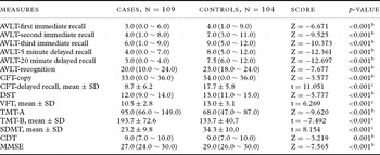
Among those identified as having aMCI, about 38.5% had isolated memory impairment, whereas the remaining 61.5% had memory impairment with lesser impairments in at least one non-memory cognitive domain. The cognitive assessments for all subjects are given in Table 1. The measurement of general and domain-specific functions differed significantly between groups. Cases displayed lower scores compared to healthy controls, with the largest impairment on 20-minute delayed recall of the AVLT (<3.0 SD of normal mean). Beyond the episodic memory, trail making test B showed the greatest impairment, deviating from normal by 1.5 SD.
Comparison of cognitive performance between aMCI patients and controlsa

a Values expressed as median (interquartile range) except where noted.
b Mann-Whitney U test.
c Independent-Samples t test.
AVLT = Auditory Verbal Learning Test; CFT = Rey-Osterrieth Complex Figure Test; DST = Digit Span Test; VFT = Verbal Fluency Test; TMT = Trail Making Test A and B; SDMT = Symbol Digit Modalities Test; CDT = Clock Drawing Test; MMSE = Mini-mental State Examination.
Genotype effects on risk for aMCI
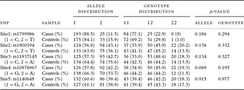
For each SNP analyzed, no deviation from the Hardy-Weinberg equilibrium was seen for any polymorphism in the LRP1 gene for the cases or the controls (p > 0.05). Table 2 shows the allele and genotype frequencies of the five polymorphisms in the LRP1gene for aMCI patients and control subjects. No differences in genotype or allele frequencies in the LRP1gene were detected between patients and controls.
Distributions of alleles and genotypes in LRP1 gene

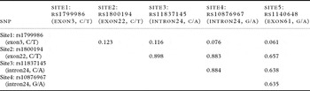
More detailed statistical analyses were performed based on SNP haplotypes. In the whole study group, three SNPs (Site2: rs1800194, Site3: rs11837145, Site4: rs10876967) form a comparatively strong LD in the LRP1 gene (Table 3) and this LD block was not composed of Site1: rs1799986 and Site5: rs1140648. Subsequently, we conducted haplotype analysis among the other three sites and evaluated the difference in the frequency distributions between aMCI patients and control subjects. Three marker haplotypes within the LD block were assessed (Table 4). The SNPs defined three haplotypes which were common (frequencies >5%), cumulatively accounting for about 90% of the observed haplotypes in patients and 96% in controls. We found these haplotypes had been described as CCG, TAA and TAG, respectively. Haplotype analysis of the LRP1 gene in our population showed a marked over-representation of the TAA haplotype in aMCI patients (p = 0.002).
Pairwise disequilibrium coefficients between SNPs in the LRP1 gene

Estimated haplotype frequencies and association with aMCI

Notes: Only the haplotypes with frequency greater than or equal to 5% in either patients or control subjects are shown. Haplotype: rs1800194 (exon22, C/T), rs11837145 (intron24, C/A), rs10876967 (intron24, G/A). Empirical 5% quantile of the best p-value: 0.02072.
Genotype effects on cognitive domain scores
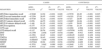
To test the hypothesis that the LRP1-risk haplotype TAA would be associated with poor performance on cognitive measure, we examined the relationship between TAA haplotype and neurocognitive function, as listed in Table 5. In patients with aMCI, the risk haplotype TAA was associated with all cognitive tests, excluding DST and CDT, with carriers of TAA in aMCI subjects demonstrating significantly poorer performance than non-carriers of aMCI groups. The results also revealed that in controls the effects on the cognitive domains of AVLT, CFT-delayed recall, DST and TMT-B were driven by TAA carrier status, especially in AVLT-delayed recall, while no differences emerged on the other tests.
Association between TAA risk haplotype and cognitive performances

TAA risk Haplotype: T (rs1800194, exon22), A (rs11837145, intron24), A (rs10876967, intron24).
P *-value: Empirical 5% quantile of the best p-value.
AVLT = Auditory Verbal Learning Test, CFT = Rey-Osterrieth Complex Figure Test, DST = Digit Span Test, VFT = Verbal Fluency Test, TMT = Trail Making Test A and B, SDMT = Symbol Digit Modalities Test, CDT = Clock Drawing Test, MMSE = Mini-mental State Examination.
Discussion
In the present study, we have identified genetic association of a possible AD risk gene haplotype with both aMCI and with severity of a specific memory dysfunction. It is plausible to conclude that the risk haplotype of the LRP1 gene affects episodic memory and is associated with a risk of aMCI.
Mild cognitive impairment is a heterogeneous disease. aMCI tends to progress to Alzheimer's type dementia, whereas non-aMCI may have a higher likelihood of progression to a non-AD dementia (Petersen et al., Reference Petersen2004). In general, the diagnosis of aMCI implies a presumed degenerative etiology, the most likely outcome being AD (Petersen et al., Reference Petersen2004). Our study only recruited aMCI subjects of degenerative etiology subtype, although we recognized the inhomogeneity in a sample including aMCI of single and multiple domains. As shown, replication of published association signals in genetically complex diseases is a difficult task (Ioannidis et al., Reference Ioannidis, Ntzani, Trikalinos and Contopoulos-Ioannidis2001). Population differences in both allele frequencies and linkage disequilibrium between genetic markers might contribute to failures. Recent mutational events may vary in different populations, thus leading to different correlation structures and complicating replication findings within the block (Ioannidis et al., Reference Ioannidis, Ntzani, Trikalinos and Contopoulos-Ioannidis2001). In this study, subjects were all Chinese from JiangSu province located in southeast China, which limited the influence of heterogeneity of population in producing a false positive result in the case-control study.
Our present study employed multi-dimension neuropsychological scores to assess cognitive function extensively in all subjects and obtain the neuropsychological characteristics of aMCI subjects. The neuropsychological battery was grouped into six cognitive domains: episodic memory, semantic memory, attention, executive function, perceptual speed and visuospatial skills. Overall cognitive function was measured using the MMSE. The AVLT represented verbal episodic memory and was used to establish the presence of episodic memory impairment (Guo et al., Reference Guo, Lv and Hong2001). In a MCI patients’ study, we identified that disproportionately severe episodic memory impairment was usually experienced earliest in the disease and was quite a strong predictor of progression to AD in MCI (Gainotti et al., Reference Gainotti, Ferraccioli, Vita and Marra2008). Semantic memory was statistically reduced in aMCI relative to cognitively intact older adults, indicating subtle changes in the quality of the semantic store and slowing of retrieval (Nutter-Upham et al., Reference Nutter-Upham2008). Although a decline in memory is consistently found, studies also often reveal additional deficits in executive functions, language, perceptual speed, verbal ability, visuospatial skill, attention and overall performance during the prodromal phase of AD (Gainotti et al., Reference Gainotti, Ferraccioli, Vita and Marra2008). Our study observed that patients in aMCI groups performed significantly poorer than those in control groups on the neuropsychological battery, both in terms of overall performance and cognitive profiles, specially the delayed recall task of the AVLT, in line with previous reports.
We first undertook a detailed survey of variations about the LRP1 gene for the association with aMCI patients in our Chinese population. We found that none of the polymorphisms was associated with aMCI in single-locus analysis. Among the five polymorphisms we analyzed, a silent C766T change in exon3 (rs1799986) has also been widely investigated. Hatanaka et al. reported that this polymorphism influenced the splicing efficiency of exon3, leading to a small, but significant decreased in full length LRP1 mRNA of the T allele, indicating that it could indeed be a functionally active variant (Hatanaka et al., Reference Hatanaka2000). Association between this polymorphism and AD has been reported, with an increased frequency of the C allele, although with conflicting results (Kang et al., Reference Kang1997; Hatanaka et al., Reference Hatanaka2000; Panza et al., Reference Panza2004; Bian et al., Reference Bian2005). The meta-analysis performed by Deng showed that the C766T polymorphism was not a major risk factor for AD, although a small effect of the polymorphism for AD risk could not be excluded (Deng et al., Reference Deng, Sun, Shi and Zhang2006). In the investigation of one polymorphism in the ligand-binding region (exon22, rs1800194), two in adjacent sequences (intron24, rs11837145, rs10876967) and one in exon61 (rs1140648) demonstrated that definite polymorphic combinations occurred more frequently even over a genomic stretch of over 25kb. The result also showed healthy blood donors were significantly more frequently carriers of the homozygous mutation variant TT-AA-AA-AA than patients suffering from myocardial infarction (Van et al., Reference Van2001).
In comparing our data with those from the previous studies, we identified some factors that may contribute to the negative results found in our study. First, we found the positive association results were more likely to be observed in Caucasian subjects. Ethnic differences, such as differences in allele frequencies and LD between marker allele and true disease allele, can be a source of heterogeneity which may have a distinct impact on these studies. They can lead to population-specific validity of genetic markers and to inverse directionality of associations when the investigated markers are not the functional variants underlying the observed risk effects. Secondly, it is possible that the polymorphisms investigated do not have an important influence on the function of LRP1. A moderate effect associated with the polymorphisms in the LRP1 gene may be due to its LD with a functional mutation present somewhere in the gene. It is possible that the gene is in equilibrium with the real causative gene in the Chinese population. Finally, the controversial data from the genetic disease-related studies evaluated could also underline the complex polygenetic background of the diseases investigated. The heritability of disease may derive from a number of genes and each gene studied conveys only a small effect on the risk of disease.
Haplotype analysis, which can be used to combine information from several polymorphisms, can capture more useful LD information and has greater statistical sensitivity when searching for novel complex disease genes. The LRP1 gene is large and thus analysis of the LD patterns within the gene is necessary. Our focus was a haplotype-based approach to test the association with aMCI, as this provides more comprehensive information about the underlying genotype in the population than does single-marker association. Haplotype analysis for the LRP1 gene in our population showed a marked over-representation of the TAA haplotype in aMCI subjects (cases 0.408 vs controls 0.255, p = 0.002), which suggested a risk association between the TAA haplotype and aMCI. Taken together, our study is the first to highlight a potential role for the LRP1 gene as a susceptibility gene contributing to the risk for aMCI in the Chinese population. Sporadic AD is a genetically complex disease. The positive association is probably due to a combination of specific genetic and environmental susceptibility factors. In this present study, LRP1 may interact with some unknown factors to promote aMCI development in the Chinese population. It is important to bear in mind that the real aMCI-risk variants in these genes are still unknown; these numbers should therefore be interpreted with caution. These variants might be nonfunctional polymorphisms, likely in LD with a biologically active variant. Further studies in larger samples, as with all genetic associations, and independent replication studies in other ethnic groups, particularly large family-based association studies, are needed to confirm the association result.
Over the past decade, there has been an increasing interest in the effect of genetic variation on cognition with the assumption that several sets of genes may differentially contribute to individual differences in how cognitive systems operate (Egan et al., Reference Egan2003). These data are consistent with the notion of gene action via an intermediate phenotype that is shared by both disorders (Gabrieli and Preston, Reference Gabrieli and Preston2003). Cognitive dysfunction appears to be a valid and heritable phenotype, specifically in the domains of working memory, verbal learning and executive function (Egan et al., Reference Egan2003). In the respect, we tested whether the presence of the TAA-risk haplotype might affect cognitive phenotype. The most striking result of the present study was that the LRP1 genetic risk factor for aMCI similarly influenced cognitive performance. We found that the TAA haplotype was associated with poor performance in terms of some cognitive profiles, especially the episodic memory task in both the aMCI and control groups. An association with episodic memory is interesting for a number of reasons. First, episodic memory has consistently been shown to be at least partly influenced by genetics on the basis of molecular studies (e.g. the association with the ApoE and BDNF Val66Met polymorphism). Secondly, regions of the medial temporal lobe, including the hippocampus, are generally considered to play critical roles in episodic memory formation and retrieval, and pathology in these regions is typically associated with memory loss. Hippocampus-based episodic memory is most vulnerable and tended to decline constantly in patients with aMCI (Gainotti et al., Reference Gainotti, Ferraccioli, Vita and Marra2008). Furthermore, LRP1 is abundantly expressed in hippocampal neurons and has a role in hippocampal LTP (Zerbinatti et al., Reference Zerbinatti2006). LRP1 also affects neuronal function, synaptic plasticity and regeneration and these effects will impact cognition (Harris-White and Frautschy, Reference Harris-White and Frautschy2005). An APP and LRP minigene overexpressing transgenic mouse model showed impairments in spatial learning and memory (Zerbinatti et al., Reference Zerbinatti2006). The existing evidence from the post-mortem study indicated that there was robust LRP1 immunoreactivity in the AD hippocampus (Herz and Strickland, Reference Herz and Strickland2001). Therefore, an effect on this cortical region may be one mechanism by which LRP1 is influencing episodic memory, and possibly also increasing the risk for aMCI. We hypothesize that the effect of this haplotype on gene expression leading to significant changes in neuronal plasticity that may produce alterations in cognitive abilities and possibly also increase the risk for aMCI. Similarly, it is possible that the LRP1-risk haplotype represents a cognitive risk factor, so that subjects with this genetic risk factor demonstrate greater cognitive burden. However, future biological verification will be necessary to examine whether this expression exists in brain tissue and/or is present in aMCI patients. Further, identification of the functional causative variants underlying the associations may help to clarify the role of LRP1 in the pathogenesis of aMCI. In the future, four steps should be taken to clarify this result. First, the functional impacts of genetic variants of LRP1 should be identified at the cellular and synaptic level. Second, these changes should be correlated with behavioral parameters. In this respect, it is critical to elucidate how different genetic variants may modulate sensitivity for positive and negative feedback. Third, using functional imaging techniques, brain activation patterns associated with different genetic variants should be identified, with a special reference to different regions of hippocampus, entorhinal cortex, medial temporal lobe and frontal lobe. Finally, it should be determined how these subtle changes contribute to the risk of aMCI, when they first appear during the presymptomatic period, how they interact with environmental factors and how measurement of these changes can be used for the early recognition of the disease.
In conclusion, this study confirmed that aMCI groups performed significantly poorer than did control subjects in the neuropsychological battery, especially the delayed recall task of the AVLT. At the same time, we found that neither the alleles nor the genotypes of the LRP1 gene conferred any increased risk for aMCI in this population. In a haplotype-based investigation of this gene, we reported a marked over-representation of the TAA haplotype in aMCI subjects and found a significant impact of this haplotype on a measure of episodic memory. Our study provided evidence that LRP1 may mediate the risk for aMCI at least partly through a deleterious effect on episodic memory. We expect that further functional verification on this leading susceptibility gene for aMCI will continue to elucidate its potential role in the pathogenesis of the disorder.
Conflict of interest
None.
Description of authors’ roles
Yong M. Shi, Hong Zhou and Zhi J. Zhang designed the study, supervised the data collection and wrote the paper. Yong M. Shi, Hong Zhou, Hui Yu and Ling L. Deng recruited the participants. Yong M. Shi and Hong Zhou were responsible for the statistical design of the study and for statistical analysis. All the authors participated in interpreting the results and drafting and reviewing the text.
Acknowledgments
This study was supported by National Basic Research Program of China (973 Program) (No. 2007CB512308 Zhang Zhijun), National Hi-Tech Research and Development Program of China (863 Program) (No. 2007AA0200Z435 Zhang Zhijun), National Natural Science Foundation of China (No. 30825014 Zhang Zhijun), and National Key Technologies R&D Program during the 11th Five-year Plan Period (No. 2006BAI02B01 Jia Jianping).