Introduction
Chronic suppurative otitis media (CSOM), adhesive otitis media and the presence of cholesteatoma in the middle ear may lead to ossicular chain defects. Reconstruction of ossicular chain defects is essential in middle-ear surgery to ensure adequate transmission of sound energy to the inner ear. Different ossiculoplasty techniques, and the partial ossicular replacement prosthesis (PORP) and total ossicular replacement prosthesis (TORP), have been recommended to reconstruct ossicular chain defects.Reference Gardner, Jackson and Kaylie1–Reference Luers, Huttenbrink, Mickenhagen and Beutner4
Austin–Kartush type B was defined as erosion of incus, intact malleus, and absence of stapes suprastructure.Reference Austin5,Reference Kartush6 The TORP is described as a good ossiculoplasty method that provides successful hearing outcomes in patients with Austin–Kartush type B ossicular defects.Reference Gerard, De Bie, Franceschi, Deggouj and Gersdorff7,Reference De Vos, Gersdorff and Gerard8 However, destabilisation or extrusion of the prosthesis may result in unsatisfactory hearing outcomes in the long term.Reference House and Teufert9
Ashraf et al.Reference Ashraf, Ghonim, Eladl and Elsisi10 stated that the malleus head can be removed and the malleus relocated, to eradicate cholesteatoma and provide satisfactory hearing outcomes. We have used manubrio-incudo-stapedioplasty as an ossiculoplasty technique for the reconstruction of Austin–Kartush type B ossicular defects. In this study, we compared hearing outcomes of manubrio-incudo-stapedioplasty with autologous incus and TORP ossiculoplasties.
Materials and methods
Patients
We reviewed 42 patients who underwent canal wall down tympanomastoidectomy for CSOM with acquired cholesteatoma in a tertiary referral centre between January 2015 and December 2017. Only patients with Austin–Kartush type B ossicular defects were included in this retrospective study. Revision cases were included if they had Austin–Kartush type B ossicular defects. The exclusion criteria included an intra-operative fixed footplate, re-perforation of the tympanic membrane and extrusion of the titanium prosthesis. The follow-up time was at least one year.
Pre-operative hearing thresholds of the patients were assessed by pure tone audiometry (using an Interacoustic clinical audiometer, model AC 40; Assens, Denmark) at frequencies of 0.5, 1, 2 and 4 kHz. The average of the four frequencies was considered as the hearing threshold for bone conduction and air conduction measurements. The hearing thresholds were reassessed at one year post-operatively.
A total of 42 patients were divided into 3 groups according to the ossiculoplasty technique; namely, manubrio-incudo-stapedioplasty, autologous incus and TORP groups.
Surgical procedure
Patients were provided with information about canal wall down tympanomastoidectomy with ossiculoplasty, including its possible risks. All surgical procedures were performed by the same surgeon, under general anaesthesia. Canal wall down tympanomastoidectomy was performed using the post-auricular approach. Intra-operative ossicular findings included erosion of the long process of the incus, the absence of stapes suprastructure, and a mobile and intact malleus. The mobility of the stapes footplate was checked with a pick.
The autologous incus or TORP were placed on the mobile footplate, as indicated by the senior surgeons.Reference Zakzouk, Bonmardion, Bouchetemble, Lerosey and Marie11
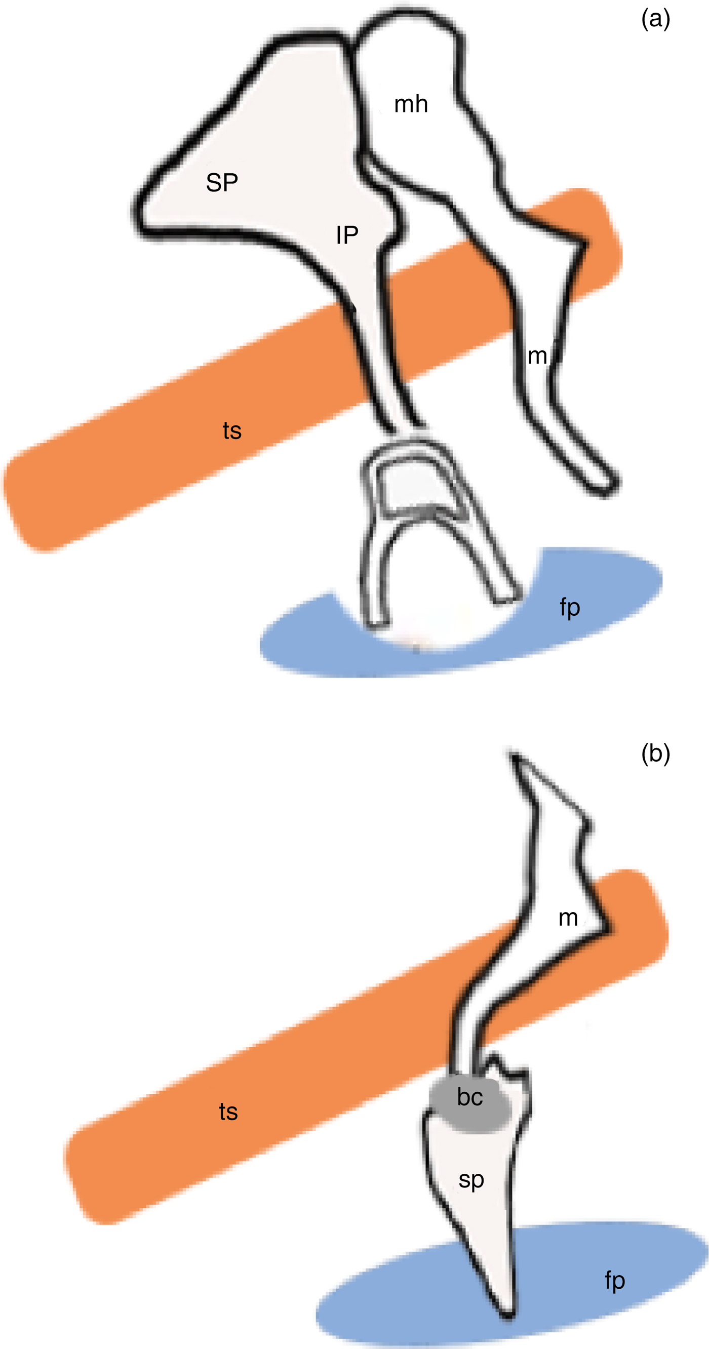
For the manubrio-incudo-stapedioplasty technique, the incudomalleolar joint was separated with a pick and the incus was extracted. The eroded incus was cleaned with a burr to prevent implantation of a cholesteatoma, and the short process was shaped and fitted to the stapes footplate. The point between the neck and head of the malleus was cut with a malleus nipper, and the head of the malleus was removed. The connection between the tensor tympani tendon and malleus was transected using a pick and microscissors. The manubrium was relocated posteriorly, and the inferior tip of the manubrium was placed in the groove that was formed on the incus body (Figure 1). The incus was positioned between the footplate and manubrium mallei, without touching the tympanic segment of the facial nerve. Bone cement (Ketac-Cem; 3M ESPE, Seefeld, Germany) was applied on the manubrium–incus junction and left for 10 minutes to allow fixation, to stabilise these ossicles.
(a) Normal anatomy of the middle-ear ossicles. (b) The head of the malleus was removed, the manubrium was shifted posteriorly, the short process of the incus was placed on the footplate, and bone cement was applied between the manubrium and incus body. Sp = short process of incus; mh = head of malleus; lp = long process of incus; ts = tympanic segment of facial nerve; m = manubrium mallei; fp = stapes footplate; bc = bone cement

Functionality of the reconstructed ossicular system was assessed based on the presence of a round window reflex. Meatoplasty was then performed. Conchal cartilage of the auricle was harvested as graft material. The cartilage–perichondrium composite graft was placed over the manubrium mallei. The graft was supported with gel sponges to provide better contact between the graft and manubrium.
Microscopic examination of the tympanic membrane was performed on days 7, 14 and 21 and months 3, 6 and 12.
Statistical analysis
The mean and standard deviation values were calculated. One-way analysis of variance was used to compare the hearing thresholds of patients among groups. The differences between pre-operative and post-operative hearing thresholds were compared using the paired t-test. The chi-square test was used to compare categorical variables. Statistical analysis was performed with SPSS software (version 16.0; IBM, Chicago, Illinois, USA). P-values of less than 0.05 were considered statistically significant.
Results
There were 13 patients in the manubrio-incudo-stapedioplasty group, 19 in the autologous incus group and 10 in the TORP group. The age and sex of the patients in the groups were similar (Table 1).
Demographic information of patients

*n = 13; †n = 19; ‡n = 10. **One-way analysis of variance test. TORP = titanium ossicular replacement prosthesis; SD = standard deviation
In two patients, who had previously undergone canal wall down tympanomastoidectomy, the manubrio-incudo-stapedioplasty technique was performed because of hearing deterioration. The findings in these two patients were of incus replacement between the footplate and graft.
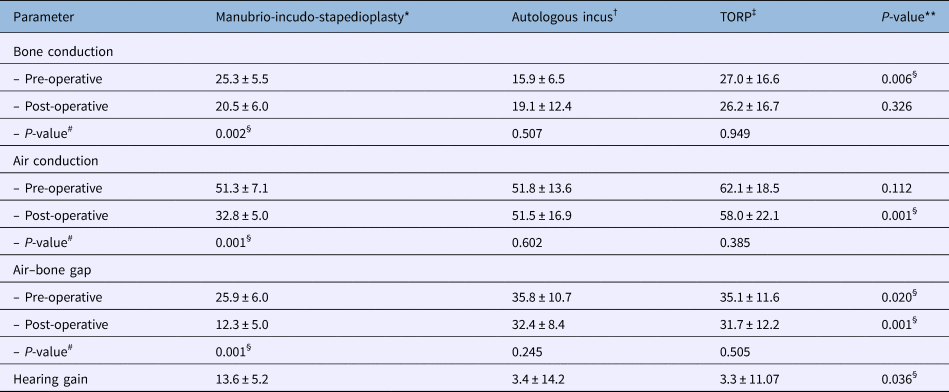
Pre-operative bone conduction thresholds were 25.3 ± 5.2 dB in the manubrio-incudo-stapedioplasty group, 15.9 ± 6.5 dB in the autologous incus group and 27.0 ± 16.6 dB in the TORP group (p < 0.05) (Table 2). Pre-operative air conduction levels were 51.3 ± 7.1 dB in the manubrio-incudo-stapedioplasty group, 51.8 ± 13.6 dB in the autologous incus group and 62.1 ± 18.5 dB in the TORP group (p > 0.05). The pre-operative air–bone gap (ABG) was 25.9 ± 6.0 dB in the manubrio-incudo-stapedioplasty group, 35.8 ± 10.7 dB in the autologous incus group and 35.1 ± 11.6 dB in the TORP group. The post-operative bone conduction and air conduction thresholds significantly decreased only in the manubrio-incudo-stapedioplasty group (p < 0.05).
Average hearing thresholds in study groups

Data represent mean ± standard deviation values, unless indicated otherwise. *n = 13; †n = 19; ‡n = 10. **One-way analysis of variance test. §Statistically significant. #Paired t-test. TORP = titanium ossicular replacement prosthesis
The hearing gain was significantly higher in the manubrio-incudo-stapedioplasty group compared to the autologous incus and TORP groups. Hearing gain was observed in: 13 patients in the manubrio-incudo-stapedioplasty group, 10 patients in the autologous incus group and 5 patients in the TORP group.
The number of patients who had an ABG lower than 20 dB was similar in each group pre-operatively (p > 0.05). In contrast, there was a significant increase in the manubrio-incudo-stapedioplasty group post-operatively (p < 0.05) (Table 3).
Patients with pre- and post-operative air–bone gaps of less than 20 dB

Data represent numbers of patients, unless indicated otherwise. *n = 13; †n = 19; ‡n = 10. **Chi-square test. §Statistically significant. TORP = titanium ossicular replacement prosthesis
Discussion
In this report, an incus bridge between the relocated manubrium and footplate was used as an ossicular chain reconstruction method, and its functional outcomes were demonstrated. The ABG significantly decreased after surgery, and no tympanic membrane perforation or retraction was observed during follow up. Manubrio-incudo-stapedioplasty ossiculoplasty provided better hearing outcomes compared to autologous incus and TORP ossiculoplasties.
Autologous or allograft ossicles, biocompatible middle-ear prostheses, and bone cement are used to reconstruct the ossicular chain system. In patients with Austin–Kartush type B ossicular defects, the TORP, PORP and autologous incus are used to provide sound transmission. However, the extrusion rates of TORP or PORP were between 4 per cent and 9 per cent in previous studies.Reference House and Teufert9,Reference Emir, Kizilkaya Kaptan, Gocmen, Uzunkulaoglu, Tuzuner and Bayiz12,Reference Vassbotn, Moller and Silvola13 Displacement and/or extrusion of the TORP was the most common reason for hearing impairment over the long term.Reference Smyth14 Surgeons have addressed this issue by using cartilage grafts to prevent prosthesis extrusion. It was shown that a cartilage graft used with a TORP resulted in increased success rates over the long term.Reference House and Teufert9
Different techniques have been shown to stabilise the TORP on the footplate for satisfactory hearing outcomes.Reference Beutner, Luers and Huttenbrink15–Reference Berenholz, Burkey and Lippy17 Vincent et al.Reference Vincent, Rovers, Mistry, Oates, Sperling and Grolman18 described a malleus relocation technique with Silastic banding to stabilise the TORP in the middle ear, and demonstrated better hearing outcomes with that technique compared to placement of the PORP on the stapes head. In addition, placement of the prosthesis between the malleus and footplate provided better hearing outcomes, compared to placement between the malleus and stapes head.Reference Murugasu, Puria and Roberson19
A previous study showed a 56 per cent failure rate in autologous incus ossiculoplasty.Reference Zakzouk, Bonmardion, Bouchetemble, Lerosey and Marie11 The loss of incus–footplate contact, lateralisation of the incus, fall of the incus into the middle ear, and contact of the incus with the bony annulus were reported as reasons for failed autologous incus ossiculoplasty.Reference Zakzouk, Bonmardion, Bouchetemble, Lerosey and Marie11
The manubrio-incudo-stapedioplasty appears to be a more stable ossiculoplasty technique than TORP or PORP placement on the footplate. Stabilisation between the incus and manubrium was strengthened with bone cement. Thus, using this technique, extrusion or displacement of the reconstruction material is less likely. When using an autologous incus, supporting this ossicle with the manubrium and bone cement provides a significant benefit, as compared to our results in the autologous incus group and those of previous reports. In addition, the incus will remain stable in the middle ear and the risk of hearing deterioration will decrease. In support of this hypothesis, we did not observe any hearing deterioration over a one-year follow-up period in patients who underwent manubrio-incudo-stapedioplasty.
• Austin–Kartush type B ossicular defects are defined as: intact and mobile malleus, and ossicular destruction in incus and stapes suprastructure.
• Allograft or autograft ossicles, partial or total ossicular replacement prostheses (TORP), and cartilage have been used to transmit sound energy to the footplate in Austin–Kartush type B damage
• Manubrio-incudo-stapedioplasty provided satisfactory hearing outcomes compared to autologous incus or TORP ossiculoplasties for Austin–Kartush type B ossicular defects
• This technique may be considered inexpensive and useful for reconstructing Austin–Kartush type B damage
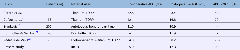
Functional outcomes of different ossiculoplasty techniques for the reconstruction of Austin–Kartush type B ossicular defects have been presented in previous reports (Table 4).Reference Gerard, De Bie, Franceschi, Deggouj and Gersdorff7,Reference De Vos, Gersdorff and Gerard8,Reference Stankovic20–Reference Redaelli de Zinis22 Gerard et al.Reference Gerard, De Bie, Franceschi, Deggouj and Gersdorff7 mentioned in their study that the use of a titanium prosthesis with bone cement in Austin–Kartush type B cases provided an increase in hearing of 9.1 dB. In that study, ABG improved from 32.5 dB to 23.4 dB post-operatively, and an ABG of less than 20 dB was observed in 50 per cent of patients.Reference Gerard, De Bie, Franceschi, Deggouj and Gersdorff7 De Vos et al.Reference De Vos, Gersdorff and Gerard8 reported a post-operative ABG of 16.6 dB in cases of Austin–Kartush type B erosions reconstructed with a titanium prosthesis. In a study comprising 200 patients, the reconstruction of Austin–Kartush type B ossicular defects with autologous bone and cartilage resulted in a post-operative ABG of 10.9 dB.Reference Stankovic20 In a different study, the post-operative ABG was 11.9 dB in 46 patients.Reference Dornhoffer and Gardner21 Post-operatively, Zakzouk et al.Reference Zakzouk, Bonmardion, Bouchetemble, Lerosey and Marie11 reported an ABG of 24 dB in their TORP group and 30 dB in their incus group. Using manubrio-incudo-stapedioplasty, the present study found that hearing gain was 13.6 dB, post-operative ABG was 12.3 dB, and 100 per cent of cases had an ABG of less than 20 dB.
Reconstruction of Austin–Kartush type B damage in different studies

ABG = air–bone gap; TORP = titanium ossicular replacement prosthesis
The presence of the manubrium mallei is important to provide satisfactory hearing outcomes and prevent lateralisation of the tympanic membrane.Reference Vincent, Oates, Sperling and Annamalai23 Direct placement of the manubrium over the stapes head has also been proposed as an ideal reconstruction method for improving hearing outcomes.Reference Vlaming and Feenstra24 Using this reconstruction method, vibrations could be transmitted to the stapes footplate with minimal energy loss. Asai et al.Reference Asai, Roberson and Goode25 reported that removal of the malleus head and section of the tensor tympani muscle may improve hearing outcomes after placement of the prosthesis between the malleus and stapes footplate. Ashraf et al.Reference Ashraf, Ghonim, Eladl and Elsisi10 stated that malleus head removal and malleus relocation can be used to eradicate cholesteatoma and improve hearing. Sennaroglu et al.Reference Sennaroglu, Gungor, Atay and Ozer26 described manubrio-stapedioplasty as a reconstruction method in patients with incus–malleus fixation due to tympanosclerosis, and demonstrated satisfactory hearing outcomes using this technique. The disadvantages of manubrium relocation include the potential risk of medialisation of the tympanic membrane. As structures supporting the malleus are dissected, the manubrium can shift towards the middle ear. Nevertheless, the incus on the footplate acts as a lever to prevent medialisation of the manubrium.
Cost-effectiveness is an important consideration in hearing reconstruction when comparing different materials. The TORP is the most common material used in the reconstruction of Austin–Kartush type B ossicular defects. In the present study, we used bone cement to stabilise the manubrium–incus connection, which cost approximately $10 USD per case. In comparison, the cost of one TORP is approximately $150 USD.
One limitation of this report is the small number of patients, as Austin–Kartush type B ossicular defects are not common in our clinical practice. However, the power of the study was high, at greater than 0.8.
Conclusion
In this report, we compared different ossiculoplasty techniques for the reconstruction of Austin–Kartush type B ossicular defects. Placement of the incus between the footplate and manubrium mallei, and stabilisation with bone cement, provided satisfactory hearing outcomes compared to autologous incus and TORP techniques. This technique may be considered a cost-effective and stable reconstruction method in cases with ossicular chain damage. However, long-term follow up is required to validate our results.
Competing interests
None declared