Introduction
The Diplostomidae (Digenea) comprise a large and cosmopolitan group of parasites, the adults of which infect a wide range of fish-eating birds (Chappell, Reference Chappell1995; McKeown & Irwin, Reference McKeown and Irwin1995; Valtonen & Gibson, Reference Valtonen and Gibson1997). Fish serve as second intermediate hosts while gastropods are the first intermediate hosts (Hoffman & Hundley, Reference Hoffman and Hundley1957; Chappell, Reference Chappell1995; Niewiadomska, Reference Niewiadomska1996; Morozińska-Gogol, Reference Morozińska-Gogol2007). In fish, diplostomid metacercariae occur in a variety of organs, particularly the eye and central nervous system, where they commonly occur free in tissues. In African freshwater fishes, diplostomid metacercariae have usually been reported in the vitreous humour of eyes or brain cavity (Musiba & Nkwengulila, Reference Musiba and Nkwengulila2006, Florio et al., Reference Florio, Gustinelli, Caffara, Turci, Quaglio, Konecny, Nikowitz, Wathuta, Magana, Otachi, Matolla, Warugu, Liti, Mbaluka, Thiga, Munguti, Akoll, Mwanja, Asaminew and Tadesse2009; Chibwana & Nkwengulila, 2010; Otachi et al., Reference Otachi, Wathuta, Magana, Fioravanti, Florio and Konecny2011; Migiro et al., Reference Migiro, Matolla, Ouko and Victor2012). Morphological identification of diplostomids has long been recognized to be problematic worldwide. Despite attempts based on infection site, chaetotaxy and multivariate analysis of morphometric data (Chappell, Reference Chappell1995; Niewiadomska & Niewiadomska-Bugaj, Reference Niewiadomska and Niewiadomska-Bugaj1995; Gibson, Reference Gibson, Margolis and Kabata1996; Cavaleiro et al., Reference Cavaleiro, Pina, Russell-Pinto, Rodrigues, Formigo, Gibson and Santos2012), considerable confusion remains. Taxonomic studies of diplostomids from the African continent are mainly limited to a few species described in the 1930s to 1960s (King & Van As, Reference King and Van As1997). In two recent surveys (Mwita & Nkwengulila, Reference Mwita and Nkwengulila2010; Chibwana et al., Reference Chibwana, Blasco-Costa, Georgieva, Hosea, Nkwengulila, Scholz and Kostadinova2013) of African diplostomids in siluriform fishes, molecular data revealed three species of Tylodelphys in the brain of the North African catfish Clarias gariepinus in Tanzania and one species of Diplostomum in the lens of the catfish Synodontis nigrita from Nigeria, all of which were genetically distinct from the growing number of diplostomid species sequenced from Europe and North America (Galazzo et al., Reference Galazzo, Dayanandan, Marcogliese and McLaughlin2002; Niewiadomska & Laskowski, Reference Niewiadomska and Laskowski2002; Locke et al., Reference Locke, McLaughlin, Dayanandan and Marcogliese2010a, Reference Locke, McLaughlin and Marcoglieseb; Rellstab et al., Reference Rellstab, Louhi, Karvonen and Jokela2011; Cavaleiro et al., Reference Cavaleiro, Pina, Russell-Pinto, Rodrigues, Formigo, Gibson and Santos2012; Behrmann-Godel, Reference Behrmann-Godel2013; Georgieva et al., Reference Georgieva, Soldánová, Pérez-del-Olmo, Dangel, Sitko, Sures and Kostadinova2013).
Here we provide further morphological, molecular and ecological data on African diplostomids. We compare our material from the eyes of fishes in Lake Naivasha, Kenya with data from diplostomids from the brain of C. gariepinus collected approximately 700 km away in Tanzania (Chibwana et al., Reference Chibwana, Blasco-Costa, Georgieva, Hosea, Nkwengulila, Scholz and Kostadinova2013). Secondly, we assess host specificity of the diplostomids encountered in five host species in three families from which no molecular data have been obtained for African diplostomids (Mwita & Nkwengulila, Reference Mwita and Nkwengulila2010; Chibwana et al., Reference Chibwana, Blasco-Costa, Georgieva, Hosea, Nkwengulila, Scholz and Kostadinova2013), namely common carp Cyprinus carpio (L. 1758) and straightfin barb Barbus paludinosus (Peters 1852), both Cyprinidae; blue spotted tilapia Oreochromis leucostictus (Trewavas 1933), redbelly tilapia Tilapia zillii (Gervais 1848), both Cichlidae, and largemouth bass Micropterus salmoides (Lacepède 1802), Centrarchidae.
Molecular analyses have proved to be powerful tools in identification and discrimination of animal species. The mitochondrial cytochrome c oxidase subunit 1 (COI) gene is useful both for population genetic studies (Avise, Reference Avise1994) and for discriminating species of animal congeners (Hebert et al., Reference Hebert, Ratnasingham and deWaard2003). The utility of the ≈ 650 bp at the 5′ end of COI in distinguishing closely related species has led to the designation of this fragment as the core target in DNA barcoding (Hebert et al., Reference Hebert, Ratnasingham and deWaard2003), and it has provided useful information in diplostomids and other helminths (Locke et al., Reference Locke, McLaughlin and Marcogliese2010b; Behrmann-Godel, Reference Behrmann-Godel2013; Chibwana et al., Reference Chibwana, Blasco-Costa, Georgieva, Hosea, Nkwengulila, Scholz and Kostadinova2013; Désilets et al., Reference Désilets, Locke, McLaughlin and Marcogliese2013; Georgieva et al., Reference Georgieva, Soldánová, Pérez-del-Olmo, Dangel, Sitko, Sures and Kostadinova2013; Sereno-Uribe et al., Reference Sereno-Uribe, Pinacho-Pinacho, García-Varela and Pérez-Ponce de León2013).
Materials and methods
Collection and examination of fish
Fish were caught from Lake Naivasha (00°45′S and 36°20′E), which covers approximately 160 km2 in a closed basin 1890 m above sea level in the eastern Rift Valley of Kenya. It is the only freshwater lake in the Rift Valley without a surface outlet but with a substantial exchange with groundwater (Gaudet & Melack, Reference Gaudet and Melack1981). It is shallow (approximately 6 m mean depth) with a volume of 4.6 km3 (Campbell et al., Reference Campbell, Osano, Hecky and Dixon2003). All the fish present in the lake have been introduced and, as such, none of the five fish species studied herein are native to the lake (Gherardi et al., Reference Gherardi, Britton, Mavuthi, Pacini, Grey, Tricarico and Harper2011). The only native and endemic fish in the lake was black lampeye Aplocheilichthys antinorii, which was last recorded in 1962 and is believed to be extirpated from the lake (Harper et al. Reference Harper, Morrison, Macharia, Mavuthi and Upton2011). Barbus paludinosus was introduced in the 1920s and restocked in 1982; O. leucostictus and T. zillii were introduced in 1956 and thereafter restocked from populations in Lake Victoria, Kenya (Gherardi et al., Reference Gherardi, Britton, Mavuthi, Pacini, Grey, Tricarico and Harper2011). Micropterus salmoides was introduced in 1929 and restocked in 1940 and 1951 as a sport fish, and C. carpio was accidentally introduced in 1999. Neither M. salmoides nor C. carpio are within their native ranges (Gherardi et al., Reference Gherardi, Britton, Mavuthi, Pacini, Grey, Tricarico and Harper2011).
A total of 288 fish were collected using a fleet of gill nets with mesh sizes 2, 2.5, 3 and 4 inches between February and August, 2011. These comprised the two cyprinids, C. carpio (n= 145) and B. paludinosus (n= 67), two cichlids, O. leucostictus (n= 56) and T. zillii (n= 18), and the centrarchid M. salmoides (n= 2). The fish were transported alive in a fish tank with lake water to the laboratory of the Department of Biological Sciences, Egerton University, Njoro, where they were killed by cervical dislocation (Schäperclaus, Reference Schäperclaus1990), weighed and measured. All parasites in the eyes were counted. Parasites were preserved in either 4% formalin or absolute ethanol. Prevalence, mean intensity and mean abundance were determined as described (Bush et al., Reference Bush, Lafferty, Lotz and Shostak1997).
Morphometrics
Using an approach previously recommended for distinguishing species in diplostomid metacercariae (Niewiadomska & Niewiadomska-Bugaj, Reference Niewiadomska and Niewiadomska-Bugaj1995), 14 measurements were made on 111 metacercariae isolated from C. carpio, M. salmoides, O. leucostictus and T. zillii. An Olympus BX40 compound transmitted light microscope with phase contrast (Olympus, Tokyo, Japan), fitted with a 35-mm camera connected to a computer with digital software was used to measure body length (BL), body width (BW), oral sucker length (OL), oral sucker width (OW), length of pharynx (PHL), width of pharynx (PHW), ventral sucker length (AL), ventral sucker width (AW), Brandes' length (HL), Brandes' width (HW), length from centre of ventral sucker to end of oral sucker (AD), length of pseudosucker (LL), width of body at level of bifurcation of intestine (WaBI) and at the mid-length of oral sucker (WaO). In addition, the following indices were calculated: BW/BL (in %), (BL × BW)/(HL × HW), (BL × BW)/(AL × AW), (OL × OW)/(AL × AW), (HL × HW)/(AL × AW), (OL × OW)/(PHL × PHW), AD/BL (in %) and WaO/WaBI. Multivariate analysis of these characters was performed using principal components analysis (PCA) extraction method (Flores & Baccala, Reference Flores and Baccala1998), while employing a Varimax with Kaiser Normalization rotation technique. The appropriateness of PCA was ensured in several ways. More than 100 metacercariae were measured, which ensured that the ratio of cases to variables exceeded 5:1 (Osborne & Costello, Reference Osborne and Costello2004). In addition, we iteratively eliminated variables with correlation coefficients |r| < 0.3 in the correlation matrix, examined the individual variables' Kaiser–Meyer–Olkin Measure of sampling adequacy (KMO)>0.5 in the anti-image correlation matrix, as well as the overall KMO>0.5 (Hutcheson & Sofroniou, Reference Hutcheson and Sofroniou1999), Bartlett's Test of Sphericity (significance < 0.05) and ensured that communalities in initial extractions were >0.5. Outliers were identified on the basis of factor scores, i.e. metacercariae scoring>3.0, and excluded from the analysis. From the extracted components, the major characters were used as inputs in a two-step cluster analysis (CA) using Euclidean distances and the Schwarz's Bayesian Information Criterion (BIC) as the clustering criterion. Cluster quality was assessed with average Silhouette width, which is the ratio of each specimen's average morphometric distance to others in the same cluster to its smallest distance to specimens in other clusters (Rousseeuw, Reference Rousseeuw1987). Statistical analysis was performed using predictive analytics software (PASW statistics 18, SPSS, Chicago, Illinois, USA).
Molecular analysis
Specimens used for molecular analysis included representatives from M. salmoides, T. zillii, O. leucostictus, and C. carpio. Total DNA was extracted from 16 single specimens (for protocols, see Moszczynska et al., Reference Moszczynska, Locke, McLaughlin, Marcogliese and Crease2009), each of which was matched with a morphologically similar specimen collected from the same tissue of the same individual host. The latter specimens were stained with acetocarmine, mounted on slides in Canada balsam, and deposited as paragenophores (Pleijel et al., Reference Pleijel, Jondelius, Norlinder, Nygren, Oxelman, Schander, Sundberg and Thollesson2008) at the Canadian Museum of Nature (catalogue number CMNPA 2013-0001). The DNA barcode region of cytochrome c oxidase I (COI) was amplified and sequenced at the Canadian Centre for DNA Barcoding in Guelph, Canada, using previously described procedures, diplostomid-specific primers and cycling conditions (Moszczynska et al., Reference Moszczynska, Locke, McLaughlin, Marcogliese and Crease2009). Chromatograms were assembled using Geneious (Biomatters, New Zealand) and sequences were aligned and analysed using MEGA 5.05 (Tamura et al., Reference Tamura, Peterson, Peterson, Stecher, Nei and Kumar2011). The resulting sequences were compared with data retrieved using BLAST searches (Altschul et al., Reference Altschul, Gish, Miller, Myers and Lipman1990) and aligned with 215 published contiguous sequences from diplostomids in North America, Europe and Africa (Moszczynska et al., Reference Moszczynska, Locke, McLaughlin, Marcogliese and Crease2009; Locke et al., Reference Locke, McLaughlin, Dayanandan and Marcogliese2010a, Reference Locke, McLaughlin and Marcoglieseb; Behrmann-Godel, Reference Behrmann-Godel2013; Chibwana et al., Reference Chibwana, Blasco-Costa, Georgieva, Hosea, Nkwengulila, Scholz and Kostadinova2013; Georgieva et al., Reference Georgieva, Soldánová, Pérez-del-Olmo, Dangel, Sitko, Sures and Kostadinova2013). The mutation of COI nucleotides was best described by the general-time-reversible model with five categories of gamma-distributed rates and by assuming that a fraction of nucleotides are invariable. Published sequences of Apatemon (Digenea: Strigeidae) species (Moszczynska et al., Reference Moszczynska, Locke, McLaughlin, Marcogliese and Crease2009; Locke et al., Reference Locke, McLaughlin and Marcogliese2010b) were used to root a phylogeny inferred using maximum likelihood, with 1000 bootstrap replicates, as strigeids are closely related and, in some cases, basal to diplostomids (Olson et al., Reference Olson, Cribb, Tkach, Bray and Littlewood2003). For comparative purposes, outgroup sequences were the same as those used by Chibwana et al. (Reference Chibwana, Blasco-Costa, Georgieva, Hosea, Nkwengulila, Scholz and Kostadinova2013). Molecular (chromatograms, sequences) and other (specimen images, morphological voucher accessions) data are published in project DIPLO at www.barcodinglife.com (see also GenBank accessions KF809484–KF809494).
Results
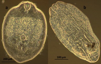
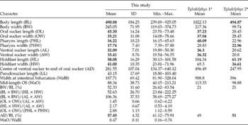
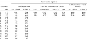
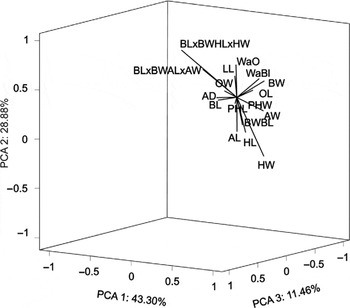
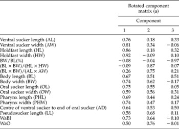
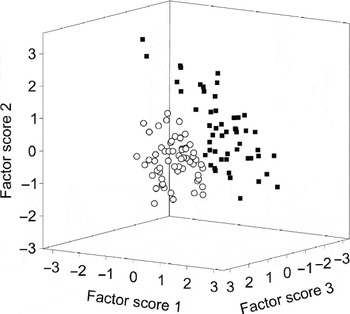
Among the four infected species, prevalence ranged from 50.0 to 88.9%, mean intensity from 4.7 ± 5.0 to 10.2 ± 8.1, and mean abundance from 2.5 to 9.1 ± 8.3 (table 1). Diplostomid metacercariae were the only parasites observed in fish eyes, and all were unencysted and found exclusively in the vitreous humour. No infection was detected in B. paludinosus. The metacercariae resembled Tylodelphys morphologically. The shapes varied from a short and ovate form (fig. 1a) with offset pseudosuckers, which appeared to be distinct from an elongate and rounded form with recessed pseudosuckers (fig. 1b). In all metacercariae, the oral sucker was circular and terminated in a conical pharynx. Posterior to the ventral sucker was a comparatively large holdfast organ. Although there were size and shape variations among the examined metacercariae, the dimensions of many features overlapped with morphometrics from diplostomids from C. gariepinus in Tanzania (Chibwana et al., Reference Chibwana, Blasco-Costa, Georgieva, Hosea, Nkwengulila, Scholz and Kostadinova2013); for example, in the BL, OL, OW, PHL, PHW, AL, AW, HL, HW and AD/BL (%) (table 2). In PCA, three principal components with eigenvalues greater than one were extracted (table 3), and they together accounted for 83.64% of the total morphometric variance in the specimens from Lake Naivasha (fig. 2). Along the first PCA axis, which accounted for 43.30% of the total variance, BL, BW, PHW, PHL, AL, AW, OL, OW, HL, HW, AD and WaBI loaded strongly. The second PCA accounted for 28.88% of the total variance, and WaO, BL × BW/HL × HW, BL × BW/AL × AW and LL were associated with it, while the third PCA, which accounted for 11.46% of the total variance, had BW/BL (%) loading on it strongly (table 4). Two groups were apparent in CA, indicating a possibility of two morphotypes with the larger cluster having 58 individuals (52.3%) and the smaller cluster having 53 individuals (47.7%). The most important variables in discriminating the two clusters were as follows: BL, AD, HL, PHW, AW, WaO, LL, OL, WaBI and BW; all other features contributed little to the discrimination of the two putative morphological types. The overall cohesion and separation of these clusters, according to the average Silhouette coefficient, was 0.6, which is fair but not conclusive support for the presence of two morphotypes (fig. 3).
The occurrence of metacercariae of Tylodelphys sp. in five fish species from Lake Naivasha, Kenya in 2011; N, number of fish examined; TL, mean total body length (cm); P (%), prevalence of infection; MI, mean intensity of infection; SD, standard deviation, for N=2 not calculated; MAI, maximum intensity of infection; MA, mean abundance of infection.

Whole mounts of representatives of two morphological types of Tylodelphys sp. from the common carp Cyprinus carpio in Lake Naivasha: (a) short and ovate, (b) elongate and rounded.

Morphometric dimensions (μm) of characters and indices (mean, standard deviation (SD) and range) in 111 Tylodelphys sp. metacercariae isolated from the vitreous humour of the eyes of fish from Lake Naivasha, Kenya, as compared to the averages of two other species (values in bold in a row indicate measurement overlaps).

*Data source: Chibwana & Nkwengulila, Reference Chibwana and Nkwengulila2010.
Eigenvalues, number of components extracted, percent variance and the cumulative variance explained for the components from principal components analysis of 14 morphometrics in 111 diplostomid metacercariae recovered from four fish species in Lake Naivasha, Kenya. Extraction method: principal component analysis. Cumul.: cumulative proportion of the variance explained (%).

Principal components analysis biplot of measurements of 17 morphological features which loaded strongly (component factor loadings) in 111 specimens of Tylodelphys sp. collected from four fish species in Lake Naivasha, Kenya. Highly and positively correlated morphological features point in the same direction, along the same axes. The proportion of total morphometric variance explained by the principal components (83.64%) are shown on the three axes. BL, body length; BW, body width; OL, oral sucker length; OW, oral sucker width; PHL, length of pharynx; PHW, width of pharynx; AL, ventral sucker length; AW, ventral sucker width; HL, Brandes' length; HW, Brandes' width; AD, length from centre of ventral sucker to end of oral sucker; LL, length of pseudosucker; WaBI, width of body at level of bifurcation of intestine; and WaO, the mid-length of oral sucker.

Factor loadings of 14 morphometrics in 111 diplostomid metacercariae recovered from four fish species in Lake Naivasha, Kenya on the first three principal components extracted in principal components analyis. Extraction method: principal component analysis. Rotation method: Varimax with Kaiser Normalization. (a) Rotation converged in six iterations.

Cluster analysis of morphometrics for 111 specimens of the Tylodelphys sp. with two initially distinguished morphotypes indicated by black squares (short and ovate morphotype) and white circles (elongate and rounded morphotype).

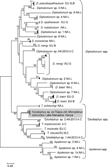
Sequences of COI of length 200–463 bp were obtained from 11 of 16 specimens, including representatives from all four infected fish species. The 11 COI sequences differed from each other by a mean of 0.3% (range 0–1.0%, uncorrected p-distance), and there was no variation in amino acid translations. Among sequences published from 51 other specimens of Tylodelphys from Africa, Europe and North America, the most similar are from Tanzanian isolates of T. mashonensis (Chibwana et al., Reference Chibwana, Blasco-Costa, Georgieva, Hosea, Nkwengulila, Scholz and Kostadinova2013), which differed from our material by an average of 9.3% (range 7.2–11.2%) and in three amino acids. In the phylogeny estimated using maximum likelihood, the two sequences of length 463 bp were nested within other Tylodelphys species (fig. 4), although statistical support for the Tylodelphys clade as whole was not strong. The variable length of the sequence precluded including all of them in phylogenetic analysis; however, in separate analyses all fell within the same clade (not shown). Given the reciprocal monophyly and low divergence within our samples, and the high divergence from the most similar samples, lack of strong morphological distinctions, and phylogenetic placement within Tylodelphys, we consider the material collected here to represent a single species of Tylodelphys distinct from those previously sequenced. Under this hypothesis, the overall mean divergence in CO1 sequences among Tylodelphys species is 10.1% (range 7.2–15.8%), and mean intraspecific divergence is 0.6% (range 0–2.4%).
Relationships among sequences of cytochrome c oxidase I including representative sequences obtained from two specimens of Tylodelphys collected from two fish species in Lake Naivasha, Kenya in the present study (selected from 11 total similar sequences in order to maximize length and overlap with other data) and 215 sequences from diplostomids published in GenBank, inferred using maximum likelihood with 1000 bootstrap replicates, and pairwise deletion of gaps. The alignment was 463 bp long, and all sequences shared at least 297 sites. The vertical height of black triangles is proportional to the number of sequences in a clade. The width of black triangles and horizontal branch lengths are both proportional to the scale = substitutions per site. GenBank accessions of data used are: KF809484–KF809494 (this study), FJ477183, FJ477185, FJ477186, FJ477194–6, FJ477198–200, FJ477223, GQ292475, GQ292476, GQ292478–502, HM064610, HM064636, HM064637, HM064644, HM064647, HM064660, HM064662–68, HM064670–80, HM064682–97, HM064699, HM064700, HM064702, HM064704–11, HM064909–15, JQ639170–204, JX986859–909, KC685327–60. NA, North America; EU, Europe; A, Africa; B, sequence from Behrmann-Godel (Reference Behrmann-Godel2013); C, sequence from Chibwana et al. (Reference Chibwana, Blasco-Costa, Georgieva, Hosea, Nkwengulila, Scholz and Kostadinova2013); G, sequence from Georgieva et al. (Reference Georgieva, Soldánová, Pérez-del-Olmo, Dangel, Sitko, Sures and Kostadinova2013); L, sequence from Locke et al. (Reference Locke, McLaughlin, Dayanandan and Marcogliese2010a, Reference Locke, McLaughlin and Marcoglieseb).

Discussion
The diversity and ecology of diplostomids infecting fish from Kenya are poorly known. Our initial observation of two morphotypes led us to suspect the presence of two species in Lake Naivasha and, while this distinction received fair support in quantitative morphological analysis, the presence of two species was effectively ruled out by molecular data. Molecular data have also revealed other apparently morphologically distinct diplostomid metacercariae to be single species (Cavaleiro et al., Reference Cavaleiro, Pina, Russell-Pinto, Rodrigues, Formigo, Gibson and Santos2012) and in other cases have confirmed separation of species in metacercarial morphotypes (Chibwana & Nkwengulila, Reference Chibwana and Nkwengulila2010; Chibwana et al., Reference Chibwana, Blasco-Costa, Georgieva, Hosea, Nkwengulila, Scholz and Kostadinova2013). Together with other published data (Chibwana & Nkwengulila, Reference Chibwana and Nkwengulila2010), our measurements also confirmed interspecific morphometric overlap in Tylodelphys. Most characters and indices were within the range of those in other Tylodelphys spp. from the cranial cavity of C. gariepinus in Tanzania (Chibwana & Nkwengulila, Reference Chibwana and Nkwengulila2010). Many factors cause phenotypic plasticity and morphological similarities between diplostomid metacercariae, including the species, size and age of hosts, and the age, population size and density of metacercariae (Chappell et al., Reference Chappell, Hardie, Secombes, Pike and Lewis1994; Chappell, Reference Chappell1995; Gibson, 1996; Niewiadomska, Reference Niewiadomska1996; Chibwana & Nkwengulila, Reference Chibwana and Nkwengulila2010). In retrospect, the morphological variation observed in our specimens was likely due to differences in contraction. Cavaleiro et al. (Reference Cavaleiro, Pina, Russell-Pinto, Rodrigues, Formigo, Gibson and Santos2012) hypothesized that similar phenotypic variation in two genetically identical but morphologically distinct metacercarial forms of Diplostomum arose from differences in specimen age. Age structure seems unlikely to be the cause of the morphotypes in diplostomids in our study, for the following reasons. First, Rauch et al. (Reference Rauch, Kalbe and Reusch2005) found almost no clones in metacercariae of Diplostomum pseudospathaceum in the eyes of sticklebacks, which shows that cercariae are encountered singly, not in simultaneous exposures to batches of clonal cercariae emerging from single snails. Second, seasonality of recruitment (e.g. Marcogliese et al., Reference Marcogliese, Compagna, Bergeron and McLaughlin2001a), which could also give rise to an age structure in metacercariae, is unlikely to have played a role in our system because of both the local climate and the six-month interval over which we sampled fish. Thus, the two main sources of age-related parasite recruitment (acquisition of same-aged clones and seasonal recruitment of same-aged parasites) are unlikely to apply in the present study, and fixation effects are a more plausible explanation for the two morphotypes.
The single species encountered here is genetically distinct from all those for which contiguous sequences have been published, including three Tylodelphys species from Tanzania. Whether this is a new species can only be determined through morphometric characterization of additional developmental stages, particularly the adult. Our results present an interesting contrast to a recent molecular survey in which three species of Tylodelphys were found in the brain of C. gariepinus in Tanzania (Chibwana et al., Reference Chibwana, Blasco-Costa, Georgieva, Hosea, Nkwengulila, Scholz and Kostadinova2013). It may have been expected that we would recover some or all of the three species of Tylodelphys as these authors, and/or that we would observe similar or higher diversity, particularly because we sampled five host species in three families and two orders; instead, a single, oioxenous Tylodelphys species was recovered. The small number (n= 11) of specimens from which sequences were obtained may have been a factor in not detecting additional diversity. However, we note comparable sample sizes in other studies. For example, Chibwana et al. (Reference Chibwana, Blasco-Costa, Georgieva, Hosea, Nkwengulila, Scholz and Kostadinova2013) sequenced COI in 33 diplostomids, but only n= 2, 3, 11 or 17 were sequenced in any particular species. Facing a similar situation (two genetically identical morphotypes), Cavaleiro et al. (Reference Cavaleiro, Pina, Russell-Pinto, Rodrigues, Formigo, Gibson and Santos2012) sequenced internal transcribed spacers (ITS) in 38 specimens.
The metacercaria of Tylodelphys sp. appears to be a true generalist (Poulin & Mouillot, Reference Poulin and Mouillot2005), as it occurred in similarly high intensities in four fish species in three families and two orders. Previous reports of diplostomid metacercariae in Kenya are from Nile tilapia Oreochromis niloticus and African catfish C. gariepinus (Florio et al., Reference Florio, Gustinelli, Caffara, Turci, Quaglio, Konecny, Nikowitz, Wathuta, Magana, Otachi, Matolla, Warugu, Liti, Mbaluka, Thiga, Munguti, Akoll, Mwanja, Asaminew and Tadesse2009; Otachi, Reference Otachi2009; Gichohi, Reference Gichohi2010; Migiro et al., Reference Migiro, Matolla, Ouko and Victor2012). Indeed, diplostomids are frequently found in tilapias from all aquatic systems (Florio et al., Reference Florio, Gustinelli, Caffara, Turci, Quaglio, Konecny, Nikowitz, Wathuta, Magana, Otachi, Matolla, Warugu, Liti, Mbaluka, Thiga, Munguti, Akoll, Mwanja, Asaminew and Tadesse2009). Infection levels were higher than previously reported in Kenya from farmed tilapia (Florio et al., Reference Florio, Gustinelli, Caffara, Turci, Quaglio, Konecny, Nikowitz, Wathuta, Magana, Otachi, Matolla, Warugu, Liti, Mbaluka, Thiga, Munguti, Akoll, Mwanja, Asaminew and Tadesse2009; Otachi, Reference Otachi2009; Gichohi, Reference Gichohi2010; Migiro et al., Reference Migiro, Matolla, Ouko and Victor2012). As discussed by Otachi et al. (Reference Otachi, Magana, Jirsa and Fellner-Frank2014), the factors influencing variation in infection levels among the fish species may include fish microhabitat and behaviour, and subsequent variation in exposure, rather than variation in susceptibility (Valtonen & Gibson, Reference Valtonen and Gibson1997; Karvonen et al., Reference Karvonen, Hudson, Seppälä and Valtonen2004). For example, B. paludinosus is a riverine species (Welcomme, Reference Welcomme1979; Mutia et al., Reference Mutia, Ntiba, Mavuti and Omondi2010) that migrates into River Malewa for breeding, and spends a considerable time there (Welcomme, Reference Welcomme1979). Gastropods have not been observed in River Malewa (Barnard & Biggs, Reference Barnard and Biggs1988), although they have been found in Lake Naivasha (Clarke et al., Reference Clarke, Beeby and Kirby1989), and this lack of contact with the first intermediate hosts of Tylodelphys could explain why B. paludinosus was uninfected. The other fishes sampled, such as T. zillii, prefer shallow, vegetated zones within the lake, where gastropod densities are higher (Styczynska-Jurewicz, Reference Styczynska-Jurewicz1959). This overlap in habitat use by the first and second intermediate hosts of Tylodelphys sp. is likely to increase the probability of infection (Styczynska-Jurewicz, Reference Styczynska-Jurewicz1959; Sweeting, Reference Sweeting1974; Marcogliese et al., Reference Marcogliese, Dumont, Gendron, Mailhot, Bergeron and McLaughlin2001b; Otachi et al. Reference Otachi, Magana, Jirsa and Fellner-Frank2014). Additionally, innate immunity could also be important, as it has been suggested to play a major role in defence in sympatric hosts against the eye fluke D. pseudospathaceum (Kalbe & Kurtz, Reference Kalbe and Kurtz2006).
This was the first study to combine morphological, molecular and ecological analysis of Tylodelphys from Kenyan fishes. The results herein demonstrate the importance of obtaining molecular data to discriminate among diplostomatid metacercariae in fish hosts, because not only are larvae difficult to differentiate morphologically, but they may appear different when they actually are not. Quantitative morphometric analysis somewhat supported the distinct status of metacercarial types, but molecular data confirmed that both morphotypes represented a single species of Tylodelphys.
The oioxenous nature of this species was clear from its high and equitable distribution among four distantly related lacustrine fishes, and its absence in B. paludinosus is attributable to the riverine habits of this host. This may reflect different patterns of habitat use and/or interactions with host immunity in the different fish species. Clearly, much remains to be learned about the diversity and distribution of diplostomids in African fishes, and molecular data, ideally in combination with appropriate morphological data from adult worms, provide an efficient way of moving forward, particularly in ecological studies.
Acknowledgements
We acknowledge the National Council of Science and Technology, Kenya, for issuance of research permit number NCST/RR1/12/1/BS011/46.
Financial support
We thank the Austrian Agency for International Cooperation in Education and Research (OeAD-GmbH), Centre for International Cooperation and Mobility (ICM) for funding this study through a scholarship/grant award to E.O.O. within the Austrian Partnership Programme in Higher Education and Research for Development (APPEAR). We also acknowledge the Canadian Genomics Research and Development Initiative for financial support.
Conflict of interests
The authors declare that there are no conflicts of interest.