Introduction
Patent ductus arteriosus (PDA) remains a significant clinical challenge in preterm infants, particularly in resource-limited settings where access to advanced surgical and catheter-based interventions may be restricted. There are substantial potential technical challenges in attempting catheter closure of preterm PDA, which include the need for relatively large introducers, haemodynamic instability during patent ductus arteriosus crossing, and potential for encroachment of the occlusive device on vital adjacent structures such as branch pulmonary arteries.Reference Francis, Singhi and Lakshmivenkateshaiab1,Reference Bentham, Meur and Hudsmith2 The advent of percutaneous patent ductus arteriosus closure using dedicated devices, such as the KONAR-MF Occluder (MFO), offers a safer and effective alternative to surgical ligation.
Objective
Our study aimed to evaluate the short-term outcomes of preterm patent ductus arteriosus closure using the KONAR MFO device in a resource-limited centre in North-East India.
Methodology
This case series presents a prospective observational study involving two patients conducted at the Division of Paediatric Cardiology, Health City Hospital, Guwahati, Assam, India, during the period from September 2023 to June 2025. The study included patients exhibiting failure of conventional pharmacological therapy, haemodynamically significant patent ductus arteriosus as determined through clinical (elevated ventilator requirements (mean airway pressure > 12, ventilator dependence, oxygenation difficulty, failed extubation, haemodynamic instability requiring inotropic agents) and echocardiographic evaluation (unrestrictive patent ductus arteriosus flow, severe left heart volume, and pressure loading), or deemed unfit or very high risk for surgical ligation. Conversely, patients with pre-existing left pulmonary artery stenosis, coarctation, right-to-left shunting PDA, and active infections were excluded from this case series. Transcatheter closure is preferred over surgical ligation, considering low weight, lack or limitation of specialised neonatal equipment, and presence of multiple comorbidities in both babies.
Comprehensive documentation was conducted to capture all relevant demographic information, including the age at the time of the procedure, body weight, height, results from transthoracic echocardiography, associated lesions, and procedural details such as fluoroscopy time, procedural duration, size of patent ductus arteriosus as determined by angiography, QP/QS ratio, and mean pulmonary artery pressure. Outcome and follow-up documentation were conducted at 1, 2, 6, 12, and 18 months, encompassing clinical information and echocardiographic details.
Ethics
The Ethical Committee of the Health City Hospital, Guwahati, India, approved this observational study with the number HCH/EC/25-10. In both patients, informed consent from their parents regarding intervention and regular follow-up was obtained in the study. All data were anonymised.
Outcome and follow-up definition
-
• A satisfactory result is defined by an intraductal device position, no residual para-device shunt, and no device-related obstruction (LPA and arch peak gradient <10 mmHg).
-
• Regular transthoracic echocardiography follow-up was done to exclude any delayed device protrusion and to monitor LPA and descending aortic flow velocities.
-
• The threshold for significant or progressive obstruction is defined as any time LPA peak gradient >25 mmHg or descending arch gradient >20 mmHg or a progressive increase in V max >0.5 m/sec from baseline.
-
• Both patients are planned for computed angiography after 2 years for better delineation of the arch and LPA.
Case 1
Clinical detail
Our first patient was a preterm baby, from a remote northeastern state with a Down’s syndrome phenotype, born at 29 weeks of gestation with a birth weight of 1.68 kg to a primigravida with gestational diabetes through a lower uterine caesarean section. Based on the initial evaluation, he was suspected of having tracheobronchomalacia and congenital heart disease after birth. He was managed in the NICU on a high-flow nasal cannula for respiratory distress. However, as the distress progressed over the next week, he was electively ventilated and referred on day 10 of life to our NICU with a paediatric cardiology referral. Clinically, the child had signs and symptoms of heart failure, a SpO2 of 92%, and a soft systolic murmur. Chest X-ray showed cardiomegaly and increased pulmonary blood flow. An echocardiogram showed multiple cardiac defects—a PDA measuring 4 mm, with flow reversal in the aorta with laminar Doppler flows across the defect, suggesting near systemic PA pressures (Figure 1 a ). There was a 6 mm mid-muscular ventricular septal defect with a bidirectional flow predominantly left-to-right shunt and a 6 mm ostium secundum ASD. As medical therapy was ineffective and echocardiography was suggestive in favour of transcatheter closure, and open-heart surgery was deemed high risk considering the weight of this baby, after a multidisciplinary heart team meeting, it was decided that transcatheter closure may be preferable to open-heart surgery. It was decided to close off the shunts serially, one by one, starting with the patent ductus arteriosus. Tracheobronchomalacia was confirmed on bronchoscopy. Karyotyping confirmed Down’s syndrome in our first case.
(a) Showing DTA angiogram done via hand through venous access after crossing PDA showing 4 mm PDA (arrow). (b, c) Showing deployment of the device by an antegrade route. (d) Showing small intra-device flow (arrow), (e, f) arrow showing DTA angiogram before releasing the device with no narrowing in arch and good flow in LPA.

Procedure details
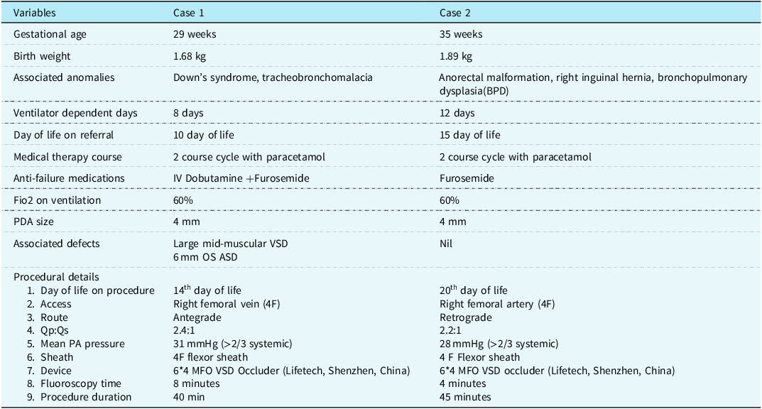
The procedure was undertaken on the 14th day of life. Informed consent from the parents was obtained after explaining the potential risks versus benefits of catheter treatment and surgical alternatives. The procedure was done under general anaesthesia as the baby remained ventilated after two failed attempts. A 4 French (Fr) right femoral venous access and right femoral artery were obtained under USG guidance. With the 4 Fr Judkins right catheter in the right atrium, a 0.018-inch straight tip Terumo wire (Terumo Corporation, Japan) was manoeuvred across the PA. The wire was placed across the patent ductus arteriosus and positioned in the descending aorta and a 4F flexor 45 cm introducer sheath (Cook Medical, Bloomington, IN, USA) was tracked over this wire into the proximal descending aorta. The mean aortic and PA pressures were 41 mmHg and 31 mmHg, respectively. The patent ductus arteriosus was angiographically profiled by injecting contrast into the side arm of the introducer, which measures 4 mm at the PA end (Figure 1a). Initially, a 5/6 Piccolo device was placed across the defect, keeping the aortic disc intraductal, but it slipped out easily. Hence, a 6 mm × 4 mm Konar MFO device (Lifetech, Shenzhen, China) was used to occlude the patent ductus arteriosus, and the device position was confirmed by echocardiography and fluoroscopy (Figure 1b-f). The total time required for the procedure was 40 min, and the fluoroscopy time was 8 min (Table 1). The pulmonary end of the device was seen protruding into the main PA with no obstruction in the LPA and arch on the angiogram.
Demographic and procedural details of cases

PDA= patent ductus arteriosus; OS ASD = ostium secundum atrial septal defect; LPA =left pulmonary artery ; Qp:Qs= pulmonary blood flow : systemic blood flow; PA = pulmonary artery.
Result and follow-up
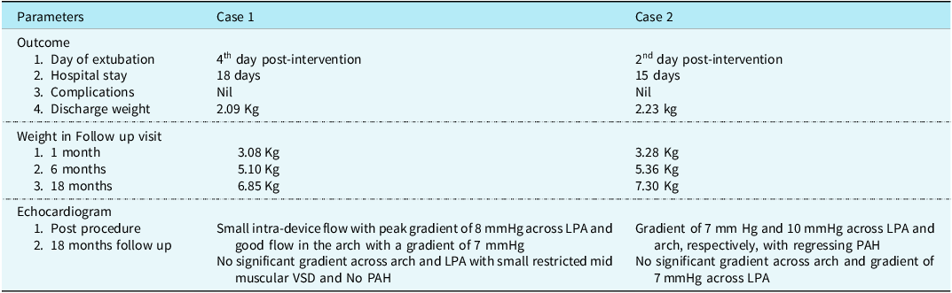
An echocardiogram performed 48 hours post-procedure showed the device in a good position with no residual flows with a gradient of 8 mmHg across LPA and good flow in the arch with a gradient of 7 mmHg. The baby was weaned off the ventilator over the next 4 days and discharged on the 28th postnatal day with a discharge weight of 2.09 kg. The ventricular septal defect flow became low velocity systolic left to right over the next 2 weeks. At the 1-month follow-up visit, he still had some noisy breathing but had gained weight, 3.08 kg. An echocardiogram at 2 month of follow-up showed no residual flows across the defect, with unobstructed flow in the LPA with a gradient of 14 mmHg. The muscular ventricular septal defect had become restricted, measuring 3 mm, with a PFO at the atrial level (Figure 2). He was regularly followed up at 2, 6, 12, and 18 months of age. At 6 months, he still had mild flow acceleration across the LPA with a gradient of 9 mmHg; however, the last echocardiography at 18 months, showed laminar flow with no gradient across the LPA and arch with no significant gradients, small restrictive mid-muscular ventricular septal defect, and intact IAS with no PAH (Table 2). He is on regular follow-up with the paediatric division for Down’s syndrome and developmental assessment.
Echocardiogram. (a) Wide open arch in 2D with unobstructed flow in aortic arch at 18 months follow-up (arrow). (b) Showing unobstructed in branch pulmonary arteries with. (c) Restricted mid-muscular VSD shunting left to right (arrow).

Outcome and Follow-up analysis

Case 2
Clinical details
Our second patient was a preterm baby born at 35 weeks of gestation with a birth weight of 1.89 kg to a primigravida through a lower segment caesarean section. Based on the initial evaluation, anorectal malformation with right inguinal hernia was diagnosed at birth. He was managed in the NICU, and after an imperforate anus was confirmed on imaging, he underwent colostomy at 48 hours of life and was electively ventilated thereafter. Two attempts at weaning from mechanical ventilation were unsuccessful, and there was no weight gain. Hence, a paediatric cardiology consultation was sought, and was referred to us on the 15th day of life. A chest X-ray showed cardiomegaly and increased pulmonary blood flow. An echocardiogram showed a patent ductus arteriosus measuring 4 mm, with flow reversal in the aorta with laminar Doppler flows across the defect, suggesting near systemic PA pressures (Figure 3a). Despite medical therapy, no improvement in haemodynamics was observed, and after discussion with the family child was taken up for transcatheter closure.
Procedural details
The procedure was undertaken on the 20th day of life. The procedure was done under general anaesthesia. A 4 French (Fr) right femoral venous access and right femoral artery were obtained. With the 4 Fr Judkins right catheter in the DTA, a 0.018-inch angled Terumo wire (Terumo Corporation, Japan) was manoeuvred across the patent ductus arteriosus into the MPA. A 4 Fr Flexor 45 cm introducer sheath (Cook Medical, Bloomington, IN, USA) was tracked over this wire into the MPA. The mean aortic and PA pressures were 32 mmHg and 28 mmHg, respectively.
A 6 mm × 4 mm Konar MF device (Lifetech, Shenzhen, China) was used to occlude the patent ductus arteriosus retrogradely, and the device position was confirmed by echocardiography and fluoroscopy (Figure 3). The total time required for the procedure was 45 minutes, and the fluoroscopy time was 4 minutes (Table 1). The pulmonary end of the device was seen protruding into the main PA with small intra-device flow on the angiogram.
(a) 900 lateral angiogram showing 4 mm PDA from DTA. (b, c) Deployment and positioning of 6*4 MFO KONAR device across PDA retrogradely. (d, f) Showing device in situ unobstructed flow in LPA (arrow) and arch (arrow) post deployment of device.

Results and follow-up
An echocardiogram performed 48 hours post-procedure showed the device in a good position with no residual flows. Branch PA flows and aortic arch flow were unobstructed with a gradient of 7 mm Hg and 10 mm Hg across LPA and arch, respectively, with regressing PAH. The baby was weaned off the ventilator over the next 2 days and discharged on the 30th postnatal day with a discharge weight of 2.23 kg. At the 1-month follow-up visit, he was in good health and weighed 3.28 kg with a peak gradient across the arch of 8 mmHg. Hence, we keep the baby on regular follow-up at 2,6,12, and 18 months with an echocardiogram showing no residual flows across the defect, unobstructed flow in LPA with a gradient of 7 mm Hg and pulsatile flow across the arch with no PAH at the latest follow-up (Figure 4 and Table 2). At present the baby is gaining weight and is asymptomatic.
Echocardiogram. (a) Arrow showing device in situ with no residual shunt and unobstructed flow across arch with no luminal narrowing at 18-month follow-up. (b) Arrow showing good flow across left pulmonary artery with no gradient.

Discussion
Our case series demonstrates that the KONAR MFO device is a viable and can be a safe option for PDA closure in preterm infants, even in settings with constrained resources, with a high procedural success rate, with both cases achieving complete occlusion without any significant residual shunting and complications. In our study, both babies were sick and ventilator-dependent. The main reasons for the failure of these ventilator-dependent cases were bronchial issues, with bronchopulmonary dysplasia developing after mechanical ventilation for a longer period. Studies have shown evidence of air-trapping in the expiratory high-resolution computed tomography images of bronchopulmonary dysplasia survivors, with a significant increase in the incidence of major airway lesions, such as tracheobronchomalacia and iatrogenic tracheobronchial stenoses.Reference Bentham, Meur and Hudsmith2,Reference Zahn, Nevin and Simmons3 In all cases, early intervention should be considered for ventilator-dependent preterm neonates with significant patent ductus arteriosus to minimise mechanical ventilation to reduce the incidence of bronchopulmonary dysplasia, bronchomalacia, and bronchial stenosis and improve their prognosis.Reference Lin, Huang and Lien4–Reference Occhipinti, De Carolis and De Rosa6
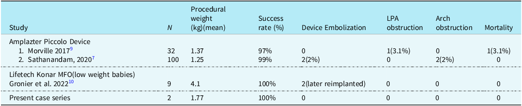
Amplatzer Piccolo is an FDA-approved device for preterm PDA closure. Transcatheter PDA closure in preterm infants weighing <2,000 g using the Amplatzer Piccolo device has been reported to be highly effective, with a success rate exceeding 98% in recent large-scale series, including the multicentre American prospective trial (success rate: 99%) and the multicentre French cohort study (success rate: 99%).Reference Sathanandam, Gutfinger and O’Brien7–Reference Morville and Akhavi9 However, as mentioned in our cases, the Amplatzer Piccolo device slipped out of the aortic end due to a large ductal diameter, a small retention disc, and high PA pressure related to associated comorbidities, making it unsuitable for such cases. Other devices, such as ADO II and Amplatzer Vascular Plug II–IV, can be used for long patent ductus arteriosus; however, size limitation with the requirement of larger guiding catheters or delivery sheaths, with a high risk of embolisation in high pulmonary artery pressure scenarios, are major limiting factorReference Malekzadeh-Milani, Akhavi and Douchin8,Reference Morville and Akhavi9 (Table 3). Our results align with previously published studies that have highlighted the effectiveness of the KONAR MFO device in low weight populations.Reference Francis, Singhi and Lakshmivenkateshaiab1–Reference Zahn, Nevin and Simmons3 Grunenwald Gronier et al.Reference Gronier, Benbrik, Romefort, Prigent, Hauet and Baruteau10and Banpurkar A et al.Reference Banpurkar, Koneti, Thakur, Kulkarni, Prabhu and Doniparti11 reported the closure of large patent ductus arteriosus using KONAR-MF™ occluder in low-weight babies with no major adverse events with a comparable success rate as of Amplatzer Piccolo. One of the notable advantages of the KONAR MFO device is its versatility, its profile allowing for patent ductus arteriosus closure across a wide range of ductal sizes and morphologies. The device’s self-expandable nature and ease of deployment make it particularly useful in preterm infants, where anatomical variations may present technical challenges. The double-disc device design with retention screws on either side and smaller sizes (5/3, 6/4, 7/5, and 8/6) does not contain any fabric and makes it much softer and easier to negotiate through small delivery or guide catheters in long preterm ductus, where we prefer to have a disc on the aortic and pulmonary end and because of the length of the device, closure is feasible without risk of embolisation.Reference Gronier, Benbrik, Romefort, Prigent, Hauet and Baruteau10–Reference Mukherji, Chattopadhyay, Ghosh, Mohapatra, De and Singhi12 Despite the overall positive outcomes, some challenges remain. No major adverse events such as device embolisation, significant bleeding, or mortality were observed during the short-term follow-up period. Longer-term follow-up studies will be essential to assess potential complications such as late device migration, residual shunting, or pulmonary artery stenosis.
Comparative analysis of various devices with current case series

In a resource-limited setting, cost-effectiveness is a critical consideration. Our case series insights on KONAR MFO as a feasible device in ventilated dependent preterm, even in a setup with a lack of or limited access to neonatal cardiac surgery.Reference Lenoir, Wanert and Bonnet13 While the KONAR MFO device may have a higher upfront cost than conventional medical therapy, its ability to facilitate early patent ductus arteriosus closure offers long-term economic benefits, with the cost of the procedure less than the Amplatzer Piccolo device and nearly half compared to surgical ligation. Additionally, ELBW infants did not experience post-PDA ligation syndrome and morbidity, like the risk of vocal cord palsy, bleeding, pneumothorax, infection, chylothorax, or haemodynamic instability after transcatheter PDA occlusion, demonstrated faster respiratory improvement and shorter mechanical ventilations, well as less overall mortality when compared to surgical PDA ligation by posterior thoracotomy.Reference Lenoir, Wanert and Bonnet13,Reference Tashiro, Wang and Sola14
Limitations
Our case series is limited by its short-term follow-up period, a relatively small sample size, and the lack of a direct comparison with other patent ductus arteriosus closure techniques. Future studies with larger cohorts and extended follow-up durations are necessary to validate these findings and assess the long-term safety and efficacy of the KONAR MFO device in preterm populations.
Conclusion
In conclusion, our case series highlights that percutaneous PDA closure using the KONAR MFO device is a safe, effective, and feasible option in preterm infants in difficult scenarios, even in resource-limited settings. With further experience and continued research, this technique has the potential to become a standard of care for preterm patent ductus arteriosus closure in similar healthcare environments. However, longer-term follow-up is essential to assess potential complications such as late device migration, residual shunting, or pulmonary artery stenosis.
Acknowledgements
We thank all nursing medical staff and parents of patients for their cooperation and support in the present study.
Financial support
The author(s) received no financial support for the research, authorship, and/or publication of this article.
Competing interests
The author(s) declared no potential conflicts of interest concerning the research, authorship, and/or publication of this article.
Ethical standard
The authors assert that all procedures contributing to this work comply with the ethical standards, and the study has been approved by the institutional committees.