Introduction
Tetralogy of fallot is associated with anomalous coronary arteries in 2–9% of total cases.Reference Kalfa, Serraf and Ly1 Among all coronary anomalies in tetralogy of fallot, anomalous left coronary artery connection to pulmonary artery is very rare in incidence. Prompt diagnosis of this condition and single-stage successful repair is challenging. We describe, here, a successfully treated association of tetralogy of fallot and anomalous left coronary artery connection to main pulmonary artery with a giant intercoronary steal collateral crossing right ventricular outflow tract in a 9-year-old boy.
Case report
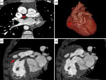
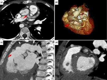
A 9-year-old boy presented with 4 months history of episodes of cyanosis and effort intolerance. On clinical examination, he was in sinus rhythm, a heart rate of 90/minute, an oxygen saturation of 80%, single second heart sound, grade 3/6 ejection systolic murmur in pulmonary area, and no cardiomegaly. Chest X-ray showed boot-shaped heart with near normal pulmonary vascularity. Electrocardiogram showed sinus rhythm, right-axis deviation, right ventricular hypertrophy, and no ST segment or T wave ischemic changes. Transthoracic echocardiogram showed severe infundibular and pulmonary valvar stenosis (a peak gradient of 85 mmHg), large subaortic ventricular septal defect, right aortic arch, normal biventricular function, a suspected right coronary artery to pulmonary artery fistula, left coronary artery origin in aorta, and course that was not clearly visualised. Conventional cardiac angiogram was suggestive of tetralogy of fallot with hypoplastic pulmonary annulus, retrograde filling of left coronary artery through large intercoronary collaterals from the dilated dominant right coronary artery, and normal biventricular function (videos 1 and 2). Computerised tomographic angiogram confirmed tetralogy of fallot with anomalous left coronary artery arising from main pulmonary trunk posteromedially 1 cm above the sinotubluar junction level corresponding to right posterior cusp. It also showed a pulmonary annulus diameter of 12 mm (←2Z score) and giant (8 mm diameter) steal intercoronary collateral connecting proximal right and left coronary artery coursing over right ventricular outflow tract 5 mm below the pulmonary annulus (Fig 1). Surgery was performed through midline sternotomy under cardiopulmonary bypass using aorto-bicaval cannulation. After snugging both branch pulmonary arteries, cardioplegia was given to both the coronaries by two separate cannulas in aorta and pulmonary trunk. Left coronary button was prepared from pulmonary trunk after dividing the pulmonary trunk at confluence. After adequate mobilisation of the left coronary artery button and confirming the lie, anastomosed to the ascending aorta. Pulmonary annulus was opened longitudinally and extended carefully 3 mm below the annulus leaving margin for suturing the transannular patch. After infundibular resection, secure ventricular septal defect closure was done with dacron patch, and transannular patch was sutured meticulous, thereby sparing the nearby giant collateral. Patient’s hemodynamics was good after coming off cardiopulmonary bypass, and transesophageal echocardiogram showed good left coronary artery antegrade flow, no regional wall motion abnormalities in anterolateral wall of left ventricle, no residual ventricular septal defect, and peak gradient across right ventricular outflow tract that was 28 mmHg. Postoperative recovery was uneventful and discharged after 1 week. On follow-up after 6 months, patient was asymptomatic, and computerised tomographic angiogram showed patent left coronary artery flow from ascending aorta, decreased size of the giant collateral to 2 mm, and completed repair of tetralogy of fallot (Fig 2).
Preoperative computerised tomographic angiogram image (a) coronal oblique view showing anomalous connection of left main coronary from main pulmonary artery (arrow), (b) volume rendering technique image showing giant collateral (*) connecting the dilated left main (+) and RCA (arrow), (c) sagittal oblique view showing infundibular stenosis (arrow), and (d) sagittal oblique view showing ventricular septal defect and aortic override.

Postoperative computerised tomographic angiogram image (a) axial oblique view showing left main coronary (arrow) arising from ascending aorta, (b) volume rendering technique image showing the collapsed collateral (*) connecting the dilated left main (+) and right coronary artery (arrow), (c) sagittal oblique view showing no residual infundibular stenosis (arrow), and (d) sagittal oblique view showing no residual ventricular septal defect (*).

Discussion
Incidence of anomalous left coronary artery connection to pulmonary artery is 1 in 300,000 of live births or 0.5% of all congenital heart diseases.Reference Heifetz, Robinowitz and Muller2 Rarely, this condition can be associated with ventricular septal defect, patent ductus arteriosus, coarctation aorta, truncus arteriosus, or tetralogy of fallot.Reference Vlodaver, Neufeld and Edwards3 Very few single-stage successful surgically treated cases of this association have been reported.Reference Bitar, Kveselis and Smith4–Reference Morell, Feccia and Cullen8 Unlike previous reported cases, which were from neonate to 8 years, our patient is 9 years of age, the oldest reported case till now.
Only 10% cases of anomalous left coronary artery connection to pulmonary artery survive infancy because of modest left coronary perfusion either antegrade or by intercoronary collaterals through circle of Vieussens from right coronary artery. When tetralogy is associated with this anomalous left coronary artery connection, clinical presentation depends on adequacy of left coronary and lung perfusion. The supply demand mismatch of right ventricle in tetralogy due to severe hypertrophy causing subendocardial ischemia is aggravated by coronary steal here. Not only left ventricle is compromised, onset of right ventricle dysfunction and failure is aggravated. When associated with tetralogy of fallot, clinical presentation depends on intercoronary collaterals, severity of pulmonary stenosis, and coronary artery dominance.Reference Kurup, Daniel and Kumar9 The intercoronary collaterals develop gradually from the right coronary system to provide a decent left coronary perfusion retrogradely and contribute a good amount to lung perfusion by drainage through the anomalous left coronary ostium into the pulmonary trunk. In our case, the rich intercoronary collaterals from the dominant right coronary artery allowed adequate left coronary perfusion along with underloaded left ventricle to maintain good left ventricular function and delayed the clinical presentation. Even though pulmonary stenosis was severe, significant intercoronary collaterals and few aortopulmonary collaterals provided near adequate pulmonary perfusion to delay the clinical manifestation. Pre- or intraoperative pulmonary arterial oxygen saturation measurement could give an idea of left-to-right shunt fraction even though it was not measured in this case. If the right coronary artery is dominant, it helps to maintain adequate left coronary and pulmonary artery perfusion, thereby delaying the clinical presentation. The right coronary artery, which is commonly hypertrophied in tetralogy of fallot, is exaggerated here because of increased distal runoff to both left coronary and pulmonary artery through collaterals to maintain adequate perfusion.
Misdiagnosis of anomalous connection of left coronary artery from pulmonary artery with the coronary to pulmonary artery fistula by transthoracic echocardiogram is possible. Poor lateral resolution of left coronary sinus to visualise left coronary origin, difficult visualisation of pulmonary origin, retrograde flow in left coronary artery, and intraseptal collaterals leads to a possible misdiagnosis of coronary artery to pulmonary artery fistula. Computerised tomographic angiogram is of immense use in such complex case, by providing excellent resolution of images, post-processing ability, and three-dimensional reconstruction for clear delineation of anatomy. Undoubtedly, two coronary system establishment is the treatment of choice, as left ventricle has to cope with the increase in work post-intracardiac repair of tetralogy of fallot. As the left coronary artery anatomy was suitable, direct translocation to ascending aorta was done. As it was possible to relieve the right ventricular outflow tract obstruction and also to reconstruct it without the division of the giant collateral, it was preserved in this case. But, one should not compromise on the completeness of repair of right ventricular outflow tract obstruction to preserve an intercoronary collateral. The incidence of late coronary stenosis following button technique varies from 5 to 8%, so by not dividing the collateral, there exists a theoretical advantage of maintaining the collateral circulation.Reference Agnoletti, Bajolle and Bonnet10
To conclude, the association of anomalous left coronary artery from pulmonary artery with tetralogy of fallot is very rare, so high index of suspicion is needed for accurate preoperative diagnosis and proper plan to execute successful single-stage.
Acknowledgements
The authors would like to thank Dr Sreenivas and Dr Sakshi Sachdeva for their help in image acquisition.
Supplementary Material
To view supplementary material for this article, please visit https://doi.org/10.1017/S1047951120001730
Financial Support
No financial support availed from any funding agency.
Conflicts of Interest
None.