Introduction
Mucormycosis is a rare opportunistic fungal infection caused by fungi belonging to Mucorales order and Mucoraceae family. Various clinical presentations include rhino-orbito-cerebral, pulmonary and cutaneous forms, and less frequently, gastrointestinal, disseminated and miscellaneous forms.Reference London and Chong1
Immunocompromised patients are at the greatest risk for mucormycosis. Diabetes mellitus is the most common predisposing factor associated with mucormycosis.Reference Yohai, Bullock, Aziz and Markert2,Reference Ferry and Abedi3 A state of diabetic ketoacidosis further increases the risk of developing mucormycosis. Persistent hyperglycaemia impairs polymorphonuclear cell chemotaxis and intracellular phagocytosis ability. The acidic environment also leads to reduction in the binding of iron to transferrin, increasing free iron concentration which enhances fungal growth.Reference Artis, Fountain and Delcher4 Other risk factors include patients with haematological malignancies, transplantation, neutropenia, corticosteroid therapy and use of deferoxamine.Reference Yohai, Bullock, Aziz and Markert2,Reference Teixeira, Medeiros, Leushner and Almeida5,Reference Jiang, Zhao and Yang6
Factors critical for management of mucormycosis include: rapidity of diagnosis, reversal of the underlying predisposing factors, appropriate aggressive surgical debridement of infected tissue and appropriate antifungal therapy. Early diagnosis is important because small, focal lesions can often be surgically excised before they progress to involve critical structures or disseminate.Reference Nithyanandam, Jacob, Battu, Thomas, Correa and D'Souza7
Applied anatomy of palate and its blood supply
The palate divides the nasal cavity and oral cavity with the hard palate positioned anteriorly and soft palate posteriorly. The palatal mucosa is strongly adhered to the underlying periosteum, which is subsequently attached to the bone via fibrous tissue pegs known as Sharpey's fibres. The hard palate comprises the palatine process of the maxilla and the horizontal palatal lamina of the palatine bones. A longitudinal suture separates the maxilla in the midline; the palatal aponeurosis attaches to the posterior margin of the hard palate and is continuous with the tensor veli palatini laterally.Reference Jamali8
The major blood supply of the hard palate comes from the greater palatine artery, which emerges from the greater palatine foramen located on the hard palate between the second and third maxillary molars. Further behind, there are the lesser palatine foramina where the branches of the lesser palatine artery emerge. These arteries supply the majority of the hard palate together with the soft palate. The branches of the greater palatine artery travel within the palatal bony groove divided into medial and lateral palatine grooves by the palatine spine. The medial palatine groove contains the greater palatine nerve, whereas the greater palatine artery lies in the lateral groove to supply the mucosa, periosteum and palatal gingiva before entering the incisive foramen to form an anastomosis with the nasopalatine artery. The nasopalatine artery enters the incisive canal to supply the anterior region of the hard palate.Reference Shahbazi, Grimm, Feigl, Gerber, Székely, Molnár and Windisch9
The main arterial supply of the soft palate is the ascending palatine artery, which most commonly arises from the facial artery but can occasionally arise from the external carotid, the ascending pharyngeal or the maxillary artery.Reference Cho, Kim, Park, Suh, Kim and Yoon10 The ascending palatine artery runs inferomedially from the lateral pharyngeal space into the palate, dividing into the anterior and posterior branches.
Knowledge of the blood supply of the palate and the course of the palatal vasculature is important in oral surgical procedures to aid in flap design and may enable clinicians to avoid intra- and post-operative complications when planning oral surgery interventions by optimised incision and flap designs.
Case series
Sixteen patients with mucormycosis who were admitted to the ENT Department of Lok Nayak Hospital, Delhi, India, underwent inferior maxillectomy; of these patients, 6 underwent mucosa-preserving subperiosteal inferior maxillectomy after informed consent. The palatal mucosa was clinically normal in all patients, who all had a positive prick test (i.e. healthy blood supply to the palate). After thorough history and clinical examination, all patients were assessed by radiological imaging using contrast-enhanced computed tomography (CT) of the nose, paranasal sinuses and orbit (Table 1).
Case series

Covid-19 = coronavirus disease 2019
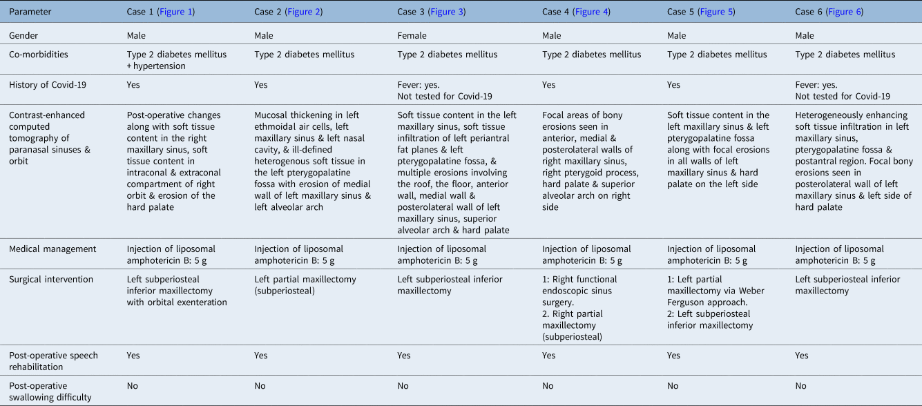
Case 1. (a) Pre-operative image. White arrow indicating palatal bulge. (b) Coronal plane contrast-enhanced computed tomography image of the nose, paranasal sinuses and orbit. Black arrow indicates right hard palate erosion. (c) Axial plane contrast-enhanced computed tomography image of the nose, paranasal sinuses and orbit. Black arrow indicates right hard palate erosion. (d) Intra-operative image showing palatal incision. (e) Intra-operative image showing gingivolabial flap. (e) Intra-operative image after closure.

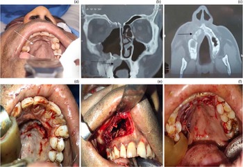
Case 2. (a) Coronal plane contrast-enhanced computed tomography of the nose, paranasal sinuses and orbit. Black dotted arrow indicates left-sided hard palate erosion. (b) Intra-operative image (edentulous patient). White arrow indicating palatal flap. Black arrow indicating gingivolabial flap. (c) Intra-operative image after closure.

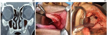
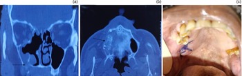
Case 3. Contrast-enhanced computed tomography of the nose, paranasal sinuses and orbit in the (a) coronal and (b) axial view. White arrow indicates left-sided hard palate erosion. (c) Intra-operative image after closure.

Case 4. Contrast-enhanced computed tomography of the nose, paranasal sinuses and orbit in the (a) coronal and (b) axial view. Black arrow indicates right-sided hard palate erosion. (c) Post-operative image.

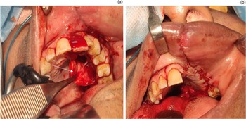
Case 5. (a) Intra-operative image. White arrow indicates palatal flap. (b) Intra-operative image after closure.

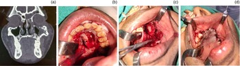
Case 6. (a) Coronal plane contrast-enhanced computed tomography of the nose, paranasal sinuses and orbit. (b) Intra-operative image. Black arrow indicates raised palatal flap. (c) Intra-operative image. (d) Intra-operative image after closure.

Surgical technique
Patients underwent debridement using subperiosteal mucosa-preserving inferior maxillectomy under general anaesthesia. Palatal incision was performed 5–6 mm away from the alveolar margin, and the mucoperiosteal flap was elevated. In cases of larger defects, the palatal flap was raised until the junction of the hard and soft palate. The ipsilateral greater palatine artery was preserved. A separate sublabial incision was given and a gingivolabial flap was raised, which was subsequently made continuous with cheek flap. The diseased tissue of the underlying hard palate was removed. Removal of hard palate bone provided access to the maxillary antrum, pterygopalatine fossa and infratemporal fossa, which was cleared of the disease if found. After adequate disease clearance and haemostasis, the palatal mucoperiosteal flap was reposited and sutured with upper gingiva-labial flap using 3-0 vicryl sutures.
Discussion
Mucormycosis often involves the hard palate, requiring debridement in the form of inferior maxillectomy. Loss of hard palate in inferior maxillectomy leads to significant morbidity to the patient in terms of speech and deglutition problems. Subperiosteal inferior maxillectomy is a modification of conventional inferior maxillectomy in which the uninvolved mucoperiosteum of the hard palate is preserved with the ipsilateral greater palatine artery. This flap is used for reconstruction of the oroantral or oronasal defect. This technique can be adopted in patients wherein the palatal mucosa is normal and not ulcerated with a good blood supply, which can be confirmed by bleeding margins of the mucosal incision and a prick test (looking for brisk bleeding on pricking).
The preservation of the palatal mucoperiosteal flap is to be considered in all mucormycosis patients, in contrast to patients with malignancy of the maxilla, where adequate oncological margins are important. In all, 16 patients with mucormycosis who had associated palatal involvement were admitted to the ENT Department of Lok Nayak Hospital and subsequently underwent inferior maxillectomy. Among them, 6 patients were taken up for mucosa-preserving subperiosteal inferior maxillectomy and were included in our case series. Other patients had extensive palatal bony and mucosal involvement; therefore, palatal flaps were not available for reconstruction of oroantral defects.
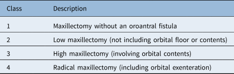
In 2000, Brown et al. classified the defect following maxillectomy into vertical and horizontal components. The vertical defect, which involves resection in the vertical plane from dentition to the skull base, is classified into class 1–4. A letter (a, b or c) is added depending on how much of the upper alveolus has been removed, which is considered to be the horizontal component of the defect (Tables 2 and 3).Reference Brown, Rogers, McNally and Boyle11
Classification of vertical defect

Classification of horizontal defect

The maxilla bears the maxillary teeth, transmits masticatory forces and provides a partition between the oral and nasal cavities and maxillary sinuses. It therefore assists in critical functions of mastication, speech and deglutition.Reference Futran and Mendez12 Loss of the maxilla is associated with significant functional morbidity. Palatal defects in isolation lead to formation of oroantral fistula, which results in difficulty in swallowing, speech and also creates cosmetic impairment and psychological consequences for patients.
• Mucormycosis is a fulminant disease of the nose and paranasal sinuses requiring aggressive surgical debridement
• Patients with sinonasal mucormycosis with palate involvement often require inferior maxillectomy
• Subperiosteal inferior maxillectomy is a modification of the conventional inferior maxillectomy wherein the healthy palate mucosa is preserved
• Oronasal separation is achieved, and speech and swallowing functions are preserved using this technique
• Mucoperiosteal palatal flap-preserving subperiosteal inferior maxillectomy has better functional and aesthetic outcomes than conventional inferior maxillectomy
Reconstruction of the maxillary is a significant challenge because the three-dimensional midface architecture has both functional and aesthetic functions. It requires the presence of a healed wound, separation of oral and nasal cavities, restoration of maxillary buttresses, restoration of functional dentition, mastication and deglutition, maintenance of a patent nasal airway, support and suspension of adynamic facial soft tissue and the restoration of an adequate and symmetrical facial form on the contralateral side, and psychological well-being.Reference Andrades, Militsakh, Matthew, Rieger and Rosenthal13 Various techniques have traditionally been used for the reconstruction of inferior maxillectomy defects, including palatal prosthesis, local flaps and microvascular tissue transfers. The choice of a specific surgical procedure mainly depends upon the site and size of the defect present and the amount and location of palatal tissues available for repair.
Various studies depicting the role of obturators and local and free flaps for maxillary reconstruction following inferior maxillectomy are described in the literature, some of which are discussed briefly here. Choung et al.Reference Choung, Nam and Kim14 described the use of ipsilateral or bilateral temporalis muscle with or without a segment of attached calvarial bone. This flap could be passed into the oral cavity by performing anterior and posterior osteotomy procedures on the zygomatic arch and mobilising it to its attachment on the coronoid process. Alternatively, Hatoko et al.Reference Hatoko, Harashina and Inoue15 reported success in reconstruction of inferior maxillectomy procedures with a fasciocutaneous radial forearm free tissue transfer in three patients who had not tolerated obturators well. Futran et al.Reference Futran, Wadsworth, Villaret and Farwell16 reported the use of the fibula free flap in 27 patients with defects, including defects of the palate that were not amenable to the use of a conventional prosthesis. All these above studies looked at malignancy of maxilla where oncological margins are to be carefully considered. On the other hand, surgical debridement for mucormycosis is aggressive only for involved areas.
In our technique of subperiosteal inferior maxillectomy, we raised a subperiosteal palatal flap along with a superior gingivolabial flap. The diseased tissue of the antrum and the palatal bone was removed, and the flaps were sutured. In cases of larger defects, the subperiosteal flaps were raised up to the hard and soft palate junction, without damaging the greater palatine artery. The complete oronasal separation was achieved in all six patients, and this resulted in better aesthetic and functional outcomes. The overall surgery time was also decreased when compared with free tissue transfer. Also, the patient does not have to bear the weight of prosthesis. The technique allowed excellent palatal function and prompt rehabilitation allowing better patient outcomes without compromising disease removal.
Conclusion
Mucoperiosteal palatal flap-preserving subperiosteal inferior maxillectomy is a desirable approach for all patients with mucormycosis and healthy palatal mucosa, leading to better functional and aesthetic outcomes for the patient.
Acknowledgements
The authors would like to thank all the faculty and staff of the Department of Otorhinolaryngology at Maulana Azad Medical College and Associated Lok Nayak Hospital for their generous help and support.
Competing interests
None declared