Introduction
Spermatogenesis is a dynamic process that is controlled by autocrine/paracrine variables in the endocrine system and, locally, by numerous interactions between developing germ cells and the environment (Caires et al., Reference Caires, Broady and McLean2010; Hai et al., Reference Hai, Hou, Liu, Liu, Yang, Li and He2014; Giudice et al., Reference Giudice, De Michele, Poels, Vermeulen and Wyns2017). The spermatogenesis process is extrinsically controlled by testicular somatic cells (Sertoli, myoid and Leydig cells) along with intrinsic regulators that include transcription factors produced by spermatogonial cells (Mei et al., Reference Mei, Wang and Wu2015). Proliferation and differentiation of spermatogonial stem cells (SSCs) is a complex and tightly regulated process that occurs in the basement compartment of seminiferous tubules (Talebi et al., Reference Talebi, Sadighi-Gilani, Koruji, Ai, Navid, Rezaie, Jabari, Ashouri-Movassagh, Khadivi, Salehi, Hoshino and Abbasi2019).
Sertoli cells are the major somatic cells of the SSC niche. They provide morphogenetic support through cell–cell interactions as well as biochemical components by secreting lactate, cytokines and hormones. Aside from mechanical and nutritional support, SCs also provide an immune-protective environment for germ cells via the blood–testis barrier (Ni et al., Reference Ni, Hao and Yang2019). Sertoli cells are thought to be the primary cellular target of testosterone signalling. In the Sertoli cell, testosterone signals can be translated directly into changes in gene expression, or testosterone can activate kinases, which may regulate processes necessary for spermatogenesis to continue (Walker, Reference Walker2011). During spermatogenesis, Sertoli cells stimulate SSC self-renewal and promote SSC differentiation; they also regulate meiosis in spermatocytes and conversion of round spermatids to spermatozoa (Ma et al., Reference Ma, Yang, Zhang, Li, Gong, Liu, Zhu, Tian, Liu, Wang, Liu, He, Liu, Yang, Li and He2013; Hai et al., Reference Hai, Hou, Liu, Liu, Yang, Li and He2014). Studies have shown that the overall efficiency and effectiveness of spermatogenesis depend strongly on the existence of the Sertoli cell–SSC niche (Spradling et al., Reference Spradling, Drummond-Barbosa and Kai2001).
Different strategies for in vitro derivation of male germ cells are mostly based on two-dimensional (2D) cultures, implementation of established medium, and feeder cells (Sousa et al., Reference Sousa, Cremades, Alves, Silva and Barros2002; Riboldi et al., Reference Riboldi, Rubio, Pellicer, Gil-Salom and Simón2012; Yang et al., Reference Yang, Ping, Ma, Li, Tian, Yang, Liu, Gong, Zhang, Li and He2014). It has been reported that during the SSC differentiation process, the spatial arrangement of testicular cells is incredibly important (Staub, Reference Staub2001). In the natural environment, meiotic cells are engulfed in Sertoli cells as large interconnected clones that have no interaction with the basement membrane; the 2D culture system cannot provide this sophisticated structure (Khajavi et al., Reference Khajavi, Akbari, Abdolsamadi, Abolhassani, Dehpour, Koruji and Habibi Roudkenar2014). According to previous studies, three-dimensional (3D) culture systems provide more spatial conditions that mimic the architecture of seminiferous tubules (Shams et al., Reference Shams, Eslahi, Movahedin, Izadyar, Asgari and Koruji2017). In a 3D culture, the cells interact with one another, the extracellular matrix (ECM), and the surrounding microenvironment. Proliferation, differentiation, cell morphology, gene and protein expression, and cell response to external factors are influenced by these interactions in 3D structures (Mohammadzadeh et al., Reference Mohammadzadeh, Mirzapour, Nowroozi, Nazarian, Piryaei, Alipour, Modarres Mousavi and Ghaffari Novin2019). To date, the behaviour of SSCs has been evaluated in cultivation systems that use 3D surfaces such as soft agar (Abu Elhija et al., Reference Abu Elhija, Lunenfeld, Schlatt and Huleihel2012), collagen (Lee et al., Reference Lee, Gye, Choi, Hong, Lee, Park, Lee and Min2007), alginate (Chu et al., Reference Chu, Schmidt, Carnes, Zhang, Kong and Hofmann2009; Jalayeri et al., Reference Jalayeri, Pirnia, Najafabad, Varzi and Gholami2017; Pirnia et al., Reference Pirnia, Parivar, Hemadi, Yaghmaei and Gholami2017), poly l-lactic acid (PLLA) (Eslahi et al., Reference Eslahi, Hadjighassem, Joghataei, Mirzapour, Bakhtiyari, Shakeri, Pirhajati, Shirinbayan and Koruji2013) and polyamide (Ultra-WebTM) nanofibres (Shakeri et al., Reference Shakeri, Kohram, Shahverdi, Shahneh, Tavakolifar, Pirouz, Shahrebabak, Koruji and Baharvand2013). According to previous research, the 3D culture system assisted by somatic cells could provide an enhanced culture system by creating both physical and paracrine support to enable SSCs to enter meiosis (Stukenborg et al., Reference Stukenborg, Schlatt, Simoni, Yeung, Elhija, Luetjens, Huleihel and Wistuba2009; Khajavi et al., Reference Khajavi, Akbari, Abdolsamadi, Abolhassani, Dehpour, Koruji and Habibi Roudkenar2014).
Although the results of numerous studies have shown the essential role of somatic testicular cells in the induction of spermatogenesis, the function of these cells in meiotic progression during co-culture with an SSC encapsulated alginate hydrogel 3D culture system remains uncertain.
Taking all this into account, the in vitro conditions for full spermatogenesis are far from routine methodology. In this study, we aimed to evaluate a co-culture of SSC encapsulated alginate hydrogel with Sertoli cells and their transplantation into azoospermic mice.
Materials and methods
Isolation and purification of SSCs
The Ethics Committee of Kermanshah University of Medical Sciences in compliance with the guidelines of Kermanshah University of Medical Sciences approved the animal experiments in this research. Testes were obtained from 80 6-day-old male neonatal BALB/c mice for testicular cell isolation and transferred to phosphate-buffered saline (PBS; Gibco, USA). A two-step enzymatic digestion process was used for tissue digestion. Prior to enzymatic digestion, the tunicae albugineae were removed from all testicles. Pieces of minced testes were digested in a solution that contained collagenase type IV (Sigma, 2 µg/ml) and DNase I (5 µg/ml) for 15 min at 37°C in a 5% CO2 incubator. After centrifugation at 800 g for 5 min, the cell pellet was re-suspended in 1 ml trypsin–EDTA (Sigma) and incubated for 5 min at 37°C with gentle pipetting to ensure suspension of the testicular cells. The cell suspension was then centrifuged for 5 min at 800 g and the cell pellet was washed in PBS. The SSCs were purified according to the Invitrogen protocol for magnetic activated cell sorting (MACS) (Jalayeri et al., Reference Jalayeri, Pirnia, Najafabad, Varzi and Gholami2017; Pirnia et al., Reference Pirnia, Parivar, Hemadi, Yaghmaei and Gholami2017).
Encapsulation of SSCs in alginate
We prepared the sodium alginate solution by dissolving 1.25 g of powdered alginate in 150 mmol NaCl at pH 7.4. This solution was mixed with the cell pellet. Next, the cell alginate solution was added slowly dropwise to 135 mmol/l of calcium chloride, which created a MicroBead cell alginate. After 10 min, the calcium chloride was extracted by washing the MicroBeads with 0.9% NaCl.
Depolymerization of the cell alginate solution
We used a solution of 119 mmol/l sodium citrate for depolymerization. The MicroBead solution was placed in an incubator for c. 30 min and then centrifuged at 1800 rpm for 8 min. Next, 1 ml of Dulbecco modified Eagle’s medium (DMEM) was added to the cell pellet.
Preparation of the Sertoli cell feeder layer
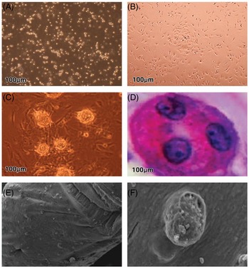
A mouse Sertoli cell line (NCBI code: C513) was purchased from the Pasteur Institute, Iran. Sertoli cells were cultured in a Datura stramonium agglutinin (DSA lectin, 10 μg/ml)-coated 25 T flask that contained DMEM supplemented with 10% (v/v) fetal bovine serum (FBS), essential amino acids (EAA), non-essential amino acids (NEAA), and an antibiotic–antimycotic solution (Invitrogen, USA). The flasks were incubated for 2 h at 37°C in a humidified atmosphere of 5% CO2 in air. When the Sertoli cells reached 90% confluency, they were treated with 10 μg/ml mitomycin C for 3 h. These cells were washed three or four times with DMEM and were subsequently used as the feeder layer (Fig. 1B).
(A) Spermatogonial stem cells (SSCs) after enzymatic digestion using trypsin–EDTA. (B) Morphology of Sertoli cells. (C) SSC colonies. (D) Microbeads after 3D long-term culture stained with haematoxylin and eosin (H&E) dye. Stained sections showed normal spermatogonia morphology. (E, F) Scanning electron microscopy micrographs from SSCs encapsulated in alginate hydrogel.

Experimental protocol
Two experimental designs were investigated. In the first group, SSCs obtained by magnetic activated cell sorting (MACS) were cultured in an alginate hydrogel 3D culture with Sertoli cells under 3D culture conditions for 30 days. In the second group, SSCs were co-cultured with Sertoli cells as the feeder layer (2D culture system) for 30 days. The culture medium consisted of 10% FBS and 10 ng/ml glial cell-derived neurotrophic factor (GDNF; Sigma). The SSCs were incubated at 37°C and 5% CO2 in a humidified atmosphere. The medium was replaced by fresh medium every 3 days. Cell growth and colony formation ability were analyzed under an invert microscope and cell viability was evaluated using the trypan blue assay.
RNA extraction, cDNA synthesis and qRT-PCR
Total RNA was extracted using a SinaPure RNA kit according to the manufacturer’s protocol. The concentration of RNA was measured using a NanoDrop 1000 spectrophotometer (Thermo Fisher Scientific). The extracted RNA was used for cDNA synthesis with a Revert Aid First Strand cDNA synthesis kit. qRT-PCR was conducted with specific primers for the integrin alpha-6, integrin beta-1, Nanog, Plzf, Thy-1, Oct4, P53 , Fas, Bax, and Bcl2 genes (Table 1). Gapdh was used as a reference gene to standardize the relative qRT-PCR results. Data were estimated using the ΔΔCt equation.
Primer sequences used for quantitative RT-PCR

5-Bromo-2-deoxyuridine (BrdU) cell labelling and transplantation into recipient mice
We added BrdU to the medium 24 h before transplantation to label and trace the cells in the recipient mice. This step was performed after depolymerization of the SSC-alginate in the 3D culture and the SSC clusters and underlying Sertoli cells were trypsinized in the 2D culture at the end of 4 weeks.
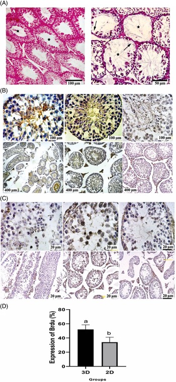
Adult male (6–8-week-old) BALB/c mice were used for recipient mice. The mice testes were treated with 40 mg/kg busulfan prior to transplantation (Fig. 3A). Then, the spermatogonial cells were transplanted into the seminiferous tubules of the recipient mice. At 6 weeks after treatment, Adult recipient mice were anesthetized with 10% ketamine and 2% xylazine. Approximately 105 of the cultured cells in 10 μl DMEM were injected into the seminiferous tubules in one testis per recipient mouse, while the other testis was used as an internal control. The cells were transplanted by retrograde injection through the efferent ducts (Gholami et al., Reference Gholami, Saki, Hemadi, Khodadadi and Mohammadi-Asl2014). The recipient testes were collected 6 weeks after transplantation, and 5–7 µm cross-sections were prepared after the cells were fixed in a paraffin block to consider spermatogenesis. We used the BrdU IHC kit according to the manufacturer’s instructions to identify the injected SSCs that were labelled with BrdU.
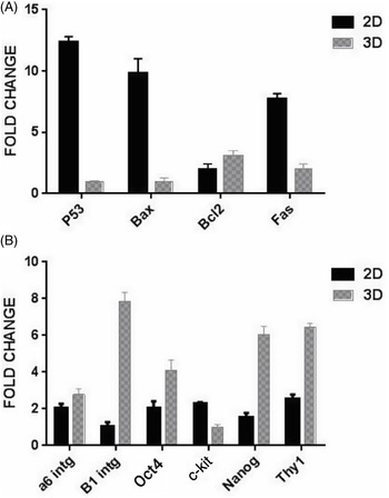
(A) Relative fold change of Fas, P53, Bax, and Bcl2 gene expression in spermatogonial cells that were cultured in 3D and 2 D systems. (B) Quantitative gene expression analysis by real-time PCR for integrin beta-1, Thy1, Oct4, integrin alpha-6, c-kit and Nanog genes in cultured mouse undifferentiated SSCs in 3D and 2D systems at the 30th day of culture. Values are mean ± standard error of the mean (SEM) and indicated statistically significant difference. Indicated statistically significant difference (P < 0.05).

Haematoxylin and eosin staining
At 6 weeks after the SSC transplantation, the mice were killed with a high dose of anaesthetic and their testicular tissues were harvested for examination. After fixation, the left testes were enclosed in paraffin, cut, and dehydrated. These tissues were then stained with haematoxylin and eosin (H&E) to evaluate the histologic changes. The findings were evaluated on the basis of a descriptive procedure. Blinded evaluation of tissue sections inside similarly staged seminiferous tubules was carried out by specialists. The results were evaluated according to the method by Johnson (Lewis-Johnes, Reference Lewis-Johnes1985).
PAS-H staining
Deparaffinized and rehydrated sections were soaked for 10 min in a 0.5% periodic acid solution and rinsed in flowing tap water. The sections were immersed in Schiff’s reagent (Merck, Darmstadt, Germany) for 15 min and then treated with sulfur water [12 ml of 10% sodium bisulfite solution, 10 ml of hydrochloric acid (1 N), and 200 ml of distilled water] three times for 3 min each. After rinsing in flowing tap water, the sections were stained with haematoxylin.
Statistical analysis
Data were analyzed and compared using one-way analysis of variance (ANOVA) followed by Tukey’s test for post-hoc analysis. Data are shown as the mean ± standard error of the mean (SEM). A P-value < 0.05 was considered to be statistically significant.
Results
Isolation and enrichment of mouse SSCs
Mouse SSCs were isolated by a procedure that involved sequential enzymatic digestion with three enzymes: collagenase IV, DNase I and trypsin. In the current research, SSCs were isolated using anti-Thy-1 antibody and MACS. The viability of the isolated cells was determined by trypan blue staining, which indicated that the vast majority (>90%) of cells were viable (Fig. 1A).
Colony formation of SSCs
The SSCs on the Sertoli cell feeder layer (Fig. 1B) were round or oval bodies with large nuclei and small cytoplasm. At 48 h, the SSCs started to form cell clusters and colonies were visible on the third day (Fig. 1C). We observed that the SSCs in the alginate capsules were circular and not attached to the surface. H&E staining results showed that the encapsulated SSCs had a healthy appearance and a well defined membrane, and nuclei were enclosed in alginate and distributed evenly inside the alginate (Fig. 1D). Electron microscopy examination of the encapsulated SSCs showed that SSCs inside the alginate scaffold formed spherical colonies and had optimal growth (Fig. 1E, F).
Molecular assessment
SSCs on the alginate hydrogel 3D culture with Sertoli cells in the 3D culture had significantly decreased expression levels of the Fas, P53, and Bax genes compared with the SSCs co-cultured with a feeder layer of Sertoli cells (P < 0.05; Fig. 2A). The expression levels of Bcl2, integrin beta-1, Thy1, Oct4, integrin alpha-6 and Nanog genes at 4 weeks after culture in the alginate hydrogel by Sertoli cells under the 3D culture of SSCs increased significantly when compared with the mSSCs co-cultured with Sertoli cells ( P < 0.05; Fig. 2A, B). There was less c-kit expression by Sertoli cells in the alginate hydrogel under the 3D culture system compared with the 2D culture system ( P < 0.05; Fig. 2B).
Fertility restoration in busulfan azoospermic mouse model by transplantation with cultured SSCs that were isolated from 6-day immature mice after culture in 3D or 2D systems. (A) Absence of any spermatogonial cells in an azoospermic mouse model seminiferous duct. (B) BrdU staining (brown stain) of 3D cultured SSCs confirms that donor-derived spermatogenesis is reconstituted. (C) BrdU staining of SSCs co-culture with Sertoli cells and transplantation. (D) Comparison of mean percentage of labelled cells of 2D and 3D cultures in transplanted testicular samples using immunohistochemistry.

Transplantation
Prior to transplantation, the SSCs were labelled with BrdU to confirm the existence of SSCs in clusters and evaluate the colonization of SSCs in the testis. At 1 month after transplantation, findings of the morphometric studies showed that c. 52% of the donor SSCs from the 3D culture combined with BrdU were visible inside the transplanted testis; c. 34% of the donor SSCs from the 2D culture combined with BrdU were visible inside the transplanted testis (Fig. 3D). BrdU cell labelling results showed that more 3D cultured cells were located in the basement membrane (Fig. 3B) compared with for 2D cultured cells (Fig. 3C). In sections of ferrous seminiferous tubules, more labelled cells were observed from the 3D culture than from the 2D cultured cells for the central part of these tubes.
PAS
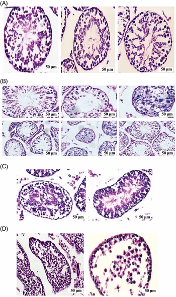
PAS staining of the transplanted samples from the 3D and 2D cultures showed that samples grafted with alginate hydrogel-based 3D cultured cells were more successful in the spermatogenesis process. In the cavities of the seminiferous tubules, the acrosome of sperm differentiated from these cells had distinct staining (Fig. 4A). However, in the samples transplanted with cells obtained from the 2D culture, the cavities of the seminiferous tubules lacked differentiated cells and, compared with the samples transplanted with cells obtained from the 3D culture, the process of cells towards differentiation was slower and the haploid spermatozoa were relatively less formed (Fig. 4B).
(A, B) PAS staining of testes tissue that received 3D or 2D cultured spermatogonial stem cell transplantation. (C, D) Testis sections stained with haematoxylin and eosin. (C) Samples that received 3D cultured spermatogonial stem cell transplantation. (D) Samples that received 2D cultured spermatogonial stem cell transplantation.

H&E staining of the transplanted tissue sections
H&E staining results indicated that the transplanted cells from the 3D and 2D cultures (Fig. 4C, D) showed the successful process of differentiation of SSCs from the 3D culture and formation of haploid cells in this group of cells compared with 2D culture transplanted cells. Fig. 4(C) shows staining of samples grafted with cells from the 3D culture. The porous compartment of the seminiferous tubules was clearly filled with floating spermatozoa cells. Fig. 4(D) shows the sample obtained from transplantation with 2D culture cells. The luminal part of the seminiferous tubules did not contain any differentiated cells.
Discussion
Long-term SCC culture under laboratory conditions can result in the loss of specific properties of these cells. A culture system that can establish conditions that more closely resemble the body can be effective in preserving, proliferating and differentiating SCCs. Various factors, including physical interaction of these cells with adjacent cells and unique molecules present in the environment, can lead to the maintenance and induction of SSCs. Therefore, neighbouring cells, growth factors and ECM compounds will modulate and regulate the differentiation, division, and apoptosis of stem cells at each time interval (Berná et al., Reference Berná, León-Quinto, Enseñat-Waser, Montanya, Martín and Soria2001). A significant body of research has been carried out on the culture, differentiation and cryopreservation of SSCs in 3D alginate hydrogel substrates (Chu et al., Reference Chu, Schmidt, Carnes, Zhang, Kong and Hofmann2009; Jalayeri et al., Reference Jalayeri, Pirnia, Najafabad, Varzi and Gholami2017; Pirnia et al., Reference Pirnia, Parivar, Hemadi, Yaghmaei and Gholami2017). The outcomes of these experiments demonstrated the role of the 3D substrate in maintenance of SSCs. In view of the lack of cytotoxicity and antioxidant properties of the alginate hydrogel, this 3D scaffold could be used for sperm stem cell culture (Jalayeri et al., Reference Jalayeri, Pirnia, Najafabad, Varzi and Gholami2017). Alginate can mimic ECM for SSCs and to promote the capacity for stemness during the cryopreservation process and to restart spermatogenesis after transplantation (Pirnia et al., Reference Pirnia, Parivar, Hemadi, Yaghmaei and Gholami2017).
In combination with somatic testicular cells, embedding SSCs in a 3D culture system can provide a structure that mimics the complex structure found in living testes. In an alginate hydrogel, somatic testicular cells and SSCs might create proper contact with the cells and stimulate the differentiation of germ cells in the culture system. In addition, alginate hydrogel and ECM similarities can provide sufficient access for cells to structural proteins and biological molecules. Alginate hydrogel in a 3D culture system can also retain growth factors that are secreted by Sertoli cells (Sargus-Patino, Reference Sargus-Patino2013). Tesarik and colleagues (Tesarik et al., Reference Tesarik, Mendoza, Anniballo and Greco2000) and Minaee Zanganeh and co-workers (Minaee Zanganeh et al., Reference Minaee Zanganeh, Rastegar, Habibi Roudkenar, Ragerdi Kashani, Amidi, Abolhasani and Barbarestani2013) have demonstrated that a SSC–Sertoli co-culture can advance spermatogenesis during short-term cultivation and provide conditions that might allow the successful in vitro differentiation of SSCs. Sertoli cells provide the critical factors required for the efficient differentiation of SSC cells into sperm cells, and their endocrine or paracrine factors play an important role in maintaining sperm cell viability and inducing animal meiosis in vitro (Sofikitis et al., Reference Sofikitis, Pappas, Kawatani, Baltogiannis, Loutradis, Kanakas, Giannakis, Dimitriadis, Tsoukanelis, Georgiou, Makrydimas, Mio, Tarlatzis, Melekos and Miyagawa2005). Our data showed that the presence of an alginate scaffold in a culture system compared with the SSCs co-cultured with Sertoli cells increased the mRNA levels of integrin alpha-6, integrin beta-1, Nanog, Plzf, Thy-1 and Oct4. Oct4 and Nanog are transcription factors essential for the preservation of embryonic stem cell pluripotency and self-renewal (Assadollahi et al., Reference Assadollahi, Fathi, Abdi, Khadem Erfan, Soleimani and Banafshi2019a, 2019b). The Oct-4 gene, a premeiotic specific marker, is expressed before spermatogenesis in male mice and is confined to spermatogonia type A (Khajavi et al., Reference Khajavi, Akbari, Abdolsamadi, Abolhassani, Dehpour, Koruji and Habibi Roudkenar2014). Studies have shown that expression of Nanog might be the cause of PTEN and TRP53 suppression (Kuijk et al., Reference Kuijk, van Mil, Brinkhof, Penning, Colenbrander and Roelen2010; Azizi et al., Reference Azizi, Skutella and Shahverdi2017). Thy-1, integrin alpha-6 and integrin beta-1 could be considered as markers of SSCs (Oatley et al., Reference Oatley, Kaucher, Avarbock and Brinster2010; Wang et al., Reference Wang, Zheng, Li, Shang, Li, Hu and Li2014). Plzf is active in the maintenance of stem cells. Loss of Plzf function changes the balance between self-renewal of spermatogonia stem cells and differentiation towards the cost of self-renewal, which leads to an increase in post-meiosis apoptotic cells (Buaas et al., Reference Buaas, Kirsh, Sharma, McLean, Morris, Griswold, de Rooij and Braun2004; Costoya et al., Reference Costoya, Hobbs, Barna, Cattoretti, Manova, Sukhwani, Orwig, Wolgemuth and Pandolfi2004). Plzf directly suppresses c-kit transcription (Filipponi et al., Reference Filipponi, Hobbs, Ottolenghi, Rossi, Jannini, Pandolfi and Dolci2007). c-kit is a marker for the lost pluripotency of SSCs and its expression continues until the initiation of meiosis (Zhang et al., Reference Zhang, Tang, Haines, Feng, Lai, Teng and Han2013). In this study, we analyzed c-kit gene expression and observed a decreased level of expression of this gene in cells cultured on alginate hydrogel with Sertoli cells under 3D culture compared with the 2D culture system in SSCs co-cultured with Sertoli cells. Increased expression of undifferentiated spermatogonial cell markers indicated that the culture conditions on the scaffold were appropriate for the expansion of SSCs; decreased expression of the c-kit gene also indicated the beneficial effects of scaffolding on self-renewal of SSCs (Talebi et al., Reference Talebi, Sadighi-Gilani, Koruji, Ai, Navid, Rezaie, Jabari, Ashouri-Movassagh, Khadivi, Salehi, Hoshino and Abbasi2019).
Apoptosis is a type of programmed cell death in which a highly complex and orderly series of biochemical (Goodman, Reference Goodman2007) apoptosis pathways are intrinsic (Bax, Bcl2, P53, caspase3 genes) and extrinsic (Fas, Fas-L genes) (Singh et al., Reference Singh, Letai and Sarosiek2019). The expression levels of apoptosis genes (Bax, P53, Bcl2, Fas) were assessed by qRT-PCR. We observed significantly lower expression levels of apoptosis genes in the group of alginate hydrogel encapsulated cells cultured with Sertoli cells compared with the group of SSCs co-cultured with Sertoli cells. Studies have shown that, by its anti-apoptotic properties, alginate prevents cell death by preventing oxidation (Król et al., Reference Król, Marycz, Kulig, Marędziak and Jarmoluk2017). Long-term encapsulation of cells in alginate microcapsules makes them non-permeable and decreases the release of insulin and cell death. Encapsulation of SSCs helps cells to detect the external environment and release small proteins such as growth factors and it does not allow large proteins such as antibodies to enter the cell. Alginate appears to provide higher biocompatibility through proper distribution of oxygen and other nutrients (Jalayeri et al., Reference Jalayeri, Pirnia, Najafabad, Varzi and Gholami2017).
Transplantation of SSCs into an azoospermic testis model has been performed on different animal models (Uchida and Dobrinski, Reference Uchida, Dobrinski, Majzoub and Agarwal2018). It was found that transplantation of seminiferous SSCs could contribute to the restoration of spermatogenesis (Brinster and Avarbock, Reference Brinster and Avarbock1994). The niche of the SSCs contains the essential structural and basement membrane of the seminiferous tubule, the Sertoli cell, peritubular myoid cells, and Leydig cells. In this research, the azoospermia model was established 2.5 months after busulfan injection (Gholami et al., Reference Gholami, Saki, Hemadi, Khodadadi and Mohammadi-Asl2014). Histological examination of the testes revealed that more tubules were devoid of germ cells and often contained a single, basal row of Sertoli cell nuclei. The Sertoli cells were naked and accessible in this state. Vacuum spaces between Sertoli cells act as hosts for transplanted SSCs and provide a suitable niche for homing cells (Rahmani et al., Reference Rahmani, Movahedin, Mazaheri and Soleimani2019). In the current research, the findings of histological studies and the trace of BrdU in the host testes 6 or 8 weeks after transplantation suggested that the transplanted SSCs localized in the base membrane of the seminiferous tubules and showed the progression of spermatogenesis to the cells in the lumen centre. The progression of spermatogenesis of transplanted SSCs was significantly higher in cells cultured on the alginate hydrogel 3D culture by Sertoli cells under 3D culture conditions than in mSSCs co-cultured with Sertoli cells. Taken together, it could be said that alginate hydrogel 3D culture by Sertoli cells under 3D culture conditions enhanced transplantation efficiency.
Our findings showed that spermatogonial cells seeded on a scaffold had a higher progression in spermatogenesis, which could be correlated with the existence of more functional Sertoli cells. The results of other studies indicated that the presence of somatic cells in the culture system increased the spermatogenesis differentiation stage (Stukenborg et al., Reference Stukenborg, Wistuba, Luetjens, Elhija, Huleihel, Lunenfeld, Gromoll, Nieschlag and Schlatt2008; Minaee Zanganeh et al., Reference Minaee Zanganeh, Rastegar, Habibi Roudkenar, Ragerdi Kashani, Amidi, Abolhasani and Barbarestani2013; Talebi et al., Reference Talebi, Sadighi-Gilani, Koruji, Ai, Navid, Rezaie, Jabari, Ashouri-Movassagh, Khadivi, Salehi, Hoshino and Abbasi2019). Finally, the potential for the alginate hydrogel with Sertoli cells to support the spread of SSCs and in vitro spermatogenesis has been demonstrated in this study. Therefore, it is possible to use the findings of this study for scientific and clinical applications.
In conclusion, we have shown that SSCs can survive and proliferate on alginate hydrogel 3D culture by Sertoli cells under 3D culture conditions up to 30 days. On this 3D alginate hydrogel with Sertoli cells under 3D culture conditions, self-renewal, colonization, and viability were improved compared with the 2D culture system in SSCs co-cultured with Sertoli cells as the feeder layer. Our study also demonstrated that transplantation of SSCs cultured in an alginate hydrogel 3D culture by Sertoli cells under 3D culture improved the quality of spermatogenesis in vivo. The process and findings of this study are new ideas for male infertility research, and they have the potential for use in the treatment of male infertility and reproduction of rare species. We hope to achieve better results in the future by applying modifications and improving culture conditions.
Data availability
Data sharing not applicable to this article as no datasets were generated or analyzed during the current study.
Acknowledgements
This study, as a MSc thesis, was funded by grants provided from Kermanshah University of Medical Sciences (IR.KUMS.REC.1397.811). We express our appreciation to all members of the Medical Biology Research Center for their helpful consultation and deliberation during this work.
Conflicts of interest
The authors declare they have no competing financial interests.
Ethics approval and consent to participate
All stages of this experiment were in accordance with the ethical standards of the Ethics Committee of Kermanshah University of Medical Sciences, Kermanshah, Iran (Approval ID: IR.KUMS.REC. 1397.811).
Funding
Kermanshah University of Medical Sciences (IR.KUMS.REC.1397.811).